Samples
Fanc-A lymphoblast cell lines and FANC-A primary fibroblast cell lines that carried out different mutations of the FANC-A gene were obtained from the ‘‘Cell Line and DNA Biobank from Patients affected by Genetic Diseases’’ (G. Gaslini Institute) - Telethon Genetic Biobank Network (Project No. GTB07001). As controls, isogenic FA-corr cell lines generated by the same FANC-A lymphoblast and fibroblast cell lines corrected with S11FAIN retrovirus (Lympho FA-corr and Fibro FA-corr) were employed [49]. None of the samples used in this study were affected by HNSCC or pre-malignant changes.
The study was conducted following the Declaration of Helsinki and approved by the regional ethics committee, protocol JS002, register number 037 − 21/01/2019. All the subjects or their legal guardians gave written informed consent to the investigation.
Cell culture and treatments
For lymphoblast cell lines, RPMI-1640 medium (GIBCO, Billing, MT, USA) containing 10% fetal bovine serum (FBS, Euroclone, Milano, Italy), 100 U/mL penicillin, and 100 µg/mL streptomycin (Euroclone, Milano, Italy) was used, and the cells were grown at 37 °C with a 5% CO2 [49]. Primary fibroblasts were cultured as a monolayer in DMEM high glucose with glutamax® (GIBCO, Billing, MT, USA), containing 10% FBS (Euroclone, Milano, Italy), 100 U/mL penicillin, and 100 µg/mL streptomycin (Euroclone, Milano, Italy) at 37 °C with a 5% CO2 [49].
FA lymphoblast and fibroblast cells transfection with miR-29a-3p
FA lymphoblast cells were transfected with miR-29a-3p mimic (Thermo Fisher Scientific, Waltham, MA, USA), using the lipofectamine RNAiMAX Transfection Reagent (Invitrogen, Waltham, MA, USA) according to the manufacturer’s instruction. In detail, for subsequent RNA extraction, 500.000 cells grown in 6-well plates were transfected with 25 pmol of miR-29a-3p mimic or negative control mimic using 7.5 µl lipofectamine. After 48 h, cells were harvested, washed with PBS, lysed, and RNA was extracted. For the subsequent biochemical analysis, 7.5 × 10⁶ cells grown in 75 cm2 flasks were transfected with 187.5 pmol of miR-29a-3p mimic or negative control mimic using 56.25 µl lipofectamine. Cells were processed 48 h post-transfection.
FA fibroblast cells were transfected with miR-29a-3p mimic (Thermo Fisher Scientific, Waltham, MA, USA) using the same transfection reagent (Invitrogen, Waltham, MA, USA). In detail, for the subsequent RNA extraction, 75,000 cells grown in 6-well plates were transfected with 25 pmol of miR-29a-3p mimic or negative control mimic using 7.5 µl lipofectamine. After 48 h, cells were harvested, washed with PBS, lysed, and RNA was extracted. For subsequent biochemical analysis, 500,000 cells grown in 175 cm2 flasks were transfected with 437.5 pmol of miR-29a-3p mimic or negative control mimic using 131.25 µl lipofectamine. Cells were processed 48 h post-transfection.
FA lymphoblast cell treatments
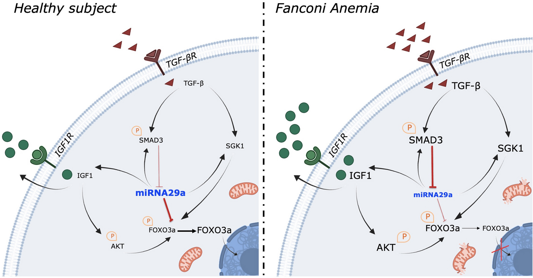
To inhibit the TGF-β pathway, lymphoblasts were treated with Luspatercept, an inhibitor acting on the SMAD2/3 signaling [50]. In detail, 500,000 cells were plated in a 2 ml culture medium in the presence of 10 µg/ml Luspatercept for 48 h.
To inhibit IGF1 signaling, 500,000 lymphoblasts were treated with 400 pM Klotho [51] for 48 h.
In silico selection of pathway-related miR-29a-3p target genes
To identify putative miR-29a-3p target genes potentially involved in Fanconi cell metabolism impairment, we utilized the miRPathDB v2.0 database (https://mpd.bioinf.uni-sb.de). A curated list of miR-29a-3p-regulated genes associated with DNA damage response, oxidative stress, mitochondrial metabolism, lipid metabolism, and apoptosis was generated by evaluating the strength of predicted miRNA-target gene interactions using TargetScan (https://mpd.bioinf.uni-sb.de). Further refinement was conducted by assessing the role and the subcellular localization of each gene with the help of the NCBI Gene database (https://www.ncbi.nlm.nih.gov/gene) and GeneCards (https://www.genecards.org).
RNA isolation to evaluate the expression of miR-29a-3p and FOXO3, SGK1, and IGF1 genes
RNA, including the small RNA fraction, was extracted using the RNeasy Plus Mini Kit (Qiagen, Hilden, Germany) according to the manufacturer’s protocol. The expression of miR-29a-3p was assessed using a specific TaqMan MicroRNA Assay (Applied Biosystems, Waltham, MA, USA). Briefly, 10 ng of RNA was reverse-transcribed using the TaqMan MicroRNA Reverse Transcription Kit and the specific RT primer. Real-time PCR was performed in triplicate with specific primers. miRNA expression levels were normalized to RNU44 expression.
To evaluate the expression of the FOXO3, SGK1, and IGF1 genes, 100 ng of RNA were reverse transcribed using the SuperScript VILO IV cDNA Synthesis Kit (Invitrogen, Waltham, MA, USA). The resulting cDNA was used for real-time PCR with primers provided in the specific TaqMan Gene Expression Assays (Applied Biosystems, Waltham, MA, USA). Gene expression levels were normalized to GAPDH expression. All experiments were performed in triplicate. Ct standard deviation values of used reference genes among the performed treatments are reported in Supplementary Table 1.
Catalase activity evaluation
Catalase (CAT) activity was assayed as a marker of cellular antioxidant defenses. For each spectrophotometric assay, 50 µg of total proteins were used, following the H2O2 decomposition at 240 nm. The assay mix contained 100 mM Tris-HCl (pH 7.4, Sigma-Aldrich, St. Louis, MO, USA) and 5 mM H2O2 (Sigma-Aldrich, St. Louis, MO, USA) [21].
Oxidative stress markers evaluation
The thiobarbituric acid reactive substances (TBARS) assay was employed to quantify malondialdehyde (MDA), an indicator of lipid peroxidation. The TBARS reagent was prepared using 0.25 M HCl, 0.25 mM trichloroacetic acid, and 26 mM thiobarbituric acid (all sourced from Merck, Darmstadt, Germany). A total of 50 µg of protein, dissolved in 300 µl of Milli-Q water, was mixed with 600 µl of the TBARS solution. The reaction mixture was incubated at 95 °C for 1 h and the absorbance was measured spectrophotometrically at 532 nm. Standard solutions of MDA, with concentrations ranging from 1 to 20 µM, were used to generate a calibration curve [21].
To measure 8-hydroxy-2-deoxyguanosine (8-OHdG), a biomarker of oxidative DNA damage, an ELISA kit (#ab201734, Abcam, Cambridge, UK) was utilized according to the manufacturer’s instructions.
Evaluation of aerobic metabolism function and efficiency
The OxPhos function was assessed by measuring the oxygen consumption rate (OCR) and FoF1-ATP synthase activity.
OCR was determined using an amperometric electrode (Unisense Microrespiration, Aarhus, Denmark) in a closed chamber. For each test, 105 cells were permeabilized for 1 min with 0.03 mg/ml digitonin and then used in the assay. To activate the respiratory pathway led by Complex I or Complex II, 10 mM pyruvate with 5 mM malate (Merck, Darmstadt, Germany) or 20 mM succinate Merck, Darmstadt, Germany) were added, respectively [21].
FoF1-ATP synthase activity was measured in 105 cells suspended in PBS plus 0.6 mM ouabain Merck, Darmstadt, Germany) and 0.25 mM di (adenosine)−5-penta-phosphate (an adenylate kinase inhibitor, Merck, Darmstadt, Germany). After 10 min of incubation, 10 mM pyruvate with 5 mM malate (Merck, Darmstadt, Germany) or 20 mM succinate (Merck, Darmstadt, Germany) were added to stimulate pathways mediated by Complex I or II, respectively. ATP production was quantified using a luminometer (GloMax® 20/20 Luminometer, Promega Italia, Milan, Italy), employing the luciferin/luciferase chemiluminescent method (ATP bioluminescence assay kit CLS II, #11699695001 Roche, Basel, Switzerland). Measurements were taken at 30-second intervals over 2 min [21].
The OxPhos efficiency was determined by calculating the P/O ratio, which represents the amount of ATP synthesized aerobically per oxygen molecule consumed. Mitochondria with optimal efficiency exhibit P/O ratios around 2.5 for pyruvate and malate or 1.5 for succinate. Ratios below these thresholds suggest incomplete oxygen utilization for ATP production, potentially reflecting increased ROS generation [21].
Assessment of ATP and AMP intracellular concentration and the consequent cellular energy status
ATP and AMP concentrations were assayed in 50 µg of total protein. ATP content spectrophotometric analysis was performed following the NADPreduction at 340 nm. The assay solution contained 100 mMTris-HCl (pH 8.0; Merck, Darmstadt, Germany), 0.2 mM NADP (Merck, Darmstadt, Germany), 5 mM MgCl2 (Merck, Darmstadt, Germany), 50 mM glucose (Merck, Darmstadt, Germany), and 3 µg of pure hexokinase and glucose-6-phosphate dehydrogenase (Merck, Darmstadt, Germany) [20].
AMP was measured spectrophotometrically following the NADH oxidation at 340 nm. The reaction medium was composed of 100 mM Tris-HCl (pH 8.0; Merck, Darmstadt, Germany), 5 mM MgCl2 (Merck, Darmstadt, Germany), 0.2 mM ATP (Merck, Darmstadt, Germany), 10 mMphosphoenolpyruvate (Merck, Darmstadt, Germany), 0.15 mM NADH (Merck, Darmstadt, Germany), 10 IU adenylate kinase, 25 IU pyruvate kinase, and 15 IU lactate dehydrogenase (Merck, Darmstadt, Germany) [20].
The cellular energy status was calculated as the ratio between intracellular concentration of ATP and AMP (ATP/AMP ratio) [20].
Evaluation of electron transfer between complex I and complex III
To evaluate the electron transfer between Complex I and Complex III, a spectrophotometric assay was performed following the reduction of oxidized cytochrome c (cytc) at 550 nm in the presence of NADH. The assay medium contained 50 mMTris-HCl (pH 7.4; Merck, Darmstadt, Germany), 5 mMKCl (Merck, Darmstadt, Germany), 2 mM MgCl2 (Merck, Darmstadt, Germany), 0.5 M NaCl (Merck, Darmstadt, Germany), 0.03% oxidized cytc (Merck, Darmstadt, Germany), and 0.6 mM NADH (Merck, Darmstadt, Germany) [20].
Western blot analysis
The expression of several proteins belonging to the FOXO3a, SGK1, IGF1, and SMAD3 (TGF-β effector) signalling pathways was assessed by western blot (WB) analysis performed on cell homogenates of FA lymphoblasts and their controls. In detail, denaturing electrophoresis (SDS-PAGE) was performed on 30 µg of proteins employing a 4–20% gradient gel (BioRad, Hercules, CA, USA). The following primary antibodies were used: phospho-H2AX (#05–636, Merck, Darmstadt, Germany). phospho-FOXO3a (#9466,Cell Signaling Technology, Beverly, MA, USA), FOXO3a (#2497S, Cell Signaling Technology, Beverly, MA, USA), GAPDH (#2118, Cell Signaling Technology, Beverly, MA, USA), Histone H3 (#4499, Cell Signaling Technology, Beverly, MA, USA), phospho-AKT (#4060S, Cell Signaling Technology, Beverly, MA, USA), AKT (#4691S, Cell Signaling Technology, Beverly, MA, USA), phospho-SGK1 (#5599, Cell Signaling Technology, Beverly, MA, USA), SGK1 (#711183, Thermo Fisher Scientific, Waltham, MA, USA), phospho-SMAD3 (#9520S, Cell Signaling Technology, Beverly, MA, USA), SMAD3 (#9523S, Cell Signaling Technology, Beverly, MA, USA), and β-Actin (#MA1-140, ThermoFisher Scientific, Waltham. MA, USA). All primary antibodies were diluted following the manufacturer’s instructions in PBS plus 0.15% Tween 20 (PBSt; Roche, Basel, Switzerland). Specific secondary antibodies were employed (Merck, Darmstadt, Germany, all diluted 1:10,000 in PBSt). Bands were detected in the presence of an enhanced chemiluminescence substrate (ECL, BioRad, Hercules, CA, USA), by a chemiluminescence system (Alliance 6.7 WL 20 M, UVITEC, Cambridge, UK). Band intensity was evaluated by UV1D software (UVITEC, Cambridge, UK). All bands of interest were normalized versus the actin signal detected on the same membrane.
Since FOXO3a function is closely linked to its translocation from the cytoplasm to the nucleus [52], FOXO3a expression has also beeen evalauted in nuclear fraction obtained from FA and FAcorr lymphoblatsts. To obtain nuclei-enriched fractions (N), cells were homogenated in a 0.25 M sucrose solution. This homogenate (H) was centrifuged at 800 g for 10 min. The supernatant was collected and used as a cytoplasmic fraction (C), and the pellet was resuspended in 0.25 M sucrose solution and centrifuged again at 800 g for 10 min to obtain N.
In addition, the expression of proteins involved in the TGF-β signalling modulation has been evaluated in samples treated with Luspatercept, a TGF-β pathway inhibitor acting on SMAD2/3 signaling [50].
Statistical analysis
Data about gene expression, western blot analysis, ATP synthesis, OCR, mitochondrial efficiency were obtained from three independent replicate experiments; the other biochemical data were obtained from six independent replicate experiments. Data were shown as means ± standard deviation (SD). Student’s t test or one-way ANOVA followed by Tukey’s multiple comparison test by Prism 9 Software (GraphPad Software Inc., Boston, MA, USA) was used to compare the differences between two or more groups. p < 0.05 was considered statistically significant. ns represent statistically non-significant, * p < 0.05, ** p < 0.01, *** p < 0.001, and **** p < 0.0001.


Comments (0)