
Remember me
The back of the original canvas has been covered in the beginning of the twentieth century by a relining canvas that was removed during conservation operations realized at the same time of this study. Also in the case of the relining canvas, it was observed the presence of several red–purple-white spots in correspondence of the same area of original canvas (Supplemental Fig. S1).
To carry out the recognition of textile fibers, two samples of threads were taken from the original canvas (TO1, TO2) and two from the relining canvas (TR1, TR2), to acquiring information on the nature of the yarn, both for the weft and warp threads. Before being positioned on the slide, the individual samples were subjected to two washes: the first carried out with a mixture of water and ethanol (ratio 1:1) and the second with a 1 M HCl solution to remove foreign substances, as well as to allow the fibers to open more easily, facilitating observation under the microscope. The samples were then observed at magnifications of 100 × and 400 ×.
Ultraviolet fluorescence photographyThe UV fluorescence analysis allows to observe the original and non-original added layers directly on the surface of the painting, starting from the most external ones, such as varnishes or protective films, up to the recognition of some pigments and binders, and the distinction of adhesives and products used in the conservation field, based on the color of the response. Furthermore, since the fluorescence of many of these materials tends to increase in intensity with aging, it is possible to carry out a preliminary study on the conservation history of the painting.
In particular, ultraviolet fluorescence photography aims at detecting the visible response generated by irradiation in the UV range 300–400 nm. The fluorescence response was recorded using a camera equipped with special filters (such as the yellow filter—UV cat), allowing the passage of only the visible radiation emitted by the object due to fluorescence, while preventing the reflected UV from being recorded.
Collection of biological samplesSampling was performed on the back of the original canvas in six distinct points (Fig. 2). Point 1 coincided with an apparently not altered area, points from 2 to 5 corresponded to lower area of the canvas that showed a greater deterioration, and finally, point 6 was located below the canvas support structure. The biological material was collected through contact plate method, using tryptic soy agar (TSA) (g L−1: casein peptone 17.0, soya peptone 3.0, NaCl 5.0, K2HPO4 2.5, glucose 2.5, agar 15.0) and Sabouraud dextrose chloramphenicol agar (SAB) (g L−1 of medium casein peptone 5.0, meat peptone 5.0, glucose 40.0, chloramphenicol 0.5, agar 15.0). The TSA medium was used for both bacteria and fungi isolation, while the SAB medium was specifical for fungi. Afterwards, the culture plates were incubated at 28 °C for 5 days and different colonies, based on their morphological differences (i.e., shape, size, color, etc.), were picked up and streaked on the same media until they were purified to homogeneity, isolating, in this way, 17 bacterial and six fungal strains, respectively. The isolated microorganisms were then characterized morphologically.
Fig. 2
Sampling points on the back of the canvas
DNA extraction, amplification, and sequencing of 16S rDNA and 18S rDNAThe DNA extraction of isolated bacteria and fungi was performed by means of REDExtract-N-Amp™ Tissue PCR kit (Merck Life Science Srl, Milan, Italy) following the supplier instructions.
The region V4-V5 of 16S rDNA gene was amplified with bacterial universal primers com1 (5′ CAGCAGCCGCGGTAATAC 3′) and com2 (5′ CCGTCAATTCCTTTGAGTTT 3′). While the region ITS2 of fungal 18S rDNA gene was amplified with primers ITS3 (5′ GCATCGATGAAGAACGCAGC 3′) and ITS4 (5′ TCCTCCGCTTATTGATATGC 3′). PCR volume (20.0 μL) contained 2.0 μL of DNA solution, 2.0 μL of 10X Red Extract-N-AmpPCR Ready Mix of the Sigma kit (Sigma, Milano-Italy), 1.0 uM of each forward and reverse primer. PCR conditions were as follows: initial denaturation for 3 min at 94 °C, 35 cycles of denaturation at 94 °C for 60 s, annealing at 55 °C for 60 s, and elongation 120 s at 72 °C and, at end of the cycles, an additional final elongation step of 7 min at 72 °C. Finally, the amplicons sequencing was performed by Eurofins Genomics (Ebersberg, Germany), and they were identified by a similarity search using the BLAST function of GenBank at the National Center for Biotechnology Information (NCBI) electronic site (NCBI. Available online: http://www.ncbi.nlm.nih.gov/, 07 March 2025). Moreover, the sequences were deposited in GenBank (05 April 2025) under accession numbers reported in Table 1.
Table 1 Microbial identificationThe isolated strains reported in Table 1 are preserved in our collection (stored at − 80 °C at the Department of Chemistry and Biology of the University of Salerno) and will be available for everyone who wants them, contacting the Dr. Gianmaria Oliva.
Measurement of painting pigments deteriorationTwo pigments were chosen for biodeterioration tests: yellow ochre 0324 (FeO(OH), CaCO3, CaSO4, CTS Conservation, Italy) and ivory black 0597 (animal bone ash and PO42−, CTS Conservation, Italy). The bacterial growth, in the presence of each pigment and their capability to induce pigment deterioration, was tested on Bushnell-Hass (BH) medium (g per 1 L of medium: MgSO4⋅7H2O 0.2, NH4NO3 1.0, CaCl2 0.02, glucose 1.0, K2HPO4 1.5, KH2PO4 1.0, pigment 1.0). Contrarily, fungal activity was evaluated on modified metal toxicity (mMT) medium (g L−1 of medium: MgSO4⋅7H2O 1.0, NH4Cl 1.0, CaCl2 0.06, Na2SO4 2.13, glucose 10.0, yeast extract 0.05, tryptone 0.5, pigment 1.0). A cell suspension of 1.0 optical density at 600 nm (OD600), for each microbial sample, was prepared in saline solution (NaCl 0.9% w/v) from fresh microbial culture. Afterwards, 3 mL of liquid medium (BH or mMT) were added in 15-ml Falcon tubes, and 30 μL of each cell suspension were inoculated into respective tubes (final OD600 = 0.01). Each sample was prepared in triplicate and was incubated for 5 days at 28 °C in orbital shaker at 200 RPM (New Brunswick Innova 43, Edison, NJ; USA).
In bacterial culture, it was evaluated; the pigment color change. In particular, the tubes were centrifugated at 6000 RPM for 10 min; the pH of supernatant was estimated with a pHmeter (S-610L, Peak Instrument, China) and then it was discarded; therefore, the precipitate was recovered and diffused homogeneously on a slide, and it was air dried. At the end, color changes were assessed with a colorimeter (Datacolor Italia s.r.l, Monza, Italy) measuring the chromaticity coordinates L*, a*, and b* and estimating the ΔE value (absolute chromatic variation) (Mao et al. 2024) defined as follows:
$$\Delta E= \sqrt^+ ^^}$$
Instead, the fungi incorporated the pigment into the mycelia; therefore, it was not possible to evaluate the colorimetric alteration as in the case of bacteria. About that, it was estimated the pigment amount incorporated in fungal biomass, measuring the difference between the initial pigment amount and that still present in solution after the fungal growth. Therefore, 1 mL of culture solution was transferred in a new tube (2.0 mL) avoiding the fungal aggregates. The tubes were centrifugated at 12,000 rpm for 5 min, the supernatant was discarded, and the samples were oven dried at 70 °C for 48 h. Finally, the dry weight of pigment was determined.
Preparation of plant extractsInitially, three plant species known for their proven antimicrobial properties were selected: rosemary (Salvia rosmarinus Spen 1835); garlic (Allium sativum L.); and lemon thyme [Thymus citriodorus (Pers.) Schreb.].
The same hydroalcoholic extracts were prepared for either rosemary or lemon thyme: 10 g of fresh leaves were homogenized in 20 mL of solution (1:1, water:ethanol 100%) in a mortar at room temperature. Instead, the garlic extract was prepared by homogenizing 10 g of fresh bulbs in 20 mL of water in a mortar. All extracts were filtered through a 0.45-μm porous membrane and then stored at 4 °C in the dark.
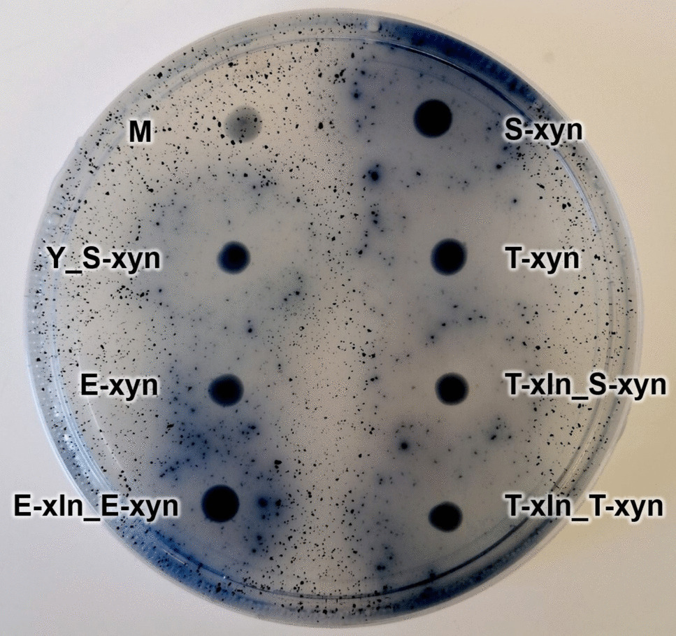
Evaluation of antimicrobial activityThe antimicrobial activity of the three natural extracts was evaluated on all isolated bacteria and fungi from the canvas. Hence, 50 μL of fresh cell suspension (0.5 OD600) of each bacterial culture, suspended in saline solution (0.9% NaCl), were diffused on Luria–Bertani (LB) agar medium. Then, 3.0 μL of each natural extract were spotted on the agar plate. A drop of extraction solvent was used as control. Finally, the plates were incubated at 28 °C for 48 h. The formation of an inhibition halo proved the effective antimicrobial of the assayed extract.
Evaluation of antimicrobial activity on canvas and different methods of applicationThe antimicrobial activity of rosemary extract was evaluated on five strains previously isolated from the original canvas and inoculated on virgin linen canvas (canvas mock-up dimension: 12 × 12 cm) (Supplemental Fig. S2A), as preliminary evaluation of the extract’s ability to inhibit the growth of selected microorganisms when applied to a textile support. In this regard, four strains sensitive to rosemary extract (1OR, 4Ea, 5Eb, 6E) and one resistant strain (4C) were selected.
A canvas sample was placed in Petri dishes (∅140 mm) and superficially sterilized with UV radiation for 15 min. Then, for each microorganism, 5.0 μL of fresh cell suspension were spotted on canvas. Suspensions were prepared from overnight grown cultures by diffusing the bacteria in saline solution (0.9% NaCl) at density of 0.5 OD600. After 1 h, microbial contamination was evaluated by means of contact plate method, using TSA as a growth medium, then plates were incubated at 28 °C for 5 and 10 days. Afterwards, the canvas was treated by means of 2.0-mL rosemary extract spraying it directly on the canvas surface (0.03 mL cm−2), repeating the application three times, 1 h apart. The canvas was air dried, and microbial survival was assessed using both TSA and SAB agar plate, as described above.
Further analyses were conducted on the old lining canvas of the painting (canvas mock-up dimension: 12 × 12 cm), removed during the conservation operations (Supplemental Fig. S2B), and the efficacy of rosemary extract against the microorganisms already present on it was evaluated. The lining canvas sample was chosen due to its close resemblance to the structure and characteristics of the original ancient canvas.
Also in this case, microbial contamination was evaluated pre- and post-treatment with rosemary extract, with contact-plate method, as indicated above.
Other two strategies of antimicrobial application were also evaluated. The first was the extract application through Evolon® tissue (Cts Conservation, Vicenza, Italy). Evolon® is an innovative technical tissue obtained from a mixture of polyester and polyamide microfilaments, which give at the material unique properties such as resistance, breathability, and lightness (Vergeer et al. 2019). Thanks to its structure without warp and weft, it is particularly suitable for several applications including material disinfection. Furthermore, it is a sustainable material, as it is recyclable and free of chemical solvents. The Evolon® tissue was immersed for 2 min in the hydro alcoholic rosemary solution, then wrung out and placed for 1 h on the canvas under a weight of about 2 kg so to have an its uniform distribution on the canvas. As previously described, biological material (microorganisms) was sampled before and after treatment in the same way as above described.
The second one was the nebulization on the pretreated canvas with cyclomethicone D5. This compound is a hydrophobic solvent used in conservation to reduce temporally the sensitivity of porous material to water. A control sampling was performed before treatment. Subsequently, 2 mL cyclomethicone D5 was sprayed on the canvas (0.03 mL cm−2) and air dried for 2 h. After that, rosemary extract was nebulized on canvas, and the application was repeated three times, 1 h apart as above. The treated canvas was air dried, and then the microbial community was collected through contact plate method, using TSA agarized media.
Finally, the possible impact of rosemary extract, on the optical properties of the cellulose support, was also evaluated through colorimetric analysis before and after the treatment. For this purpose, the extract was sprayed on a piece of virgin canvas (as above), and it was measured the ΔE value (absolute chromatic variation).
Comments (0)