2.1 Clinical tissue sample collection
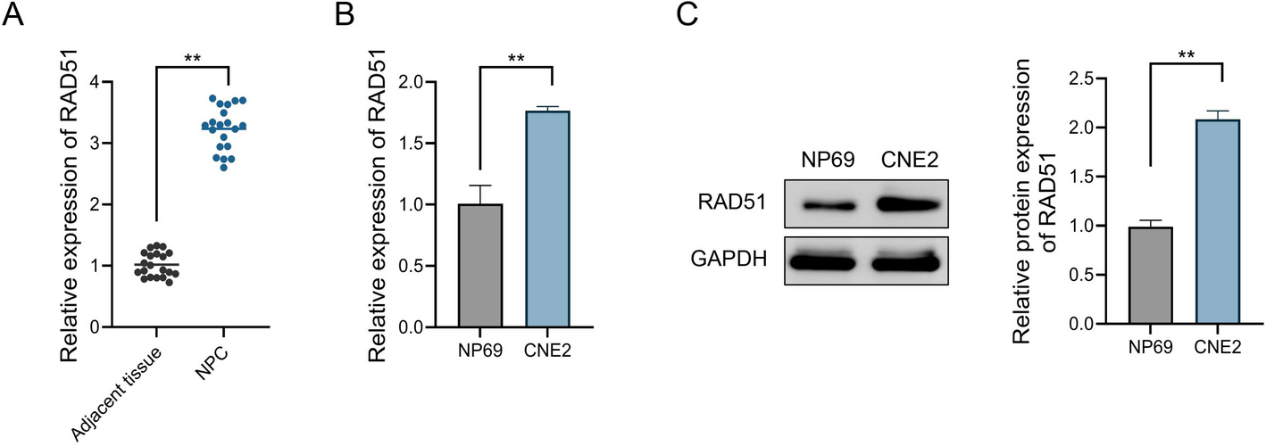
The clinical component of this study involved an observational, tissue-based comparison of RAD51 expression between nasopharyngeal carcinoma (NPC) tumors and adjacent normal tissues. A total of 20 patients with histologically confirmed NPC were enrolled at The First Affiliated Hospital of Jinan University between February 2024 and June 2024 (Table S1). The sample size was determined based on the number of eligible patients during the study period and practical constraints related to tissue acquisition. All patients provided written informed consent prior to participation. Tumor and paired adjacent normal tissues were collected during diagnostic nasopharyngoscopic biopsy and stored at − 80 °C for further analysis. The sample size was determined based on the number of eligible patients during the study period and practical constraints related to tissue acquisition.
Inclusion criteria were: (1) age between 18 and 75 years; (2) newly diagnosed, untreated primary NPC; and (3) sufficient tissue sample obtained via nasopharyngoscopic biopsy. Exclusion criteria included: (1) prior radiotherapy, chemotherapy, or surgery; (2) concurrent autoimmune or chronic inflammatory diseases; and (3) history of other malignancies. During the biopsy procedure, both primary tumor tissues and adjacent histologically normal nasopharyngeal epithelial tissues were collected from the same patients. Based on pathological examination, samples were classified into two groups: the NPC tumor tissue group (NPC group) and the adjacent normal tissue group (Adjacent group). The use of adjacent normal tissues as internal controls was justified to minimize inter-individual variability, as these tissues were anatomically and genetically matched to the corresponding tumor samples, yet devoid of malignant features. All collected tissues were immediately snap-frozen in liquid nitrogen and subsequently stored at − 80 °C until further molecular analysis. The study was approved by the Ethics Committee of The First Affiliated Hospital of Jinan University (Approval No. KY-20240254) and conducted in accordance with the Declaration of Helsinki (as revised in 2013). Prior to enrollment, all patients were fully informed of the study objectives, procedures, risks, and benefits, and provided written informed consent. All patient data were anonymized to ensure confidentiality. Patients were screened consecutively according to the inclusion and exclusion criteria. No patients were excluded due to tissue insufficiency or protocol violations. All 20 patients who met the eligibility criteria were enrolled and included in the final analysis. No patients were excluded, and no missing data occurred.
2.2 Cell culture, reagents, treatment, and grouping
Human normal nasopharyngeal epithelial cell line NP69 and the radioresistant human NPC cell line CNE2 were utilized in this study. These cells were provided by the American Type Culture Collection (Manassas, VA, USA). The NP69 and CNE2 cells were cultured in Roswell Park Memorial Institute-1640 medium (Absin, China) containing 10% fetal bovine serum (Servicebio, China) and 1% penicillin-streptomycin solution (Servicebio, China) at 37 °C with 5% CO₂.
The buffer containing recombinant human RAD51 protein (rhRAD51) was purchased from Abcam (UK). Sterile phosphate-buffered saline (PBS) was added to prepare solutions of varying concentrations for cell treatment [20].
The solid RAD51 inhibitor B02 and Caspase-8-specific inhibitor Z-IETD-FMK were both obtained from MedChemExpress (USA). According to the manufacturer’s instructions, these compounds were dissolved in 0.5% dimethyl sulfoxide solution to create stock solutions, which were then diluted with sterile phosphate-buffered saline to prepare solutions of different concentrations for cell treatment [21].
As previous described, the prepared rhRAD51, B02, and Z-IETD-FMK solutions were added to the culture medium to treat CNE2 cells for 24 h [22, 23]. After treatment, cells were collected for subsequent experiments. Briefly, in this study, CNE2 cells were divided into the following groups:
(1) Control group: CNE2 cells received routine culture for 24 h without other treatment; (2) 50 ng/mL group: CNE2 cells were treated with 50 ng/mL rhRAD51 solution for 24 h; (3) 100 ng/mL group: CNE2 cells were subjected to 100 ng/mL rhRAD51 solution for 24 h; (4) 200 ng/mL group: CNE2 cells received 200 ng/mL rhRAD51 solution treatment for 24 h; (5) 5 µM group: CNE2 cells were treated with 5 µM B02 solution for 24 h; (6) 10 µM group: CNE2 cells were subjected to 10 µM B02 solution for 24 h; (7) 20 µM group: CNE2 cells received 20 µM B02 solution treatment for 24 h; (8) Z-IETD-FMK group: CNE2 cells were exposed to 40 µM Z-IETD-FMK solution for 24 h; (9) rhRAD51 group: CNE2 cells were treated with 200 ng/mL rhRAD51 solution for 24 h; (10) rhRAD51 + Z-IETD-FMK group: CNE2 cells were subjected to 200 ng/mL rhRAD51 solution and 40 µM Z-IETD-FMK solution for 24 h; (11) B02 group: CNE2 cells underwent 20 µM B02 solution for 24 h; (12) B02 + Z-IETD-FMK group: CNE2 cells were exposed to 20 µM B02 solution and 40 µM Z-IETD-FMK solution for 24 h.
2.3 Cell counting Kit-8 (CCK-8)
The treated CNE2 cells were seeded into 96-well plates at a density of 5 × 10³ cells/well. After 24 h of incubation, the cells were treated with drug solutions as described above. Next, 10 µL of CCK-8 solution (DOJINDO, Japan) was added to each well, followed by incubation for 2 h at 37 °C with 5% CO₂. Following incubation, the optical density of each well was measured at a wavelength of 450 nm using a multimode microplate reader (Thermo Fisher Scientific, USA) to assess cell viability. In addition, a blank control (culture medium without cells) was included to account for background interference.
2.4 Real-time quantitative polymerase chain reaction (RT-qPCR)
Total RNA was extracted by uniformly mixing Trizol reagent (Sigma-Aldrich, USA) with clinical tissue samples or cell lines. Subsequently, the total RNA was reverse-transcribed into cDNA using the PrimeScript RT Reagent Kit (Takara Biotechnology, China), and RT-qPCR analysis was conducted using SYBR Premix Ex Taq (Takara Biotechnology, China) on a QuantStudio 6 Flex system (Applied Biosystems, USA) to assess the RAD51 expression levels. The PCR cycling conditions were as follows: initial denaturation at 95℃ for 30 s, followed by 40 cycles of denaturation at 95 °C for 30 s, annealing at 60℃ for 30 s, and extension at 72℃ for 30 s. GAPDH was used as an internal reference to normalize the RAD51 expression levels, and the results were quantified using the 2−△△Ct method. The primers used for RT-qPCR are listed in Table 1.
Table 1 RT-qPCR primer sequencesRT-qPCR, real-time quantitative polymerase chain reaction; GAPDH, glyceraldehyde-3-phosphate dehydrogenase.
2.5 Lactate dehydrogenase (LDH) assay
Firstly, 120 µL of the supernatant was collected from each group of CNE2 cell culture medium. Then, 60 µL of working solution from the LDH cytotoxicity assay kit (Beyotime Biotechnology, China) was added for thorough mixing, followed by 30 min of incubation in the dark at room temperature. The optical density at 490 nm was measured using a multimode microplate reader (Thermo Fisher Scientific, USA) to determine the LDH levels released by the cells.
2.6 Western blot
Total protein was extracted from treated CNE2 cells using radio-immunoprecipitation assay buffer containing protease and phosphatase inhibitors (Thermo Fisher Scientific, USA). The total protein concentration was measured using the bicinchoninic acid protein assay kit (Beyotime Biotechnology, China). Proteins were separated using 10–12% sodium dodecyl sulfate-polyacrylamide gel electrophoresis and transferred to polyvinylidene fluoride membranes (Servicebio, China). Then, the membranes were blocked with 5% skimmed milk at room temperature for 1 h. Subsequently, the membranes were incubated overnight at 4 °C with primary antibodies. The primary antibodies used were displayed as follows: Caspase-8 (1:1000, #4790, Cell Signaling Technology, USA), Cleaved-Caspase-8 (1:1000, #9496, Cell Signaling Technology, USA), Caspase-1 (1:1000, #2225, Cell Signaling Technology, USA), Cleaved-Caspase-1 (1:1000, #4199, Cell Signaling Technology, USA), Caspase-3 (1:1000, #9662, Cell Signaling Technology, USA), Cleaved-Caspase-3 (1:1000, #9661, Cell Signaling Technology, USA), gasdermin D (GSDMD, 1:1000, #39754, Cell Signaling Technology, USA), GSDMD-N (1:1000, #36425, Cell Signaling Technology, USA), interleukin-(IL) 18 (1:1000, #67775, Cell Signaling Technology, USA), IL-1β (1:1000, #12703, Cell Signaling Technology, USA), and GAPDH (1:1000, #2118, Cell Signaling Technology, USA). The following day, the membranes were washed three times with Tris buffered saline with Tween buffer. Afterward, the membranes were incubated with the Anti-rabbit IgG, Horseradish Peroxidase-linked Antibody (1:2000, #7074, Cell Signaling Technology, USA) at room temperature for 1 h. Ultimately, immunoreactive bands were detected using the Odyssey Infrared Imaging System (LiCor, USA), and ImageJ software (NIH, USA) was used for semi-quantitative analysis of each band. GAPDH was served as an internal reference to normalize the expression levels of target proteins.
2.7 Statistical analysis
Statistical analysis was performed using SPSS software (version 22.0). Normality of the data was assessed using the Shapiro-Wilk test, which confirmed that the data followed a normal distribution. Data are presented as means ± standard deviation (SD). The homogeneity of variances was evaluated using Levene’s test, and the assumption of equal variances was met. For comparisons between two groups, a two-tailed Student’s t-test was applied. For comparisons among multiple groups, one-way analysis of variance (ANOVA) was used, followed by Tukey’s post hoc test with Bonferroni correction for multiple testing. No missing data were observed in any of the experimental groups. All experiments were performed with at least three independent replicates. P < 0.05 was considered statistically significant.


Comments (0)