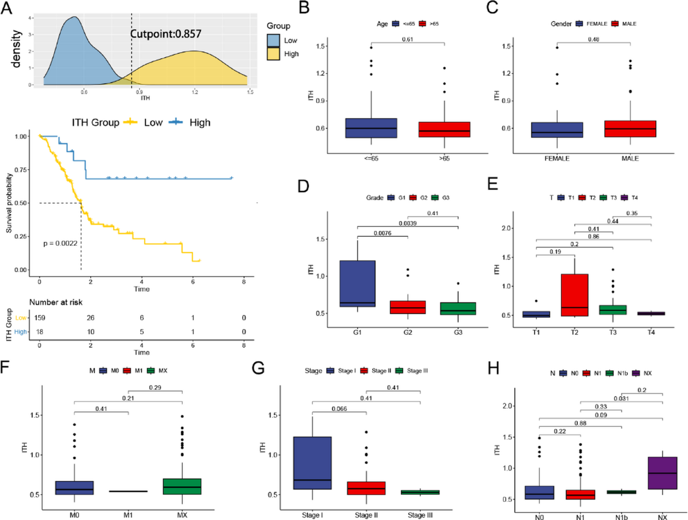
This analysis of the NCDB represents one of the largest assessments of nodal disease in SNSCC. The rate of clinical LNM at presentation of 14.3% is in accordance with many published database and retrospective studies. We discovered that among participating surgical centers, END for SNSCC is rarely performed with only 15.3% of cN0 patients receiving END. This corroborates findings by Crawford et al. who identified an END rate of 19.6% for all SNSCC patients with T3 and T4 disease [16]. Furthermore, occult LNM was relatively rare, identified in 12.0% of patients. Previous NCDB studies that only assessed patients with T3 and T4 SNSCC identified an occult LNM rate of 12.7% [16]; similarly, a SEER database study approximated the rate of occult LNM from 11–17% [17]. It is significant, albeit unsurprising given prior head and neck studies, that pathological LVI strongly increases the likelihood of occult LNM (OR 8.61, CI 2.63–30.2, p < 0.001). A retrospective analysis of 138 patients with maxillary sinus cancer demonstrated an association of LVI with nodal metastasis, although the authors did not assess specifically for occult LNM [18].
SNSCC most commonly affects patients in the 6th and 7th decades of life [9, 19] with an estimated 80% of patients affected diagnosed at 55 years and older [10]. We found advanced age (74 years old and greater) to have a significantly higher OR of occult LNM (OR 8.17, CI 1.59–64.7, p < 0.021). This finding is significant in that increasing age has been significantly associated with worse overall survival [20] and higher pathologic nodal staging [21]. The implication of higher rates of occult LNM with age suggest the multifactorial nature of age-related mortality in SNSCC and possible importance of considering END in this group of patients.
4.1 Nasal cavity
The nasal cavity is a distinct pathologic subsite for SNSCC and varies histologically and clinically from the paranasal sinuses. The literature currently reports that nasal cavity SNSCC account for as much as 25% of all SNSCC [10]. This is in contrast with our cohort for which nasal cavity SCC represents 55.7% of all surgically treated cases of SNSCC, likely related to the fact that nasal cavity tumors may be more amenable to surgical resection compared to other sinonasal subsites.
The nasal cavity subgroup had a clinical LNM rate of 7.6%, which closely aligns with a prior NCDB study by Ranasinghe et al. who identified clinical LNM at presentation to be 7.9% and a SEER database study by Ahn et al. who identified it to be 9.3% [8, 22]. The rate of clinical LNM at presentation increased from a rate of 3.1% for T1 tumors to 14.7% for T4 tumors. This aligns closely with findings by Ahn et al. who found a greater likelihood of LNM for nasal cavity tumors greater than Stage 2 [10, 22]. The nasal cavity also had a significantly lower rate of pathologic LNM false positivity when compared to all sinonasal subsites and the maxillary sinus (18.9% vs. 30.8%, and 35.3%, respectively). Fewer patients underwent END for nasal cavity subsite (7.2%) compared to any subsite (15.3%) the maxillary sinus subsite (31.9%).
4.2 Maxillary sinus
The maxillary sinus is often described as the most common site for SNSCC reportedly accounting for as much as 60% of cases [10, 18]. Maxillary sinus accounted for 37.8% of surgically treated SNSCC in our study. Clinical LNM at presentation was the highest for maxillary subsite at 25.3%. The likelihood of clinical LNM was directly proportional to increasing T stage with the exception of T3, with T1, T2, T3, and T4 having rates of 7.3%, 23.9%, 20.5%, and 30.7% respectively. This is slightly higher than previously described estimates that identified clinical LNM rates of 18.6% for T2 and 22.3% for T3/4 tumors of the maxillary sinus [22].
Rates of END were much higher for the maxillary sinus than any other subsite with 31.9% of patients receiving END overall; patients with T4 had an END rate of 35.7% and all other subsites ranged from 27.5 to 30.2%. Rates of occult LNM remained at 11.9% overall, consistent with our overall SNSCC occult LNM rate of 12.0% and prior studies assessing only T3 and T4 SNSCC of the maxillary sinus [16]. Importantly, some authors have posited that maxillary sinus tumors that invade into the hard palate may be managed as oral cavity SCC [17, 23], which may have contributed to our higher observed rates of END for this subsite.
The maxillary sinus had a false positive LNM rate of 35.3% overall, which is nearly double that of the nasal cavity. A potential explanation for higher false positivity in the maxillary sinus is the predilection for infection with maxillary sinus obstruction compared to a nasal cavity mass; sinus obstruction leading to an acute sinusitis can cause reactive lymphadenopathy mistaken for malignant lymphadenopathy, although this warrants further investigation.
4.3 Ethmoid sinus
Ethmoid sinus SNSCC historically accounts for a poorer overall survival likely secondary to a positive margin rate as high as 32.5%, either due to malignancy mistaken for sinusitis or involvement of critical structures such as the orbit and skull base [24]. Within our cohort, ethmoid sinus SCC accounted for 6.5% of all surgically treated SNSCC; of this group, only 7% (9 patients) had clinical LNM and there were no false positives. Only 8 patients received END, and none of them demonstrated occult LNM. Existing data on ethmoid sinus SNSCC is consistent with our findings of low rates of clinically evident LNM and occult LNM [23,24,25].
4.4 Scoring system for clinical implementation
Our uniquely derived scoring system (Table 6) translates our work into a practical tool for END decision-making. It helps provide risk thresholds, allowing clinicians to identify patients in whom END may be safely omitted (NPV > 95%) and those who would benefit from END. This approach offers a structured risk stratification model. Future research in external validation and integration of this scoring system as a clinical nomogram for decision making would be fruitful.
4.5 Limitations
The retrospective nature of this cohort study has several inherent limitations, including missing or incomplete data capture and inconsistencies in coding information. Although the National Cancer Database (NCDB) captures at least 72% of all newly diagnosed malignancies at an institutional level [19], there is currently no disease-free survival information available in this database. This may have introduced a loss-to-follow-up bias, which can potentially be negated by maintaining high follow-up rates (> 80%) but is not possible in this study.
It should also be noted that the NCDB does not include all high-risk pathologic variables, such as perineural invasion, which may increase the risk of LNM, but could not be included in our multivariate model of occult metastasis.
Another limitation is the use of histology codes 8070 (squamous cell carcinoma, not otherwise specified), 8071 (keratinizing), and 8072 (non-keratinizing). With newer diagnostic criteria, the broad classification of 8070 may encompass cases that would now be more precisely categorized under newer histological subtypes introduced in recent classification updates. However, given the constraints of the database, the histological coding available at the time of diagnosis, the retrospective nature of our study, we applied the best available classification to capture the relevant cases.
Other limitations include the lack of generalizability given the low proportions of non-academic centers and non-White patients, potential selection biases, such as some patients not receiving therapeutic neck dissections for clinically apparent nodal disease. Additionally, some subgroup analyses, particularly those involving elderly patients or rare subsites like the ethmoid sinus, are limited by small sample sizes and may produce wide confidence intervals that reduce statistical precision and should be interpreted with caution.
Our definition of neck dissection as removal of ≥ 3 lymph nodes, though consistent with prior NCDB studies [14, 15], may include limited or inadequate dissections, potentially affecting estimates of nodal positivity and false negative rates. Additionally, while the NCDB does distinguish between radiation to the primary site versus the neck, the granularity and completeness of this variable are limited, and we cannot reliably determine the specific intent or overlap of radiation fields in all cases. This may still introduce some confounding in the interpretation of occult metastasis rates.
We also chose not to perform a survival analysis for END, due to concerns with comparisons to a group that did not receive END. Specifically, those who did not receive END may have received other treatment to the neck including radiation therapy which is not clearly described in the NCDB. Finally, there were no cases of primary sphenoid or frontal sinus SCC included in the NCDB for the years queried. Naturally, this analysis cannot predict LNM in malignancies originating from these locations. Finally, it is important to understand that reliance on histologic evaluation of the primary tumor for lymphovascular invasion may underestimate its presence, and future studies incorporating advanced imaging modalities may provide a more accurate assessment of nodal metastatic risk.


Comments (0)