
Remember me

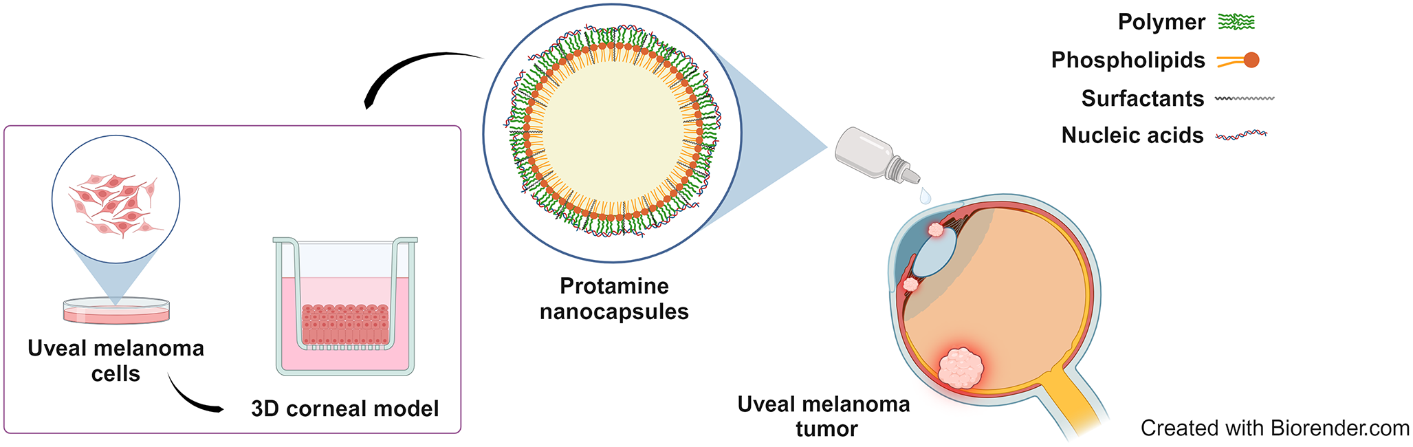
Protamine NCs were prepared using a solvent-displacement method developed by our group [36, 37]. The addition of the oily phase to the aqueous phase under stirring precipitates the lipid components as the polar solvent diffuses into the aqueous phase, where the cationic polymer interacts electrostatically with the oil nanodroplet of vitamin E, forming a polymeric shell around it [44]. In addition, this core structure is further stabilized by surfactants such as sodium cholate and Tween® 80. The formulation was composed of a homogeneous population of blank particles with a size below 250 nm, low PDI, and positive surface charge (Table 1). Regarding their morphological characteristics, STEM images showed homogeneous individual particles with spherical shape (Fig. 1A). The negative staining observed revealed a dark inner portion corresponding to the oily core of vitamin E, and the polymeric outer layer as a line surrounding the circular droplets, which is the capsular structure typically observed for this type of carriers [44,45,46,47].
Table 1 Mean particle size, polydispersity index (PDI) and zeta potential of blank protamine NCs, and loaded with 1% and 2.5% (w/w) of pDNA, with respect to the total mass of the NCs (Mean ± SD (n > 3))The pEGFP-Luc was associated to protamine NCs selecting 1% and 2.5% (w/w) of pDNA payload, with respect to the total mass of the NCs. The results collected in Table 1 showed a slight increase in the size of loaded NCs compared to the blank formulation. This same result was observed associating other nucleic acids, such as 1.5% (w/w) of miRNA payload, with respect to the total mass of the NCs (Table S1). This could be explained by a rearrangement of the polymeric protamine shell upon the incorporation of the nucleic acids. For the same reason, important changes in the surface charge of the formulation were observed. The NCs loaded with both pDNA and miRNA acquired negative zeta potential, indicating the surface association of both nucleic acids. In the case of plasmid DNA, the surface charge became increasingly negative as more pDNA was associated: -31 mV with 1% (w/w) of pDNA and − 42 mV with 2.5% (w/w) of pDNA. The STEM images of pDNA-loaded NCs also showed a homogenous population of spherical particles with similar structure to blank protamine NCs, but with a more compact conformation, especially for 1% of pDNA-loaded NCs (Fig. 1B and C) [48].
Fig. 1
STEM images of blank protamine NCs (A) and loaded with 1% (w/w) of pDNA (B) and 2.5% (w/w) of pDNA (C), with respect to the total mass of the NCs (scale bar = 100 and 200 nm, magnification 100,000 and 200,000 KX)
Protamine nanocapsules for the association and release of nucleic acidsThe nucleic acid association was analyzed by agarose gel electrophoresis in the presence/absence of heparin, which is a sulfated glycosaminoglycan with strong negative charge and high affinity for protamine, capable of displacing the polynucleotides from the NCs [41]. In addition, considering the envisaged administration route, the potential of the protamine NCs to associate nucleic acids was also determined incubating the pDNA-loaded formulation in SLF medium. At time zero, an effective binding of the genetic material could be observed in Milli-Q water (lane 1) (Fig. 2A), as well as in SLF medium (Fig. 2B, C and D), in comparison to the formulation mixed with heparin (lane 2). In this case, the lane was more marked suggesting gradual release of the encapsulated polynucleotide. However, the nucleic acid association was most effective using a loading of 1% (w/w) of pDNA than 2.5% (Fig. S1) and, especially, in SLF medium. The salt composition of this medium could favor the electrostatic interactions between the components of the NCs and the nucleic acids. Moreover, the binding of the genetic material to protamine NCs was stable over time in SLF medium, mainly, using low pDNA-loads, remaining intact during this time. In the case of 2.5% (w/w) of pDNA formulation, the bands were less intense after 4 h than at the other time points (Fig. S1). This could be due to pDNA not being completely associated with the NCs, which could result in partial degradation over time. Overall, there is an efficient and reversible association with the possibility of sustained release in biological media.
On the other hand, a greater efficiency was observed by associating other nucleic acids such as miRNA (Fig. S2). miRNAs are shorter-stranded RNA, approximately 22 nucleotides in length [49], which allowed a stronger interaction with nanocapsule components.
Fig. 2
Agarose gel images of protamine NCs loaded with 1% of pDNA with respect to the total mass of the NCs (lane 1) at t = 0 h (A), and incubated in simulated lacrimal fluid at time zero (B), and after 30 min (C) and 4 h (D) at 37ºC (lane 1). A displacement assay upon incubation of protamine NCs with heparin using the mass ratio 1:25 (w/w) allowed the migration of the associated nucleic acids (lane 2). The amount of pDNA per lane was 0.135 µg. Naked pDNA was lane 0
Stability of protamine nanocapsulesIn the present work, the stability of blank and nucleic acid-loaded protamine NCs was analyzed by measuring the size, PDI, surface charge and DCR in aqueous suspension for 30 days at 4 °C (Fig. 3). The optimization of the storage conditions of the NCs is highly important due to their influence on biocompatibility and their physicochemical characteristics [50]. After one month, blank (Fig. 3A) and pDNA-loaded (Fig. 3B and C) formulations maintained their physicochemical characteristics showing a particle size below 250 nm and 300 nm, respectively, with no significant difference from initially stored NCs, and without losing homogeneity in the population. Regarding the zeta potential, a positive and negative surface charge were maintained over time for blank (above + 40 mV) and loaded-NCs (above − 28 mV for 1% (w/w) of pDNA and − 41 mV for 2.5% (w/w) of pDNA), respectively. In addition, the count-rate was also maintained within the same range throughout indicating the absence of significant aggregation phenomena (data not shown). These results reflect the excellent stability of the formulation, consistent with similar results obtained by our research group where protamine NCs also showed no significant differences in the particle size and zeta potential in long-term stability [18, 36, 37].
Fig. 3
Stability of aqueous suspensions of blank protamine NCs (A) and loaded with 1% (B) and 2.5% (w/w) (C) of pDNA, with respect to the total mass of NCs, measuring the particle size (bars) and polydispersity index (PDI) (dots), and zeta potential (ZP) at storage conditions for 30 days (Mean ± SD (n = 9))
To determine the feasibility of the formulation for in vitro testing, the stability of protamine NCs was also studied in cell culture medium. In biological media, the possible interactions between particles and serum proteins could give rise to the presence of aggregates. Blank protamine NCs experienced a slight increase in particle size and PDI when they were diluted in cell culture medium at time zero, due to the loss of charge-induced stability in buffered media (Fig. 4A). In addition, this size increase was more evident when NCs were diluted in medium with fetal bovine serum. This effect could be attributed to the electrostatic interaction between positively charged protamine with negatively charged serum proteins [51]. As expected, a decrease in the DCR values was observed in culture media confirming the possible presence of aggregates (data not shown). In contrast, no significant change in particle size was observed after 4 h in supplemented or non-supplemented medium compared to the initial sizes. Regarding the stability of nucleic acid loaded NCs (Fig. 4B and C), the results were similar to blank protamine NCs. After 4 h of their incubation in cell culture medium with and without serum, the pDNA-loaded NCs were stable in terms of size and PDI, compared to the initial sizes. In this case, when they were diluted in biological medium at time zero, the physicochemical characteristics of protamine NCs loaded with 2.5% (w/w) of pDNA did not have a significant modification in size in comparison with their dilution in Milli-Q water (Fig. 4C). This superior stability in physiological environment could lead to a lower interaction of serum proteins when a larger amount of genetic material is associated to the protamine NCs, and therefore, the increase in particles size is not observed as loading 1% (w/w) of pDNA (Fig. 4B) [46].
Fig. 4
Stability of blank (A) and loaded protamine NCs with 1% (B) and 2.5% (w/w) (C) of pDNA, with respect to the total mass of NCs, measuring the particle size (bars) and polydispersity index (PDI) (dots) at 37 °C for 0, 2 and 4 h in supplemented and non-supplemented RPMI cell culture medium (Mean ± SD (n = 9))
Finally, in order to use these nanocapsules as a potential eye drop formulation, the stability of protamine NCs associated with different percentages of plasmid DNA was also studied in the administration media and upon contact with the lachrymal fluids. The incubation of pDNA-loaded NCs was performed in SLF at different time points (0 h, 30 min, and 4 h) at 37 °C. Particle size is known to be an important parameter related to the capacity of the nanoparticulate system to interact with mucosal surfaces in general, and with the ocular mucosa in particular [52]. Figure 5 showed similar results to those obtained when diluting the loaded-NCs in cell culture medium. Briefly, at time zero, the size and PDI of NCs loaded with 1% (w/w) of pDNA increased when they were diluted in simulated lacrimal fluid (Fig. 5A). Ophthalmic formulations typically consist of solutions composed mainly of different salts without proteins. The positive ions (monovalent and divalent cations) of the organic salts that constitute the SLF buffer could stabilize the protamine NCs with higher plasmid DNA loading, such as 2.5% (w/w) of pDNA (Fig. 5B), due to greater electrostatic interactions with the negatively charged phosphate groups of nucleic acids arranged on the NC surface. No significant changes in particle size and PDI were observed at least during 30 min in SLF, although some ionic cross-linking was observed between the monovalent and divalent positive ions of the salts in the SLF with the negative charges of the phosphate groups of the pDNA, especially with 1% (w/w) of pDNA. These data were consistent with previous studies carried out by our research group [18] and with results obtained when protamine NCs were incubated also in simulated gastrointestinal media, where no degradation or aggregation was observed [53]. Topically administered formulations generally disappear from the ocular surface within a few minutes due to the blink reflex and the rapid renewal of the tear film. Considering this, the stability of this nanosystem for at least 30 min is enough, as this duration aligns with the theoretical time required for the interaction of the NCs with the corneal epithelium [18].
Fig. 5
Stability of protamine NCs loaded with 1% (A) and 2.5% (w/w) (B) of pDNA, with respect to the total mass of NCs, measuring the particle size (bars) and polydispersity (PDI) (dots) at 37 °C for 0 h, 30 min and 4 h in SLF (Mean ± SD (n = 9))
Cell viability in uveal melanoma cellsThe cytotoxicity of nanocarriers is one of the most important factors limiting their medical application. The physicochemical characteristics of the NCs such as size and surface charge, their hydrophobicity, and their supramolecular structure are parameters that define their biocompatibility and cellular interactions [54]. In the present work, the biocompatibility of protamine NCs was evaluated in UM cancer cells by a metabolic assay, demonstrating that this nanosystem did not compromise the cell viability either at 24 h and 48 h post-incubation (Fig. 6).
Previous studies have demonstrated the efficacy and safety of polymeric NCs. The cytotoxicity profile of this formulation is in line with previously reported data of other polymeric nanosystems for the delivery of complex macromolecules to the ocular surface. Our formulation has similar physicochemical characteristics to chitosan and/or hyaluronic acid NCs, which are the most studied formulations for the treatment of ocular diseases [17, 19, 23, 42]. In addition, their common non-toxic structure of an oily core covered by a polymeric shell makes them a potential alternative to formulations marketed for ocular field [55]. In vivo studies have demonstrated that the use of polymers, whether anionic such as polyethylene glycol or cationic such as chitosan, as surface coating of the NCs lend them properties of low or no toxicity [54]. In this aspect, the selection of protamine is an advantage due to it well-documented safety and presence of several FDA-approved formulations [56]. In addition, studies by S. Reimóndez-Troitiño et al., have shown that polymeric nanosystems, especially, protamine NCs, did not produce any evidence of ocular irritation or epithelial alterations [18].
Fig. 6
Cell viability assay after 24 h (light-grey bars) and 48 h (dark-grey bars) of the removal of increasing concentrations of blank protamine NCs from 30 to 1340 µg/mL in UM cancer cells (Mean ± SD (n = 3))
Uptake of protamine nanocapsules in uveal melanoma cellsBlank protamine NCs were labelled with the fluorochrome 5-TAMRA to study their cellular internalization in UM cells. This fluorophore is a succinimidyl ester with good reactivity and selectivity with primary and secondary aliphatic amines forming stable amides identical to natural peptide bonds [57]. Within the structure of protamine sulfate [58, 59], proline residues seem to be the most reactive for attacking this succinimidyl group [60]. The high reactivity suggested efficient labelling of the total amount of protamine. The excess of the unreacted 5-TAMRA was removed by dialysis, ensuring that only labelled protamine was the final product. The addition of the 5-TAMRA fluorochrome did not affect negatively to the formulation due to labelled NCs presented similar physicochemical properties than non-labelled one (Table S2).
Cellular uptake was evaluated by confocal microscopy after 4 h. In Fig. 7B, the maximum projection showed the intracellular localization of protamine NCs in UM cells, where the finding could be verified by observing the image including the orthogonal sections on X and Y axes. In the present work, the efficient internalization of fluorescently labelled protamine NCs might also be related to the penetration enhancing properties of protamine [61]. This cationic peptide presents high content of arginine residues, where six of them constitute the nuclear localization signal (NLS) [62, 63]. This arginine sequence allows protamine to aid has cellular internalization [64], and subsequent translocation of molecules from the cytoplasm to the nucleus.
To provide a quantitative evaluation, the NC uptake was also analyzed by flow cytometry in living cells using the LIVE/DEAD™ Fixable Aqua Dead Cell Stain reagent. After cell incubation with Pr-TAMRA NCs, the histograms corresponding to the fluorescence signal of 5-TAMRA showed a shift towards the 5-TAMRA (+) region (Fig. 7D) compared to the control (unstained cells) (Fig. 7C). This indicated that almost 36% of UM cells were positive for the presence of these NCs (Table S3). In addition, there was high cell viability, as indicated by the peak corresponding to the fluorescence signal of Aqua mostly in the live region (Aqua (-)).
Fig. 7
Representative confocal microscopy images of NC uptake in UM cells: non-treated (A) and treated with Pr-TAMRA NCs (red channel) for 4 h at 37 °C (B). Nuclei of the cells were stained with DAPI (blue channel) (magnification 20x, z1.25, scale bar = 100 μm). Flow cytometry histograms quantifying the uptake in UM cells non-treated (C) and treated with Pr-TAMRA NCs (54 µg/cm2) after 4 h post-treatment (D) (LIVE/DEAD™ Fixable Aqua Dead Cell Stain as a viability reagent)
Transfection of protamine nanocapsules in uveal melanoma cellsAs a proof of concept for protamine NCs as gene delivery carriers to treat UM, their transfection capacity was analyzed by evaluating the expression of the EGFP protein. This formulation was chosen to carry out the transfection studies due to the physicochemical properties in terms of size and PDI, good stability and profile under storage and in different biological media, and higher total pDNA load. The fluorescent image showed small green bright spots in the cytoplasm, close to cell nucleus, after 48 h of the NC incubation with the cells (Fig. 8A). This could demonstrate that the plasmid DNA was released from the protamine NCs when exposed to the mildly acidic environment of UM cells. This result, in combination with those previously obtained by our research group by evaluating the GFP expression in colorectal cancer cells, suggested that the development of protamine NCs could work as a safe and reliable nanoplatform for effective gene delivery [36].
Moreover, the expression of this protein was quantified in the largest number of UM cells. For this purpose, the expression was analyzed by flow cytometry measuring the fluorescence-positive events corresponding to cells treated with protamine NCs associated with 2.5% (w/w) of pDNA, in comparison with those treated with naked plasmid DNA and Lipofectamine® 2000 (Fig. 8B). The percentage EGFP values of cells treated with naked pDNA, and the control (cells not-treated) were similar in terms of EGFP-positive events and, thus, indicated negligible protein expression. In the case of the pDNA associated with protamine NCs, the transfection results are agreement with those of the fluorescent image. However, the low EGFP expression values may be due to the sustained release of the plasmid DNA for a long time. Several studies have been reported the pattern of sustained release over time of different biomolecules such as insulin and genes, or drugs [37, 65,66,67]. Therefore, protamine is considered promising for the design of nanocarriers for in vivo delivery, nevertheless, further studies are still needed for the optimization of the NC transfection capacity as a next step toward its preclinical development.
Fig. 8
Fluorescent microscopy image of EGFP expression (green channel) after 48 h of the treatment of protamine NCs loaded with 2.5% (w/w) of pDNA at dose 2.5 µg of pDNA incubated for 4 h at 37 °C (A). Nuclei of the cells were stained with DAPI (blue channel) (scale bar = 20 μm). Quantification of EGFP expression by flow cytometry measuring the percentage of UM positive cells after 48 h (incubated for 4 h at 37 ºC) (B) The data of the Y-axis are represented using a logarithmic scale (Mean ± SD (n = 3))
Epithelial barrier integrity and permeability assay in 3D corneal modelTEER measurement is the most widely used method to study the corneal permeability and the integrity of cell barriers [39]. Modifications in the integrity of the epithelial barrier membrane are usually associated with a reduction in TEER values, which indicates an alteration of the ocular barrier due to the opening of tight junctions [68]. It is well established that TEER values in the range of ≥ 750 and ≤ 2,500 Ω·cm2 are indicative of 3D epithelial conformation and a correct barrier function [39, 43, 69].
The QobuR-RhCE model is constituted by human corneal cells that form a stratified squamous cellular superstructure resembling a healthy human corneal epithelium [43, 69]. In the present work, TEER was measured in this 3D model before and after the treatment with non-fluorescent and fluorescently labelled protamine NCs, naked Cy3-siRNA and Cy3-siRNA-loaded protamine NCs.
Figure 9A shows that, after the standard 30-min treatment, the TEER values (Ω·cm²) of corneal epithelium exposed to protamine NCs, loaded and unloaded with nucleic acids, were comparable to the negative control value for epithelial barrier integrity (H2Od). Values of approximately 1,500 Ω·cm² remained within the range of ≥ 750 and ≤ 2,500 Ω·cm², confirming that the treatment did not compromise the integrity of the epithelial barrier. To evaluate this effect over time, the maximum observation time of the corneal model was extended up to 4 h. This epithelial model tended to show a decrease in TEER values with prolonged immersion times due to progressive epithelial degradation [39, 43, 69]. However, after 4 h of treatment, TEER values remained above 750 Ω·cm² (880 Ω·cm²), confirming that epithelial integrity was preserved and suggesting low toxicity profile for the formulation. These findings are consistent with data from studies on the permeability of similar polymeric nanosystems reported in the literature [
Comments (0)