
Remember me

Kaplan–Meier analysis was performed to evaluate the prognostic significance of eEF2 K expression in triple-negative breast cancer (TNBC) and basal-like breast cancer subtypes. In this study, survival probabilities were compared between patients with high and low eEF2 K expression, utilizing various survival endpoints, including overall survival (OS) and recurrence-free survival (RFS). The figure demonstrates that high eEF2 K expression is associated with poor patient survival in several subtypes of breast cancer. Specifically, in ER(-), PR(-), HER2(-) patients, those with high eEF2 K expression had a significantly lower OS compared to those with low expression, with a hazard ratio (HR) of 1.9 and a statistically significant log-rank p-value of 0.015. Similarly, for patients with basal-like breast cancer (PAM50 subtype), high eEF2 K expression correlated with reduced OS (HR = 1.84, log-rank p = 0.02), further emphasizing the negative impact of eEF2 K on patient prognosis.
Interestingly, while the relationship between eEF2 K expression and RFS was not statistically significant in ER(-), PR(-), HER2(-) patients (HR = 1.43, log-rank p = 0.22), a notable trend was observed. In the basal-like, lymph-node positive group (PAM50), high eEF2 K expression was significantly associated with reduced RFS (HR = 1.85, log-rank p = 0.015), suggesting that eEF2 K may play a role in promoting tumor recurrence in more aggressive forms of breast cancer. Overall, these findings highlight eEF2 K as a potential biomarker for poor prognosis in TNBC and basal-like breast cancer, particularly in terms of OS and RFS. The differences in hazard ratios and log-rank p-values across different patient subgroups suggest that the impact of eEF2 K on survival may vary depending on the specific breast cancer subtype and other clinical factors. Finally, Kaplan–Meier survival analysis and in vitro experiments consistently demonstrate the therapeutic potential of targeting eEF2 K in TNBC. The elevated eEF2 K expression correlates with worse survival outcomes in specific breast cancer subtypes, and its silencing, as part of an HNP-based therapy, may offer an innovative approach for improving prognosis in TNBC patients Fig. 3.
Fig. 3
Kaplan–Meier survival analysis of overall survival and recurrence-free survival in breast cancer patients based on eEF2 K expression levels. The survival curves illustrate the differences between patients with high and low eEF2 K expression in various subtypes of breast cancer. (a) ER(-), PR(-), HER2(-) overall survival (OS) shows a trend toward poorer outcomes in patients with high eEF2 K expression (HR = 1.9, p = 0.15). (b) ER(-), PR(-), HER2(-) RFS indicates statistically with (HR = 1.43, p = 0.22). (c) In ER(-), PR(-), HER2(-), PAM50: basal-like subtype, high eEF2 K expression correlates with worse overall survival (HR = 2.71, p = 0.054). (d) PAM50: basal subtype overall survival demonstrates a significant reduction in survival for patients with high eEF2 K expression (HR = 1.84, p = 0.02). (e) PAM50: basal subtype with lymph node positivity shows a significantly decreased RFS for patients with high eEF2 K expression (HR = 1.85, p = 0.015)
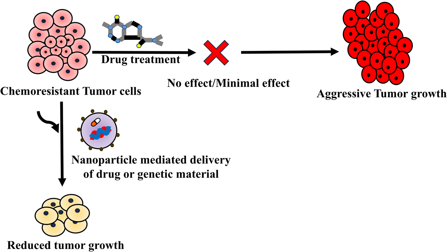
Hybrid nanoparticles leading by LbL method as a modular platform for codelivery of eEF2 K-siRNA and quercetinAs detailed in the methods section, the hybrid nanoparticles developed using the Layer-by-Layer technique were characterized using NTA, ZetaSizer, STEM, SEM and EDX (Fig. 4). The size analysis of HNPs, which increased with each layer added using the LbL method, was conducted using NTA. The AgNP + QU particles, initially 79.5 nm, increased to 117 nm after PAH coating and reached 153 nm upon complexation with eEF2 K-siRNA. After the final PSS coating, the HNPs stabilized at a final size of 133 nm (Fig. 4-a).
Fig. 4
Characterization of HNP. (a) Size change of HNP after each layer formation. (b) Zeta-potential levels after each electrostatic interaction. (c) TEM images for different layers of HNP (200 nm, scale bar). (d) Elemental analysis data for HNP from EDX. (e) SEM result for HNP (1 µm, scale bar)
The charge difference between each layer, electrostatically bound, was determined by measuring the zeta potentials. The surface charge, which was approximately − 30 mV for AgNP and QU complexes, increased to around + 40 mV after the PAH coating. Upon the addition of eEF2 K-siRNA molecules, the zeta potential decreased to approximately + 18 mV and reached a final value of − 35 mV after the PSS coating (Fig. 4-b).
To understand the morphological structure of the hybrid nanoparticles, imaging was performed using electron microscopy. STEM analysis provided two-dimensional images, allowing for the examination of the dimensional and structural changes in the HNPs. Additionally, to determine whether the particles were spherical or circular, SEM analysis was performed to obtain three-dimensional images. The resulting images confirmed the formation of spherical particles (Fig. 4-c/e) To verify the successful application of the LbL method during particle synthesis, elemental analysis of the particles was conducted. EDX spectra were collected, and the elemental composition of the particles was analyzed by mass percentage. The analysis revealed that the particles contained 54.7% silver and 27.45% carbon by mass (Fig. 4-d).
After the synthesis of the developed HNP particles, the amount of quercetin adsorbed on the AgNP core was determined. For this purpose, a calibration curve was developed by using free quercetin standards at 371 nm wavelength due to the maximum absorbance value of quercetin observed at this point. Following this, the synthesized AgNP + QU complex was centrifuged at 15,000 rpm for 30 min to precipitate the particles, and the supernatant, containing the unbound quercetin molecules, was analyzed by using the calibration curve. Finally, the adsorption efficiency of quercetin molecules was determined as 55.84% (Supplementary Fig. 1).
Similarly, the optimal ratio for complex formation between eEF2 K-siRNA molecules and HNPs was determined. In this context, the concentration of eEF2 K-siRNA was kept constant while the HNP concentrations were varied. The complexes formed at different ratios were analyzed using agarose gel electrophoresis, a method previously established in our laboratory [31]. The most efficient complex was assumed to be 5:1 (NP:siRNA) as a ratio (Supplementary Fig. 2-a).
The zeta potential values of the complexes formed at different ratios of HNP, and eEF2 K-siRNA were also examined to gain insights into the binding ratios. Free siRNA molecules had a zeta potential of − 27 mV, and with increasing HNP concentrations, the surface charge of the particles increased. The most efficient ratio, 5:1 (NP:siRNA), which was the first ratio with a positive surface charge, was measured to be approximately + 18 mV (Supplementary Fig. 2-b).
Conveniently cellular uptake of HNPs without any external stimuliIn this context, cell nuclei were stained with DAPI to allow fluorescent imaging and the eEF2 K-siRNA molecules used in the HNPs were replaced with FAM-labeled anti-GAPDH-siRNA (FAM-siRNA), allowing the particles to be tracked under the fluorescent microscope due to the FAM tag. The cell nuclei appeared as blue fluorescence from DAPI staining, while the HNPs were visualized as red fluorescence due to the FAM-siRNA. The merged image was obtained by overlaying these two fluorescence images, providing insights into the intracellular localization of the HNPs.
As seen in Fig. 5, the merged images show that the red fluorescence from the particles is located adjacent to the blue fluorescence from the cell nuclei in all three cell lines (MDA-MB- 231, BT- 549, and 4T1). This indicates that the particles were successfully internalized by the cells, and cellular uptake did not vary between different cell types/lines. Furthermore, the absence of red fluorescence in the dark areas of the merged images confirms that no particles remained outside the cells and that all delivered particles were readily internalized. This suggests that the designed system does not require any additional stimuli or external forces for cell entry.
Fig. 5
In vitro cellular internalization of HNP for MDA-MB- 231, BT- 549 & 4T1 cells. Fluorescence imaging of cells incubated with hybrid nanoparticles loaded with FAM-labeled anti-GAPDH siRNA for 6 h. Nuclei are stained with DAPI (blue), and the FAM-labeled siRNA (red) indicates successful nanoparticle uptake. Cells were fixed with 4% paraformaldehyde and imaged using a Leica & Dm Il Led Fluo fluorescent inverted microscope (100 µm, scale bar)
Delivery of eEF2 K-siRNA supporting with quercetin in HNP as potential TNBC therapeuticsThe effects of HNPs at concentrations ranging from 0.94 to 30 nM on cell viability were observed in MDA-MB- 231, BT- 549, and 4T1 cell lines over 24, 48, and 72 h. The results revealed a consistent trend across all three cell lines. After 72 h of treatment, 30 nM HNPs reduced cell viability by approximately 50% in all cell lines, while empty nanoparticles lacking eEF2 K-siRNA and quercetin did not affect cell viability. In this way, we concluded that the designed HNP has no toxic effect on the cells, and all of the results from a decrease in cellular viability arose therapeutic effects rather than toxicity. Moreover, as incubation time increased, HNPs containing only quercetin significantly decreased TNBC cell viability. However, they contain only eEF2 K-siRNA, which showed a significant effect on cell viability only in the BT- 549 cell line after 72 h. These results indicate that increased HNP concentration and treatment duration reduced cell viability across all three TNBC cell lines (Fig. 6-a).
Fig. 6
In vitro cellular treatment results. (a) Resazurin assay results of empty HNP, only eEF2 K-siRNA loaded HNP (siRNA-HNP), only Quercetin loaded HNP (QU-HNP) and different concentrations of eEF2 K-siRNA & quercetin loaded HNP (siRNA-QU-HNP) for MDA-MB- 231, BT- 549 & 4T1 cells. (b) Colony formation assay images for MDA-MB- 231, BT- 549 & 4T1 cells treated with different concentrations of HNP and Doxorubicin (DOX) as a positive control. (c-e) Quantitative results for colony formation assay retrieved from ImageJ program (in this case, more than 50 cells were considered as a colony). (*: p < 0.05, **: p < 0.01, ***: p < 0.001, ****: p < 0.0001; one-way ANOVA calculated in GraphPad program)
Then, we simulated an environment covering a small amount of cancer cells to understand how HNP treatment effects on the colony formation of the cancer cells. In this way, we expected to observe the capacity of HNPs to suppress both the initial formation of cancerous tissue from a cancer cell, and the potential relapse of the disease following treatment. As shown in Fig. 6b, colony formation was analyzed in three TNBC cell lines over a 13-day period. In untreated control wells (0 nM), cancer cells successfully proliferated, forming colonies that covered the entire well surface by the end of the incubation period. However, treatment with 30 nM HNPs completely inhibited colony formation, demonstrating the strong anti-proliferative effect of HNPs. Doxorubicin (DOX), a well-known chemotherapy drug, was used as a positive control, further validating the effectiveness of the HNP treatment.
The ability of HNPs to inhibit colony formation highlights their potential in preventing both the growth of cancerous tissue from individual cancer cells and the recurrence of cancer post-treatment. This assay provided valuable insights into the long-term therapeutic potential of HNPs and their impact on limiting the tumorigenic capacity of cancer cells (Fig. 6-c/d/e).
Slowing down the migration of TNBC type cell lines after HNP treatmentIn this study, we employed a wound healing assay to evaluate the impact of HNPs on the migratory capacity of TNBC cells. Specifically, the assay was performed using three TNBC cell lines (MDA-MB- 231, BT- 549, and 4T1) to assess the effect of different concentrations of HNPs (ranging from 0.94 to 30 nM) on cell migration over a 72-h period.
As shown in Fig. 7, untreated cells (0 nM HNPs) fully closed the wound within 72 h, indicating active cell migration. In contrast, cells treated with increasing concentrations of HNPs demonstrated significant delays in wound closure, with the highest dose of 30 nM HNP exhibiting the most pronounced inhibitory effect. Microscopic analysis (Fig. 7-a) revealed that, at this concentration, wound closure was largely hindered, suggesting that the nanoparticles significantly.
Fig. 7
(a) In vitro, wound healing (migration assay images for MDA-MB- 231, BT- 549 & 4T1 cells treated with different concentrations of HNP. (b-d) Quantitative results for wound distance in µm scale calculated with ImageJ program and analyzed with one-way ANOVA in GraphPad (*: p < 0.05, **: p < 0.01, ***: p < 0.001, ****: p < 0.0001)
impaired the migratory capacity of the cancer cells. This inhibition of migration was consistent with the trends observed in our previous cell viability and colony formation assays, where the 30 nM HNP treatment led to a substantial reduction in both cell survival and proliferation.
The results from this assay suggest that HNPs may suppress cell migration in a dose-dependent manner, which is a crucial factor in limiting TNBC metastasis. In TNBC, overexpression of eEF2 K has been shown to enhance cell migration and invasion by activating oncogenic pathways such as Src, FAK, PI3 K/Akt, and c-Myc, as well as promoting epithelial-to-mesenchymal transition (EMT) through the integrin β1/Src/FAK axis and cyclin D1 signaling. These pathways are key drivers of tumor progression, drug resistance, and metastatic spread [32,33,34]. Notably, knockdown of eEF2 K has been demonstrated to significantly inhibit cell motility by suppressing these signaling cascades. Our findings align with these reports, as we observed a concentration-dependent inhibition of migration upon treatment with eEF2 K-siRNA and Quercetin-loaded HNPs. This suggests that HNPs may attenuate TNBC metastasis by downregulating eEF2 K and subsequently impairing the activity of migration-associated pathways. As illustrated in Fig. 7, although complete inhibition of migration was not achieved, the observed delay in wound closure indicates that HNP treatment may significantly slow the metastatic potential of TNBC cells, providing a promising therapeutic strategy to mitigate tumor progression.
Necrotic activation of HNP on TNBC cell linesThe apoptosis assay, utilizing Annexin V and PI staining, is employed to determine whether treated cells undergo apoptosis or necrosis. Annexin V binds to phosphatidylserine, a phospholipid typically located on the inner side of the cell membrane. During apoptosis, the cell membrane flips, exposing phosphatidylserine to the cell's exterior, allowing Annexin V to bind and signal the occurrence of apoptosis. On the other hand, PI (propidium iodide) stains nucleic acids like DNA and RNA. In cells undergoing apoptosis or necrosis, membrane permeability increases, allowing PI to enter and bind to DNA, resulting in fluorescence. In healthy cells with intact membranes, PI is excluded due to its size, preventing the generation of a signal. This principle is utilized for analysis through flow cytometry [35].
Following a 72-h HNP treatment at different concentrations ranging from 0 to 30 nM, cells were stained using the mentioned method and analyzed via flow cytometry. The results, presented in Fig. 8-a, categorize cells into four quadrants: the lower left represents healthy cells, the lower right shows early apoptotic cells, the upper right indicates late apoptosis, and the upper left reflects necrotic cells. Gating was performed based on unstained, Annexin V-only, and PI-only controls to establish appropriate fluorescence compensation and minimize spectral overlap. A dot plot of Annexin V-FITC (x-axis) versus PI (y-axis) was generated, and distinct quadrants were used for classification: viable cells (Annexin V-/PI-), early apoptotic cells (Annexin V +/PI-), late apoptotic cells (Annexin V +/PI +), and necrotic cells (Annexin V-/PI +). This approach ensured accurate discrimination between apoptosis and necrosis while preventing misclassification due to late-stage apoptosis or secondary necrosis.
Fig. 8
(a) Apoptosis assay (Annexin V & PI staining) results for MDA-MB- 231, BT- 549 & 4T1 cells treated with different concentrations of HNP. Quantitative results of necrotic levels of (b) MDA-MB- 231, (c) BT- 549 & (d) 4T1 cells, respectively. (*: p < 0.05, **: p < 0.01, ***: p < 0.001, ****: p < 0.0001; one-way ANOVA calculated in GraphPad program)
As shown in the bar graphs illustrated in Fig. 8-b/c/d, a notable increase in the percentage of necrotic cells was observed with higher treatment concentrations. In particular, the highest dose of 30 nM HNP significantly elevated the necrosis rate across all three cell lines, resulting in approximately 35% to 40% necrotic cells. These findings indicate that the 30 nM dose is the most effective concentration, exerting a strong therapeutic effect on the cancer cells in all three TNBC lines. Furthermore, this dose significantly increased the permeability of the cell membrane, leading to cell death. A similar trend in response to treatment was observed in the MDA-MB- 231, BT- 549, and 4T1 cell lines.
Notably, the MDA-MB- 231 cell line exhibited a higher rate of necrosis compared to the other two cell lines, highlighting its greater susceptibility to HNP treatment. Breast cancer cell lines exhibit distinct biological and genetic properties. The MDA-MB- 231 cell line, a metastatic TNBC model, is derived from human breast invasive ductal carcinoma and is highly aggressive. It is characterized by the absence of estrogen receptor (ER-), progesterone receptor (PR-), and human epidermal growth factor receptor 2 (HER2-), making it a representative model for studying invasive TNBC [36,37,38]. In contrast, the BT- 549 cell line, classified as a primary TNBC model, originates from human papillary invasive ductal carcinoma and also lacks ER, PR, and HER2 expressions. However, BT- 549 cells harbor mutations in PTEN, RB1, and TP53, whereas MDA-MB- 231 cells carry mutations in BRAF, CDKN2 A, KRAS, NF2, and TP53, contributing to differences in tumorigenicity and invasiveness [37,38,39]. Given that TNBC subtypes respond differently to therapeutic agents [40, 41], we hypothesize that the higher necrosis rates observed in MDA-MB- 231 cells compared to other cell lines may be attributed to their enhanced metastatic potential and unique genetic alterations that influence cell death pathways.
Analysis of intracellular protein level changes and understanding of necroptosis and parthanatos mechanisms of cell death activation with HNP treatmentIn this study, intracellular protein level changes following the treatment with hybrid nanoparticles were analyzed using Western blot. We found that HNP treatment led to marked inhibition of eEF2 K expression in both MDA-MB- 231 cells and BT- 549 cells (Fig. 9). In addition to we found that HNP treatment significantly expressions of poly [ADP-ribose] polymerase 1 (PARP- 1) and apoptosis inducing factor (AIF) in MDA-MB- 231 cells, while suppressed PARP- 1 and AIF in BT- 549 cells compared to control cells (Fig. 9).
Fig. 9
Western blot assay results for (a) MDA-MB- 231 & (b) BT- 549 cell lines treated with higher concentrations of HNPs and their control groups which is empty nanoparticles in the same concentration. (*: p < 0.05, **: p < 0.01, ***: p < 0.001, ****: p < 0.0001; student t-test calculated in GraphPad program)
For anti-cancer therapy, the successful induction of cell death of the tumor is one of the most important objectives. Recent studies are reported that overexpression of PARP1 can promote necroptosis (a regulated form of cell necrosis). Regulated necrosis includes a wide variety of cell death pathways that share characteristic of necrotic processes. One of these pathways is PARP- 1-mediated cell death, which is named as parthanatos [42].
Under pathological conditions, PARP- 1 over-activation causes the accumulation of PAR polymers and nuclear translocation of AIF, where it causes chromatin condensation and DNA fragmentation. Therefore, AIF is a key mediator in parthanatos, which is independent of caspase activation [43,44,45]. Our results showed that mechanisms for the cell death of the two cells were different. Necrotic Cell death mechanisms varied between the cell lines, as MDA-MB- 231 were found to die by PARP overexpression, while BT- 549 cells by suppression of PARP expression, likely due to the differences in genetic background. When the overall results are evaluated, it is evident that our gene therapy agent worked effectively in all cell lines, successfully reducing eEF2 K protein levels. Moreover, our results strongly suggest that HNP treatment is mediated PARP- 1-dependent mechanism of cell death in the human tumor cell lines MDA-MB- 231.
Inhibiting spheroid formation of TNBC cell lines with HNP treatmentThe 3D spheroid assay is a necessary tool for mimicking the in vivo environment in cancer research. Unlike traditional 2D cell cultures, which grow cells in a monolayer, the 3D spheroid model enables the formation of spherical clusters of cells that closely resemble the architecture of solid tumors. This three-dimensional structure allows us to evaluate drug penetration, cell interactions, and the overall behavior of tumor cells within a more realistic tumor microenvironment. Tumor tissues in the body have layers of cells with varying access to nutrients and oxygen, leading to differences in treatment response. The spheroid assay helps replicate these conditions, making it a valuable model for preclinical studies of cancer therapeutics.
In our study, we used the 3D spheroid assay to understand how our hybrid nanoparticle treatment affects tumor-like structures compared to traditional 2D models. In 2D experiments such as cell viability, colony formation, and migration assays, cells are plated in a monolayer, allowing for direct contact with the treatment, leading to fast and effective outcomes. However, in real-life tumor scenarios, therapeutic agents primarily interact with cells at the tumor's surface, with limited penetration into deeper layers. This challenge is replicated in the 3D spheroid model, making it essential for evaluating how well a treatment penetrates and impacts the entire tumor mass.
In the 3D spheroid assay, we used TNBC (triple-negative breast cancer) cell lines to grow multicellular spheroids and tested different concentrations of HNP treatment. While the concentrations used in 2D experiments were sufficient, they proved inadequate for 3D spheroids due to their more complex and layered structure. Therefore, we increased the HNP doses to 60 nM and 120 nM and extended the treatment period to seven days to observe long-term effects on the spheroids (Fig. 10).
Fig. 10

Spheroid formation and treatment with HNPs. Spheroids images formed at the PEG-dextran interface 24 h post-seeding of 1,500,000 cells/well. They were treated with 15, 30, 60, and 120 nM concentrations of HNPs for 7 days and obtained quantitative results of spheroid diameters of (a) MDA-MB- 231, (b) BT- 549 & (c) 4T1 cells, respectively. (*: p < 0.05, **: p < 0.01, ***: p < 0.001, ****: p < 0.0001; one-way ANOVA calculated in GraphPad program) (5 µm, scale bar)
The results of our experiment, shown in Fig. 10, demonstrate that the 120 nM HNP treatment reduced spheroid diameters by half after seven days, indicating significant tumor shrinkage. The 60 nM dose also showed a slight effect, though not as pronounced, while the other doses did not produce any statistically significant changes. This indicates that a higher dose of HNP is required to effectively penetrate and reduce the size of the spheroids, which more accurately represents solid tumors.
These findings underline the importance of the 3D spheroid model in preclinical studies, as it highlights the challenges of drug delivery in a more complex tumor environment. It also shows the necessity of optimizing treatment concentrations and durations for effective tumor control in vivo. By using the 3D spheroid model, we were able to better understand the potential of HNP as a therapeutic agent for treating TNBC, providing a more accurate evaluation than could be achieved with 2D cell cultures alone. Lastly, the success of our HNP treatment in this assay highlights its potential for overcoming the challenges of drug delivery in solid tumors and opens the door for further in vivo testing.
Tracking of HNPs under Raman spectroscopy by gaining theranostic propertiesAfter all cellular experiments were performed successfully, we just wondered whether the hybrid nanoparticles gained the ability to be tracked based on their location under Raman spectroscopy. In this purpose, thanks to the Raman-active molecule 4-ATP (4-Aminothiophenol), we produced.
a theranostic nanoparticles with HNPs. Through the synthesis process, a sulfur bond was formed between the silver atoms and the -SH group of the 4-ATP molecules, allowing the system to be modified (Fig. 11-a).
Fig. 11
Theranostic properties of HNP under Raman spectroscopy results. (a) Synthesis illustration for 4-ATP conjugation of HNP. (b) Raman spectra taken from each layer of the HNP structure. (c) The images of HNPs administered to the cells under Raman spectroscopy and the spectra obtained from these images (20 µm & 50 µm scale bar for Raman image and Bright field, respectively)
In the SERS spectra of the synthesized theranostic nanoparticles, peaks corresponding to C–C bonds at 1590 cm−1 and C-S bonds at 1086 cm−1 from 4-ATP were observed. These peaks, along with the shifts they exhibit, were used to characterize the theranostic nanoparticles, and their cellular uptake was subsequently examined. After the conjugation of 4-ATP, the nanoparticles were coated with the polymers PAH (Polyallylamine Hydrochloride) and PSS (Polystyrene Sulfonate). It was observed that the 4-ATP peak at 1086 cm−1 shifted to 1075 cm−1, and the peak at 1590 cm−1 shifted to 1576 cm−1, further confirming the characterization of the nanoparticles (Fig. 11-b).
Due to the 4-ATP conjugation, the destination of HNP carriers within the body, as well as the tissues and organs where they accumulate, can be tracked using Raman spectroscopy. Based on the spectra and images obtained from the cells under Raman, it can be easily concluded that the designed particles have acquired theranostic properties (Fig. 11-c).
Comments (0)