Ethics statement
According to the guidelines of the China Council on Animal Care and Use, all animal experiments were performed. This study was approved by the Committee of Zhejiang Baiyue Biotech Co., Ltd for Animals Welfare (approval number: ZJBYLA-IACUC-20220610). Every effort was made to minimize the suffering of animals.
Animal modeling
48 male Sprague–Dawley rats (6–7 weeks age), weighing 200–225 g, were provided by Hangzhou Medical College (SCXK (Zhejiang) 2019–0002, Zhejiang, China). The rats were reared in a room with an ambient temperature of 20 ± 1 °C, a relative humidity of 50%, and a 12/12 h light/dark cycle. After 7 days of adaptive feeding, the rats showed normal feeding and drinking behavior, and the follow-up experiments were conducted.
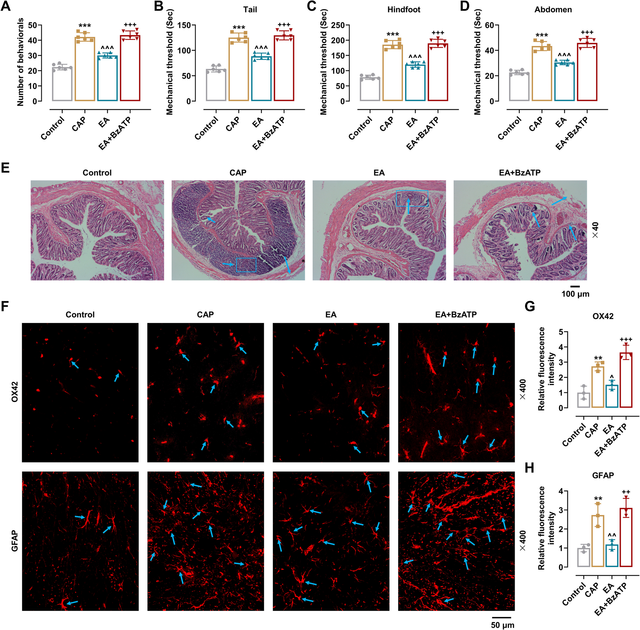
The rats were randomly divided into Control group, CAP group, EA group and EA + 3′-O-(4-Benzoylbenzoyl)-ATP (BzATP, a P2X4R agonist used to verify that EA alleviates pain by inhibiting P2X4R) group, with 12 rats in each group. Rats in CAP, EA and EA + BzATP groups were stimulated by CAP (TOP0198, Shanghai Taopu Biomedical Technology Co., Ltd, China) to establish visceral pain model. 1% (weight/volume) CAP for visceral pain induction was prepared in a solvent containing 10% anhydrous ethanol (459,836, Sigma-Aldrich, Germany), 10% TWEEN® 80 (P1754, Sigma-Aldrich, Germany), and 80% sterile saline (ZY033, Shanghai Zeye Biotechnology Co., LTD, China). Pentobarbital (50 mg/kg, P3761, Sigma-Aldrich, Germany) was used for anesthesia [15], and the exposed skin around the anus of rats was smeared with Vaseline (PF02212, PUFFE, China). In CAP, EA, and EA + BzATP groups, circular tube (diameter = 1.5 mm) was quickly inserted into the rectum through the anus of rats, and 0.5 ml of 10–4 mmol/L CAP was injected into the rectum every 5 min. In the Control group, the same volume of saline was injected into the rectum through the anus of rats [16].
EA treatment
Meanwhile, rats in the EA and EA + BzATP groups were treated with EA. Simply put, the self-made fixator was used to acupuncture the rats when they were awake and the acupuncture sites were carefully disinfected with povidone iodine (PVP1, Sigma-Aldrich, Germany). 0.35*25 mm acupuncture needle was inserted into the “Baliao” acupoints of the rat (rats have three pairs of posterior sacral foramina, where the 2nd and 3rd pairs are similar to those of humans: Ci Liao (BL 32), Zhong Liao (BL 33), and Xia Liao (BL 34). The Ci Liao (BL 32) and Zhong Liao (BL 33) are located 5–10 mm lateral to the superior border of the 2nd and 3rd intervertebral space of the rats’ tail, while the Xia Liao (BL 34) is located 5–10 mm lateral to the superior border of the 3rd and 4th intervertebral space of the rats’ tail) [17]. The needles were connected with HANS acupuncture point nerve stimulator (HANS-200A, Huawei Co., Ltd., Beijing, China). The parameters were set as follows: 2–15 Hz, and square wave current output (pulse width: 0.2 ms). Intensities ranging from 1 mA to 1.5 mA were delivered for a period of 30 min. The acupuncture was performed once a day at a fixed time for 7 days. Rats in EA and EA + BzATP groups received EA 2 h after waking up, while rats in the Control group and CAP group were inserted needles, but did not receive electrical stimulation [18].
BzATP Treatment
Afterwards, rats in Control group, CAP group and EA group received subdural injection of 0.25 ml 5% dimethyl sulfoxide (D2650, Sigma-Aldrich, Germany), while rats in EA + BzATP group received subdural injection of 0.25 ml BzATP (10.5 nmol [19], BCP32054, BioChemPartner, China) in 5% dimethyl sulfoxide [20].
Visceral pain-related behaviors
The number of visceral pain-related behaviors (licking abdomen, stretching, contractions of abdomen etc.) of rats in all the groups were observed and recorded as previously presented after 7 days of treatment [21].
Measurement of mechanical pain threshold
The visceral pain sensation usually induces changes in the rat's somatic response to mechanical stimuli. Use von Frey filaments to study the changes in the response of the skin on the rat's tail, hindfoot and abdomen to mechanical stimulation. The rats were placed in a transparent plexiglass box with a metal mesh at the bottom. The tail, hindfoot and abdomen of rats were stimulated with the electronic Von Frey system (IITC, USA) (gradient folding force: from 2.0 g/s to the cut-off force 50 g, each force was applied five times, the measurement interval was 30 s). Retracting the paws, arching the back and lifting the tail of the rat were considered as positive reactions. The strength given in response was recorded and the average value of three measurements was calculated [22, 23].
Hematoxylin/eosin (HE) staining
Following the above experiments, sodium pentobarbital (45 mg/kg, P3761, The BSZH Scientific Inc, China) was injected through the abdominal cavity to anesthetize the rats [24]. Blood samples were collected from the abdomen of rats and were centrifuged at 1000 × g for 20 min. Supernatant was removed and stored at −20℃ until use. The rats were then sacrificed by cervical dislocation. Meanwhile, rectal tissue and L4-L5 spinal cord tissue from the rats were collected and immersed in 30% sucrose solution. Then, a low-temperature slicer (KD-2850, Kohuai, China) was used to prepare 30-μm thick sections of rectal tissue. The sections were stained with Hematoxylin (C0105S, Beyotime, Shanghai, China) (5 min) and eosin (C0105S, Beyotime, Shanghai, China) (1 min). After washing, the sections were rinsed with a graded ethanol (70% 10 s, 80% 10 s, 90% 10 s, and 100% for 10 s) and transparentized with xylene (534,056, Sigma-Aldrich, Germany) for 5 min. Then, the sections were immersed in fresh xylene for another 5 min, sealed by resinene and observed under a microscope (× 40, BX61VS, Olympus, Japan).
Immunofluorescence
The spinal cord tissues obtained during the above steps were cut into 40-μm thick sections using the low-temperature slicer. The sections were fixed in 4% paraformaldehyde (158,127, Sigma-Aldrich, Germany), and then permeabilized in phosphate buffered saline (PBS) with 0.5% Triton‐X‐100 (Thermo Fisher HFH10, Thermo Fisher) for 15 min. 3% bovine serum albumin (BSA) (V900933, Sigma-Aldridge, Germany) was used for blockage at room temperature for 30 min. Then, the sections were incubated with Anti-glial fibrillary acidic protein (GFAP, ab4648, 1:10, Abcam, UK) and Anti-OX42 (Alexa Fluor® 647) (ab216524, 1:100, Abcam, UK) overnight at 4℃, and reacted with matched secondary antibody goat anti-rabbit IgG H&L (Alexa Fluor®647) (ab150079, 1:5000, Abcam, UK) at 37℃ for 45 min. The nuclei were stained with DAPI (C1005, Beyotime, Shanghai, China). The results were observed under fluorescence microscope (× 400, BX61VS, Olympus, Japan) and evaluated with Image J.
Cell culture
HMO6 cells (CVCL_5G94, American Type Culture Collection) were soaked in 89% Dulbecco's Modified Eagle's Medium (DMEM, A1451801, Thermo Fisher, USA), supplemented with 10% fetal bovine serum (FBS, 10100139 C, Gibco, USA) and 1% penicillin–streptomycin (10.000 U penicillin and 10 mg streptomycin/ml, 15140122, Gibco, USA) at 37℃ with 5% CO2.
Cell grouping and treatment
Three different ways of cell grouping were performed in this paper. First, HMO6 cells were divided into Control (no treatment), CAP (treated with CAP (100 µM) [25]), and CAP + 5-BDBD (treated with CAP (100 µM) and 5-BDBD (10 µM, as the antagonist of P2X4R [26])) groups. Second, HMO6 cells were assigned into Blank, negative control (NC) and TRPV1 groups. HMO6 cells were not treated in blank group, and transfected with NC/TRPV1 overexpression plasmid in NC/TRPV1 group. Third, HMO6 cells were distributed to CAP, CAP + 5-BDBD, CAP + 5-BDBD + NC, and CAP + 5-BDBD + TRPV1 groups. Cells in all groups were treated with CAP (100 µM), and cells in all groups except the CAP group received 5-BDBD treatment. Besides, cells in CAP + 5-BDBD + NC group and CAP + 5-BDBD + TRPV1 group were given NC or TRPV1 overexpression plasmid transfection.
Measurement of proinflammatory cytokines
The spinal cord tissue was homogenized, and then centrifuged at 4000 × g for 15 min. The supernatant was taken for the measurement. Meanwhile, serum obtained and HMO6 cells was used for measurement of proinflammatory cytokines, interleukin (IL)−1β, IL-6 and tumor necrosis factor-α (TNF-α), using IL-1β enzyme-linked immunosorbent assay (ELISA) kit (SP12225, Wuhan Saipei Biotechnology Co. Ltd (spbio), China), IL-6 ELISA kit (SP12279, spbio, China), and TNF-α ELISA kit (SP12250, spbio, China). In light of the manufacturer’s instructions, 50 μl of the sample was incubated with 50 μl of biotin-labeled antibody in the wells at 37℃ (1 h). Then, the wells were washed and dried three times. Next, 50 μl of affinity chain enzyme-HRP was added for incubation (30 min) at 37℃. The wells were washed and dried again. After 50 μl of substrate A as well as B was added, incubation was performed for another 10 min at 37℃ in the dark. Lastly, the plate was removed and 50 μl of Stop Solution was added to stop the reaction. Optical density (OD) value of each well was immediately measured at a wavelength of 450 nm by the microplate reader (HH35000310, PerkinElmer, Berlingen, USA).
Transfection
TRPV1 overexpression plasmid was amplified and inserted into the pCMV6-Entry vector (PS10000, OriGene, USA). Meanwhile, the empty vector was regarded as the NC. Lipo6000™ Transfection Reagent (Lipo6000™, C0526) purchased from Beyotime (Shanghai, China) was used for the transfection. Briefly, 3 × 10⁵ HMO6 cells were seeded to 96-well plate (CLS3922, Corning, USA). Lipo6000™ (0.2 μl) as well as 100 ng TRPV1 overexpression plasmid/NC was diluted with 5 μl DMEM without serum. Then, HMO6 cells were co-cultured with the mixed solution of diluted Lipo6000™ and NC/TRPV1 overexpression plasmid at 37℃ for 6 h. Next, the cells were incubated in the medium for 24 h with a fresh complete medium. Finally, transfection efficiency was determined by quantitative real-time polymerase chain reaction (qRT-PCR).
qRT-PCR
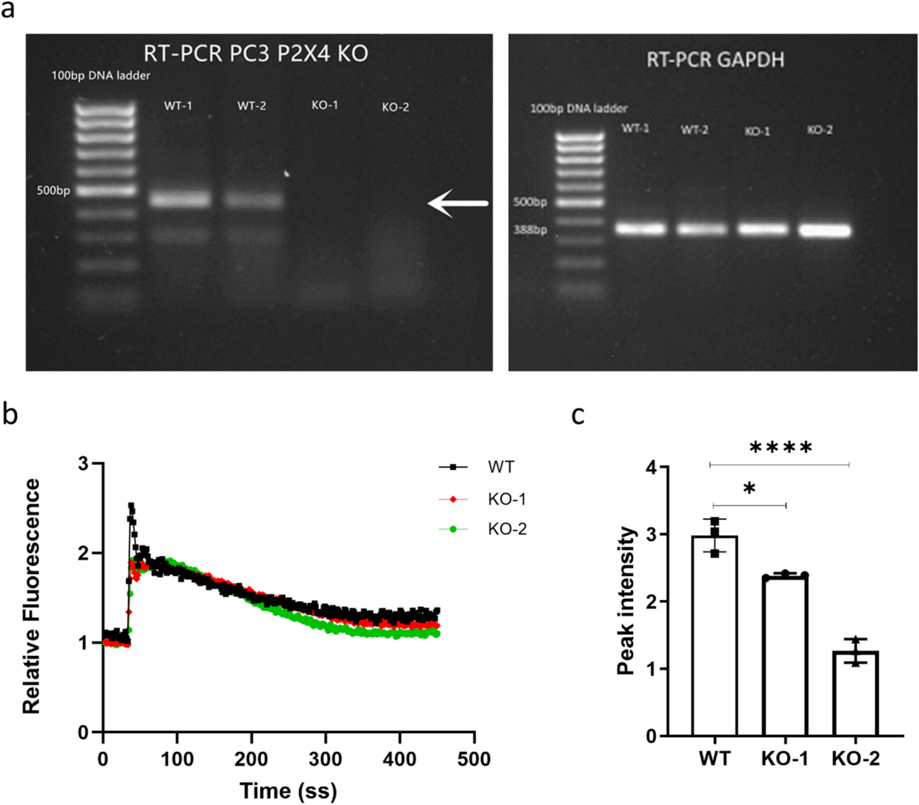
Total RNA was separated from HMO6 cells using NucleoSpin® RNA Plus kit (740,984.50, MACHEREY–NAGEL, Germany), and quantified by NanoDrop 2000 spectrophotometer (ND-2000, Thermo Fisher, USA). According to the manual of Transcriptor Reverse Transcriptase kit (C58-3,531,295,001, Roche, Switzerland), RNA was used for cDNA synthesis. With One-Step qRT-PCR Kit (4305A, GENENODE, China) and CFX384 Touch PCR detection system (1,855,195, Bio-Rad, USA), the PCR was performed with parameters as follows: 42℃, 30 min; 94℃, 2 min; 94℃ (20 s), 60℃ (20 s), 72℃ (20 s) × 40 cycles. The quantification was evaluated by 2−ΔΔCT method, where glyceraldehyde-3-phosphate dehydrogenase (GAPDH) served as the internal reference [27]. The sequences of primers were as follows: TRPV1-forward primer: ACTCTTCTCCCACACGAG; TRPV1-reverse primer: AAGATACTCCTGCGATCATA; GAPDH-forward primer: GGTGATGCTTTTCCTAGAT; GAPDH-reverse primer: CATACTTCTCATGGTTCACA.
Western blot
By lysing the HMO6 cells in RIPA Lysis Buffer (HY-K1001, MedChemExpress, USA), total cellular protein was separated. Then, the cell suspension was obtained after centrifugation (2,000 rmp, 5 min), and protein quantification was conducted using BCA Assay Kit (23,225, Thermo Fisher, USA). SDS-PAGE (89,888, Thermo Fisher Scientific, USA) was exploited for the separation of protein (20 μg), which was immediately loaded onto polyvinylidene fluoride membranes (88,518, Thermo Fisher Scientific, USA). Next, the membranes were blocked with Western Blocking Buffer (YT067, Biorab, China) for 10 min, and protein was then reacted at 4℃ with primary antibodies, including anti-P2X4 receptor (P2X4R) (ab134559, 43 kDa, 1:1000, Abcam, UK), anti-TRPV1 (ab305299, 95 kDa, 1:1000, Abcam, UK), anti-phosphorylated p38 (p-p38) (ab4822, 38 kDa, 1:1000, Abcam, UK), anti-p38 (ab170099, 42 kDa, 1:1000, Abcam, UK), anti-ionized calcium binding adapter molecule 1 (Iba-1) (ab178846, 17 kDa, 1:1000, Abcam, UK), and anti-GAPDH (ab8245, 36 kDa, 1:1000, Abcam, UK) antibodies overnight, followed by the incubation with HRP-conjugated secondary antibodies rabbit anti-goat IgG H&L (ab6741, 1:5000, Abcam, UK), anti-mouse IgG H&L (HRP) (ab6728, 1:2,000, Abcam, UK) and goat anti-rabbit IgG H&L (HRP) (ab6721, 1:2000, Abcam, UK) for 60 min. Next, the protein bands were visualized by Novex™ enhanced chemiluminescence reagent kit (WP20005, ThermoFisher Scientific, USA) and electro-chemiluminescent system (BioRad, USA), and evaluated by a semi-quantification software, ImageJ, with GAPDH as the internal reference.
Statistical analysis
Statistical analyses were performed using GraphPad Prism 8.0. Measurement data were expressed as mean ± standard deviation. Normal distribution was tested with Shapiro–Wilk test. One-way analysis of variance (ANOVA) was used for comparison between multiple groups, followed by Tukey's post-hoc test, and p < 0.05 was considered statistically significant.

Comments (0)