Animals
All animal studies adhered to the principles of the European Communities Council Directive (2010/63/EU), and relevant national licenses were approved by the Research Ethics Committee of the Royal College of Surgeons in Ireland (RCSI) (REC 1322) and the Irish Products Regulatory Authority (AE19127/P038). 8–12-week FVB/NJ wild-type male mice were sourced from the Biomedical Research Facility (BRF, RCSI, Dublin, Ireland). Mice were housed in groups of 2–5 per cage and kept in a controlled animal facility on a 12 h light/dark cycle at 22 ± 1ºC and humidity of 40–60%.
Status epilepticus mouse model
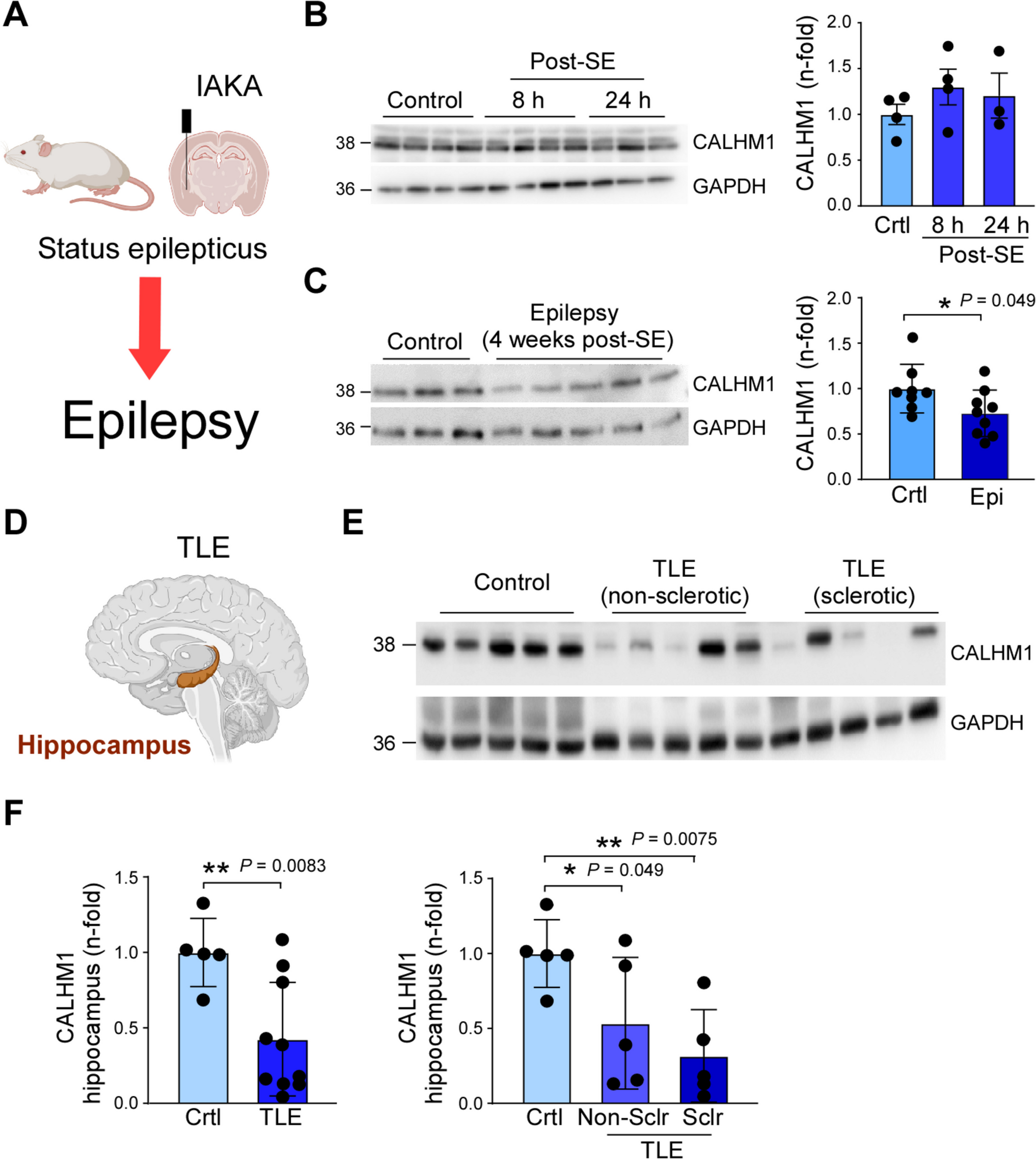
Status epilepticus (SE) was induced as described previously [18]. Isoflurane anesthetized mice (5% induction, 1–2% maintenance) were placed in a stereotaxic frame and a midline scalp incision was performed to expose the skull. To minimize pain during and post-surgery, mice were treated with buprenorphine (0.05 mg/kg) and EMLA cream (Aspen Pharma, UK). A guide cannula for intra-amygdala kainic acid (IAKA) injection (coordinates from Bregma; AP = −0.94 mm, L = −2.85 mm) and three cortical electrodes, one on top of each hippocampus and the reference electrode on top of the frontal cortex, were fixed in place with dental cement. Approximately 1 h post-EEG electrode and cannula implantation, SE was induced in awake, hand-restrained mice, by a microinjection of 0.2 µg KA in 0.2 µl phosphate-buffered saline (PBS) (Sigma-Aldrich, Dublin, Ireland) into the right basolateral amygdala. Vehicle-injected control animals received 0.2 µl of PBS (pH = 7.4) solution. The anticonvulsant lorazepam (6 mg/kg) (Wyetch, Taplow, UK) was delivered intraperitoneal (i.p.) 40 min following IAKA or vehicle to curtail seizures and reduce morbidity and mortality.
Drug administration
CGP37157 [15] was delivered by an intracerebroventricular (i.c.v.) injection (2 µl) 10 min prior to IAKA injection via a previously implanted cannula fixed with dental cement (coordinates from Bregma; AP = −0.4 mm, L = −1 mm, depth 2 mm). Animals were divided into three treatment groups: (i) control, injected with vehicle (PBS), (ii) mice receiving 1 µM of CGP37157, and (iii) mice receiving 10 µM of CGP37157.
Seizure severity analysis
An electroencephalogram (EEG) was recorded using an Xltek recording system (Optima Medical Ltd, Guildford, UK) [18]. To analyze EEG frequency and amplitude signals, EEG data were uploaded into Labchart7 software (AD Instruments Ltd, Oxford, UK). Seizure onset was analyzed offline. Seizure onset was defined as first seizure burst detectable on the EEG consisting of high amplitude (> twice baseline) high frequency polyspiking of a minimum of 5 s in duration. The duration of high-frequency (> 5 Hz) and high-amplitude (HFHA) (> 2 times baseline) polyspike discharges of ≥ 5 s duration was also counted manually by a reviewer blinded to treatment.
Human brain tissue
All subjects gave their informed consent for inclusion before they participated in the study. The study was conducted in accordance with the Declaration of Helsinki, and the protocol was approved by the Ethics Committee of Beaumont Hospital, Dublin (05/18). Briefly, patients (N = 10) were referred for surgical resection of the temporal lobe for the treatment of intractable TLE. After temporal lobe resection, cortex and hippocampi were obtained from the same patient and frozen in liquid nitrogen and stored at −80 °C until use. A pathologist assessed hippocampal tissue and confirmed the absence of significant neuronal loss. Control (autopsy) temporal hippocampi (N = 5) and cortex (N = 5) were obtained from individuals from the Brain and Tissue Bank for Developmental Disorders at the University of Maryland, Baltimore, MD, U.S.A. Brain sample and donor metadata are available in Supplementary Table 1.
Western blotting
Western blot analysis was performed as described previously [18]. Lysis buffer (100 mM NaCl, 50 mM NaF, 1% Tx-100, 5 mM EDTA pH 8.0, 20 mM HEPES pH 7.4) containing a cocktail of phosphatase and protease inhibitors was used to homogenize hippocampal brain tissue and to extract proteins. Following electrophoresis, proteins were transferred to a nitrocellulose membrane (GE Health Care, Illinois, USA) and immunoblotted with CALHM1 antibody (Proteintech Europe) (1:1000, prepared in 5% milk- tris-buffered saline-tween (TBST)). Membranes were incubated with horseradish peroxidase-conjugated goat anti-rabbit (1:5000, prepared in 5% milk-TBST, Sigma-Aldrich, Dublin, Ireland). Protein bands were visualized using Fujifilm LAS-4000 system with chemiluminescence (Immombilon Western HRP substrate, Merck Millipore, Massachusetts, USA) followed by analysis using ImageJ. Protein quantity was normalized to the loading control GAPDH (1:5000 prepared in 5% milk-TBST; anti-mouse; Sigma-Aldrich) or β-Actin (1:5000 prepared in 5% milk-TBST; anti-mouse; Sigma-Aldrich).
Statistical analysis
Statistical analysis of data was carried out using GraphPad Prism 8 and STATVIEW software (SAS Institute, Cary, NC, U.S.A). Analysis of variance (ANOVA) with post hoc Fisher’s protected least significant difference was used to analyze group data of three or more. For two-group comparisons, Student’s t-test was used to determine statistical differences between groups. Normality and lognormality test were used to verify the normal distribution between groups. Outliers were identified according to the Grubbs’ test. Significance was accepted at *P < 0.05.

Comments (0)