
Remember me
Correction to: Purinergic Signalling
Unfortunately, the original published version of the article does not contain the correct figures for this article. Below are the three correct figures with their respective captions:

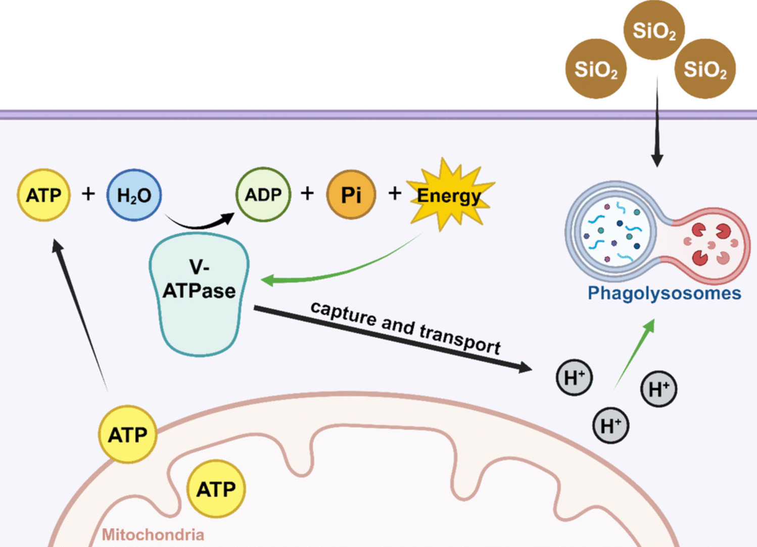
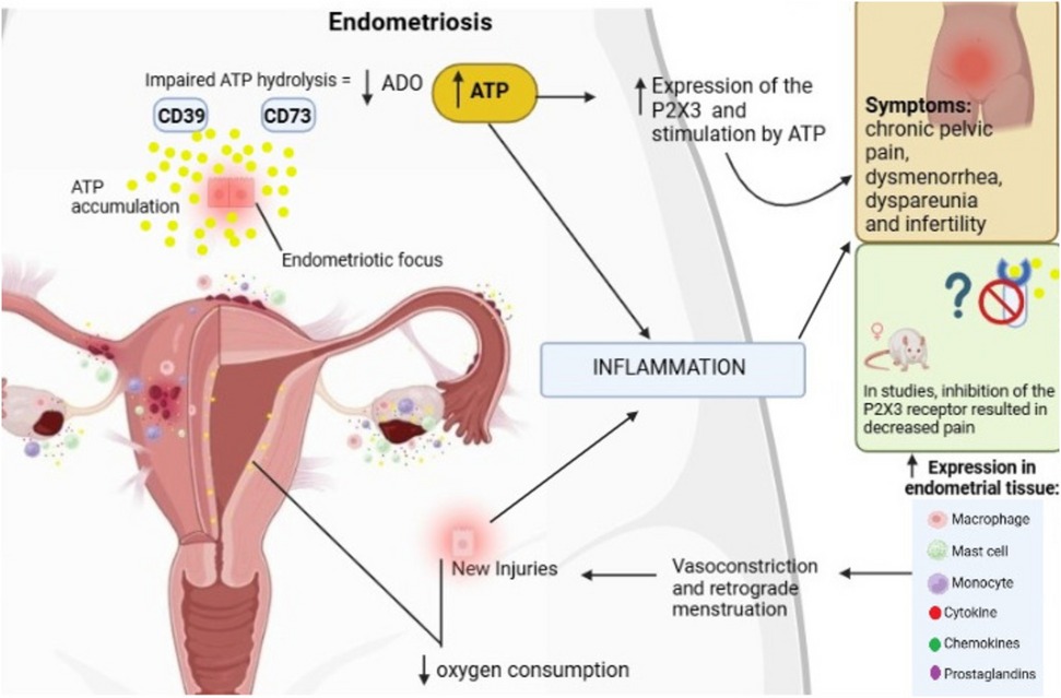
Fig. 1 In the endometrial tissue of individuals with endometriosis, the immune cells that secrete cytokines, chemokines and prostaglandins (depending on their cell lineage) increase, causing inflammation and tissue vasoconstriction, thus promoting inflammation and new endometriotic lesions, with the contribution of extracellular ATP. In addition, there is a change in the cellular respiration pathway, with a decrease in substances such as NADH, FAD, carnitine, creatine, malic acid and tryptophan, reducing oxygen consumption

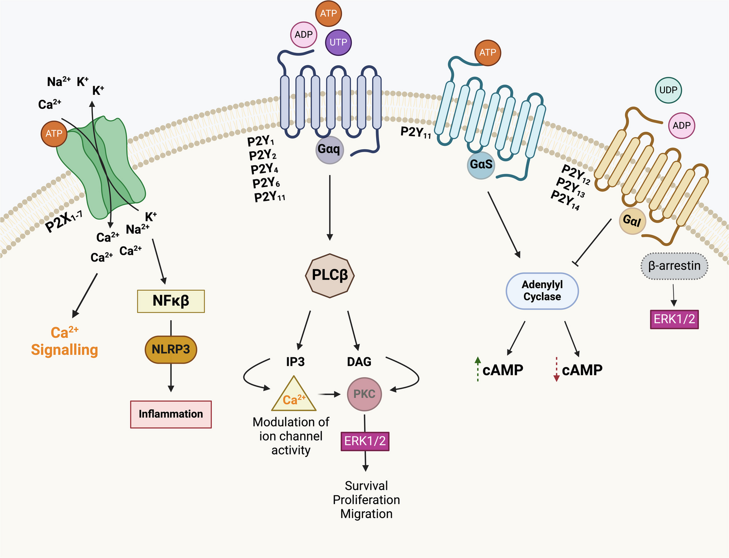
Fig. 2 In women with endometriosis, P2X3 receptors are expressed in more significant quantities in the endometrial tissue, especially in the disease foci. Inflammation and impaired expression of the CD73-CD39 axis increase the available ATP levels, which promotes constant stimulation of these receptors and causes Ca++ influx and depolarization of nociceptive fibers close to the endometriotic foci. Furthermore, it is worth noting that P2X3 receptors also have high affinity for Na+/K+ ions, which are associated with membrane potential as well

Fig. 3 The inflammation resulting from cell damage in endometriotic foci correlates with increased ATP release, P2X3 receptor expression, and consequent sensitization. In addition, it generates hyperstimulation of nerve fibers, mainly types C and Aδ, which activate the ATF3/AP-1 pathway at the dorsal root ganglion level, generating pain impulses to the CNS
The original article has been corrected.
Comments (0)