Clinical studyPatients
Our retrospective cohort study recruited 151 patients with HIV infection of more than 1 year in September 2023 from outpatient clinics at Shanghai Public Health Clinical Center. All participants who met the following inclusion criteria were included: (1) Patients’ age between 30 and 80 years old. (2) Patients who have been receiving at least 1 year of continuous HARRT therapy. Participants were excluded from the study if they ever had following diseases before medication: (1) Lower limb injury history; (2) Lower limb developmental abnormalities; (3) Lower limb deformities or history of lower limb fractures; (4) Pre-existing lower limb joint dysfunction; (5) Bilateral lower limb length discrepancy; (6) history of lower limb surgical infections; (7) History of intra-articular injections; rheumatoid arthritis or other inflammatory joint diseases; Paget’s disease; synovial chondromatosis; joint infection; osteochondroma; gout; osteopetrosis and other nervous system diseases affecting lower limb mobility such as spine degeneration or brain injury; (8) Inability to complete the questionnaire; (9) Inability to complete imaging examinations.
All patients were separated in two cohorts depending on whether they have received regimes containing lopinavir/ritonavir for more than 1 year. It is important to notice that lopinavir/ritonavir is the only available PI option in Shanghai.
The study was approved by the ethics committee of Shanghai Ninth People’s Hospital affiliated to Shanghai Jiao Tong University School of Medicine (SH9H-2022-T285-1). All patients gave informed consent.
Outcomes
Knee Injury and Osteoarthritis Outcome Score (KOOS score) was acquired through questionnaires. Radiological examinations (anterior and lateral radiographs of both knees) were done and evaluated by two experienced joint surgeons (ZZ and HL) who were blinded to the grouping of patients in Shanghai Ninth People’s Hospital. Knee radiographs were graded based on the Kellgren-Lawrence (KL) grading system43 from 0 to 4 (0, no OA; 1, doubtful OA; 2, minimal OA; 3, moderate OA; 4, severe OA). Physical examinations were done by experienced joint surgeons (ZZ and HL) and positive clinical signs were defined as presence of knee joint space tenderness, patellofemoral joint tenderness, or patellofemoral joint grinding pain, either individually or in combination. KOOS score is a knee specific instrument to assess patients’ opinions about their knees44 and it consists of 5 subscales: including Pain, other Symptoms, Activities of Daily Living (ADL), Sport and Reaction Function (Sport/Rec) and knee-related Quality of Life (QOL). The score ranges from 0 to 100, with 0 representing worse problems and 100 representing no problems for each subscale. A total score has not been validated and is not recommended.
Covariates
Baseline characteristics including demographic characteristics (i.e., age, sex, and postmenopausal status), HIV features (i.e., durations of HIV infection, CD4 level and viral load) and metabolic features (i.e., BMI, triglycerides, total cholesterol, HDL-cho, LDL-cho and glycaemia) were assessed using the nearest available data prior to the index date.
Basic studyHuman cartilage specimen collection
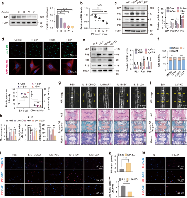
Cartilage samples were acquired with patients’ informed consent with ethnic approval from the ethics committee of the Shanghai Ninth People’s Hospital affiliated to Shanghai Jiao Tong University School of Medicine (SH9H-2021-T401-4). Human tibial plateaus were collected from patients undergoing TKA (Total knee arthroplasty) (n = 41; aged 70.6 ± 8.0 years; 10 males and 31 females). we drilled an osteochondral plug with an area of 0.5 cm × 0.5 cm in center of medial and lateral part of tibial plateau. The removed plugs were fixed in 4% paraformaldehyde for further histological assessment (Safranine O/Fast Green staining) and Zmpste24 immunohistochemistry.
Animals
Zmpste24 globally knockout mice (Zmpste24-/-) were bought from GemPharmaTech. Routine genotyping of mice tail DNA was performed according to the instruction of Jackson Laboratory with PCR kit (Takara, Japan). Homozygous mice (Zmpste24-/-) used in this study were all bred from heterozygous mice (Zmpste24+/-). Wild-type C57BL/6J mice were purchased from the animal center of Shanghai Ninth People’s Hospital. All mice were of C57BL/6J background. All mice were provided with a standard diet and housed in specific pathogen free (SPF) cages at a temperature of 24°C and a humidity of 60%. All animal experiments were approved by ethics committee of the Shanghai Ninth People’s Hospital affiliated to Shanghai Jiaotong University School of Medicine (SH9H-2022-A858-1).
For mice receiving the destabilization of the medial meniscus (DMM) surgery, we transected the medial meniscotibial ligament to enable destabilization of the medial meniscus, which is in accordance with the protocol of previous study.45
For the senile OA model, we fed C57BL/6J mice normally and sacrificed them at 24 months old, with 3-month-old mice as controls. Both lower extremities were collected and fixed for further radiological and histological analysis.
Drug administration
The mice were administered intraperitoneally PBS (control), Lopinavir/Ritonavir (100/25 or 200/50 mg/kg) every other day beginning 1 week after DMM surgery. Lopinavir/Ritonavir combination was dissolved in corn oil and the drug concentration was in accordance with the study of Alonso et al.36 In our study, for the in vitro experiments, we only used lopinavir to stimulate the chondrocytes. However, for the in vivo experiments, we combined lopinavir with ritonavir in accordance with the component ratio for patients. It is important to note that ritonavir was not used for its antiviral activities but rather as a booster for other antiviral drugs. Therefore, we only used the combination of lopinavir and ritonavir in the in vivo experiments to simulate the situation when patients take these medications in clinical practice.
Hotplate pain assay
The mice were placed on the hotplate at 55 °C.46,47 Response time was recorded as the period between hindlimbs touching the hotplate and the occurrence of response behaviors such as paw shaking, paw licking, or jumping. The hotplate pain assay was performed every 2 weeks after DMM surgery, and at least three replicative response times were recorded for each mouse. The observers were blinded to the grouping of all tested mice.
Von Frey tests
Von Frey test was carried out to measure the pain threshold in hindlimbs of mice using an electronic Von Frey filament (Xinruan Technology, China). Each mouse was allowed to acclimate to the test chamber for 30 min before the test. A positive response was recorded if the animal exhibited any nociceptive behavior such as paw withdrawal, shaking, or licking. The threshold force exerted by the electronic filament was recorded, with at least three replicative forces measured for each mouse in each test.
Micro-CT
After a 2-day fixation period, knee joints obtained from Zmpste24-/- mice were scanned using a high-resolution micro-CT scanner (Skyscan 1072, Belgium) with a pixel size of 9 μm, a 55 kVp source, and a 145 μAmp current. Osteophyte volume was quantified, and representative three-dimensional reconstructions of subchondral bone sections were generated using CT-Vox software (Bruker, Germany), as described in our previous study.14
Histologic analyses
Serial tissue sectioning of 5 μm thickness in a sagittal or coronal plane was performed and stained with Safranine O/Fast Green to evaluate cartilage degeneration and clefts. OARSI score was evaluated according to the standards of Sophocleous.45 The OARSI score for each mouse knee joint is obtained by adding the OARSI scores of the femur and tibia. Furthermore, sections were stained with Hematoxylin and eosin (HE) staining to evaluate synovial inflammation, and the synovitis score was calculated using Krenn’s principle.48,49
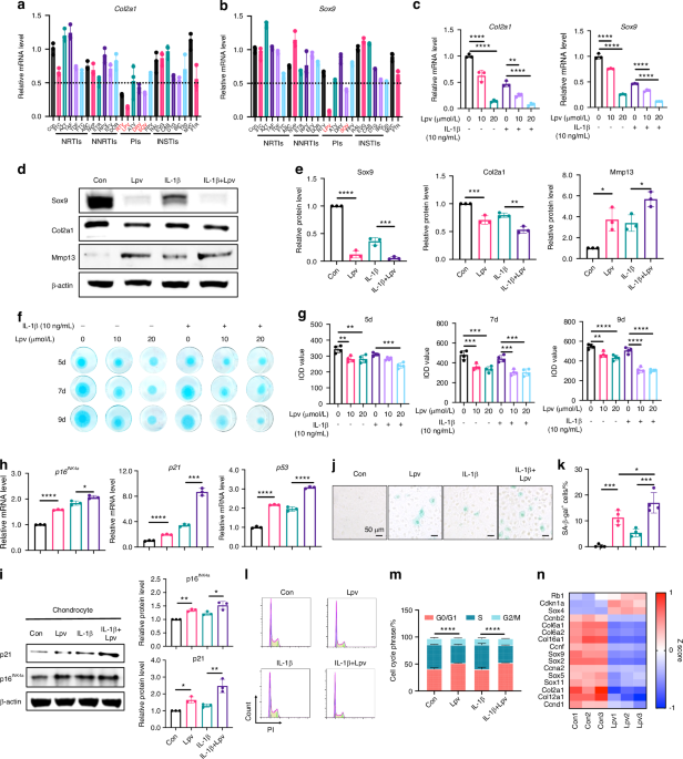
Compound library screening
A customized compound library was obtained from TopScience, which included 25 FDA-approved anti-HIV drugs from seven different categories (listed in Table S2). Each drug was added individually at a final concentration of 20 μmol/L to ATDC5 cells. Dimethylsulfoxide (DMSO) was used as a control. After 24 h of incubation, RNA was extracted from the cells for the detection of Col2a1 and Sox9 expression using RT-qPCR.
Co-immunoprecipitation (Co-IP)
For Co-IP, whole protein extracts were lysed in 1 mL of cell lysis buffer for IP (P0013, Beyotime, China) containing 1% protease inhibitor cocktail at 4 °C for 30 min. Anti-Flag or Anti-HA immunomagnetic beads (Bimake, China) were added to the proteins and thoroughly mixed for 1 h at room temperature. The immunomagnetic beads were then collected using a magnet, washed three times with tris-buffered saline and Tween 20, and mixed with 1X loading buffer (Biosharp). The mixture was denatured at 99 °C for 15 min, followed by an immunoblotting procedure.
Cell culture and reagents
Murine chondrocytes were isolated from newborn C57BL/6J mice following a previously established protocol.50 Chondrocytes were cultured in Dulbecco’s Modified Eagle Medium/Nutrient Mixture F12 (DMEM/F12) supplemented with 10% fetal bovine serum (FBS) and 1% penicillin-streptomycin (Gibco, Thermo Fisher Scientific, Waltham, MA, United States).
The immortalized mouse chondrocyte cell line ATDC5 and HEK293T cells were obtained from the Cell Bank of the Chinese Academy of Sciences (Shanghai, China). ATDC5 cells were cultured in DMEM with 4.5 g glucose/L, 5% FBS, and 1% penicillin-streptomycin. Cells were incubated in a humid environment at 37 °C with 5% CO2. Lopinavir, ritonavir, and doxorubicin were purchased from MCE (New Jersey, United States) and dissolved in DMSO. The final concentration of DMSO in the cell culture medium was maintained below 0.1%. IL-1β protein was purchased from Genscript.
Plasmid and lentiviral transfections
All plasmids and lentiviruses used for cell transfection, including HA-Usp7, Flag-Lamin A, sh-Zmpste24, and Lenti-Mdm2, were purchased from Obio Biotechnology. For transfection of 293T or ATDC5 cells with these plasmids, 10 μg of the target plasmid was mixed with 15 μL of lipofectamine 3000 (Thermo Fisher Scientific) and 10 μL of P3000. The mixture was incubated for 15 min and then added to the cells for a 48-h transfection. Lentivirus was added to the medium with an optimal multiplicity of infection (MOI) of 20 to infect the cells. Puromycin was added 48 h after infection to select for a stable Mdm2 overexpression cell line.
3D culture
For 3D culture, 2 × 10⁵ primary chondrocytes were centrifuged for 5 min at 1 500 r/min after which the pellet was cultured in chondrogenic medium (MUXMX-90041, Cyagen). Culture medium was replaced twice a week. After 21 days of chondrogenic 3D culture, the pellets were fixed in 4% PFA, dehydrated with gradient concentration of alcohol and embedded in SAKURA Tissue-Tek O.C.T. Compound. 5 μm sections were sliced with a Leica freezing microtome followed by Safranine O, Alcian Blue and Toluidine Blue staining.
Immunofluorescence (IF)
For cell IF staining, cells were washed with PBS for three times and fixed with 4% PFA. After 0.1% Triton-X permeabilization for 10 min and 5% BSA block for 30 min at room temperature, cells were incubated with primary antibodies, including anti-Lamin A (4777, 1:100, CST), anti-Usp7 (4833, 1:100, CST) and anti-Mdm2 (66511-1-lg, 1:100, proteintech) at 4 °C overnight. Cells were incubated with corresponding secondary antibody, including goat anti-rabbit IgG H&L (Alexa Fluor 488, 1:1 000, Abcam) and goat anti-mouse IgG H&L (Alexa Fluor 555, 1:1 000, Abcam) at room temperature for 1 h in the next day. Nucleus was stained with DAPI and representative images were taken with a high-resolution Leica confocal microscope. ImageJ software was utilized for quantitative analysis.
Quantitative real-time PCR (qRT-PCR)
Total RNA was isolated from cells using the Total RNA Extraction Kit (R6812-01HP, Omega Bio-tek Inc., Norcross, GA, United States). qRT-PCR was performed using an ABI 7500 Sequencing Detection System (Applied Biosystems, Foster City, CA, USA). The primer sequences used for the target genes are listed in Table S3.
Statistical analysis
Baseline characteristics and outcomes of included patients were presented as number (percentage) for categorical variables and mean (standard deviation) for continuous variables. χ2 test was used for categorical variables and two sample t-test was used for continuous variables in order to evaluate the differences between protease inhibitor and non-protease inhibitor patients.
Multivariable linear regression was used to determine the association between PI use and KOOS scores, including Pain, Symptom, DL, Sport/Rec, QOL, etc. Standardized coefficients (β) and their 95% confidence intervals were calculated. Multivariable logistic regression was used to clarify the association between PI use and radiological outcomes. Odds ratios (OR) and their 95% confidence intervals were calculated. Four models that adjusted for different variables were used to evaluate the stability of the results. Model 1 was not adjusted for any covariate. Model 2 was adjusted for age and sex. Model 3 was further adjusted for metabolic features including BMI, triglycerides, total cholesterol, HDL-cho, LDL-cho and glycaemia. Model 4 was adjusted for terms in model 3 and HIV features, including durations of HIV infection, CD4 level, and viral load (undetectable or not). All statistical analyses were performed using SPSS version 25.0 (SPSS Inc., Chicago, IL, USA). All P values for our clinical study are two-sided. A P < 0.05 indicated a statistical significance.
In basic studies, for data with a normal distribution, the unpaired Student’s t-test was used for comparisons between two groups. One-way ANOVA followed by Turkey’s post hoc tests were used for comparisons among three or more groups. The Mann-Whitney and Kruskal-Wallis tests were used for non-normally distributed data. Spearman correlation analysis was used to test the correlation between OARSI score and Zmpste24 expression level, and regression equations were obtained using simple linear regression methods. GraphPad Prism 8.0 (GraphPad Software Inc., San Diego, CA, USA) was used for designing and drawing statistical charts. Statistical significance was set at P < 0.05.


Comments (0)