AnimalsGeneration of a conditional C451A mERα knockout mouse model
To generate a transgenic mouse model facilitating a conditional amino acid shift (cysteine > alanine) at site 451 (C451A) in Esr1 by introducing the mutation TGC > GCA, a targeting vector was constructed as follows (Fig. S2a): The 5’ end of the vector contained a short homology arm (SHA) targeting the intronic region before exon 7, while the 3’ end of the vector contained a long homology arm (LHA) targeting the intronic region after exon 7, leading to excision of the original exon 7 upon homologous recombination. Between the SHA and LHA, the vector contained a floxed region (FR) flanked by two loxP sites, followed downstream by exon 7 containing the C451A amino acid substitution, and the negative selection marker thymidine kinase (TK). The FR contained the following components: two positive selection markers, Neomycin resistance (NeoR) and Puromycin resistance (PuroR), flanked by the Flp recombination sites FRT and F3, respectively; the WT exons 7–9, fused without introns; and a polyadenylation signal (hGHpA cassette) to prevent downstream transcription. After Flp recombination, the floxed allele results in transcription of the fused WT exons 7–9, leading to translation of a WT ERα protein. When a Cre recombinase is introduced, the floxed region is excised and the exon 7 containing the mutation is instead transcribed, leading to translation of the mutated ERα C451A protein. The targeting vector was generated using BAC clones from the C57BL/6J RPCI-23 BAC library and was transfected into the Taconic Biosciences C57BL/6N TacES cell line. Homologous recombinant clones were isolated using double positive (NeoR and PuroR) and negative (TK) selections. The generation of this transgenic mouse model (C57BL/6NTac-Esr1tm6116(C451A)Tac) was conducted at Taconic (Borup, Denmark), and is denoted as C451Af/f mice.
To verify that the introduction of the floxed region does not affect the phenotype, homozygous C451Af/f female mice were compared to WT littermates at 16 weeks of age. The C451Af/f female mice manifested the same phenotype as WT female littermates regarding organ weights, serum steroid concentrations, and bone parameters (Table S2).
Generation of a global C451A mERα knockout mouse model
To generate mice with a global ERα C451A mutation (C451A mice), that lack mERα signaling in all cells, Pgk1-Cre mice were used, targeting both germ cells and somatic cells.40 C451Af/f mice were bred with Pgk1-Cre mice, and insertion of the mutation was confirmed by DNA sequencing of ear clips from the C451A mice (n = 3) and WT littermates (n = 3) (Eurofins Genomics, Ebersberg, Germany), validating the C451Af/f model (Fig. S2b). The C451A mice, generated using Pgk1-Cre mice, exhibited disturbed serum steroid levels and unaffected skeletal bone mass compared to WT controls (Table S3). These results are in line with the phenotypes shown in C451A-ERα mice generated by knock-in mutation.22,28,39 C451A female mice and their WT female littermates were used as donors for the bone marrow transplant (BMT) experiment and in the in vitro experiments (see below).
Generation of an osteoblast lineage-specific C451A mERα knockout mouse model
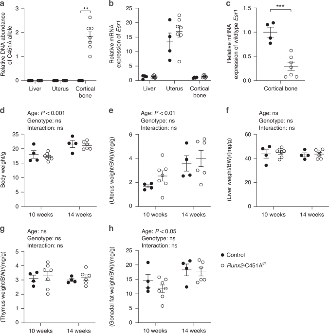
To generate mice with specific inactivation of mERα signaling in osteoblast lineage cells, C451Af/f mice were bred with Runx2-Cre mice, generating Runx2-C451Af/f mice. It has been shown previously that Runx2-Cre mice manifest the same skeletal phenotype as WT littermates.41Runx2-C451Af/f female mice were used to study the effects of lacking mERα signaling in osteoblast lineage cells, and homozygous C451Af/f female littermates were used as controls. Two independent studies were conducted, one terminated at 10 weeks, and one terminated at 14 weeks of age (n = 4–7 per group).
All animals were kept in a standard animal facility with regulated temperature (22 °C) and a 12-hour light:12-h darkness cycle. Mice were provided with a phytoestrogen-free pellet diet (Teklad diet 2016, Envigo, Indianapolis, Indiana, United States) and tap water ad libitum. Animal experiments were approved by the Ethical Committee for Animal Research in Gothenburg (Göteborgs djurförsöksetiska nämnd) and reported according to ARRIVE guidelines. Primers used for genotyping the mice are listed in Table S4.
At termination, mice were anesthetized with Ketador/Dexdomitor (Richter Pharma, Wels, Austria/Orion Pharma, Espoo, Finland). Blood samples were obtained from the axillary artery, followed by euthanasia through cervical dislocation. Soft tissues were dissected, weighed, snap-frozen in liquid nitrogen, and stored at −80 °C. Bone marrow was collected from tibia and femur by cutting the ends followed by centrifugation, snap-frozen in liquid nitrogen, and stored at −80 °C. The remaining cortical bone was collected from tibia and femur and stored at −80 °C for DNA isolation or RNA preparation (stored in RNAprotect Tissue Reagent, Qiagen, Hilden, Germany). Tibia, femur, and vertebra L5 were dissected, fixed in 4% paraformaldehyde for 2 days and then stored in 70% ethanol for further analyses. One vertebra L5 in the 10-week-old Runx2-C451Af/f group was excluded because of dissection damage. Humerus was dissected and stored at −20 °C for three-point bending analysis.
Bone marrow transplantation
Wildtype C57BL/6NTac female mice were lethally irradiated with a RS 2000 X-ray irradiator (Rad Source Technologies, Georgia, USA). The radiation dosage administrated was 4.5 Gy over a duration of 170 s per session, repeated twice with a four-hour interval of rest, reaching a total dosage of 9 Gy. After the second irradiation, BMT was performed by intravenous injection of 350 000 donor bone marrow cells obtained from either C451A female mice, which lack mERα signaling in all cells, or WT female littermates. The donor cells were purified before transplantation using EasyStepTM Mouse Hematopoietic Progenitor Cell Isolation Kit (STEMCELL Technologies, Vancouver, Canada). WT recipient mice receiving WT bone marrow (WT/WT) were used as controls for WT recipient mice receiving C451A bone marrow (WT/C451A). Antibiotic treatment with 0.6 mg/mL Baytril (Bayer, Leverkusen, Germany) in the drinking water was started one week before BMT and discontinued two weeks after BMT. All mice received radiation and BMT at 11 weeks of age, and they were sacrificed at 16 weeks of age. Since the mice were part of another experiment (unpublished data), they were sham operated at 10 weeks of age and received subcutaneous injections of vehicle treatment (Miglyol 812, OmyaPeralta GmbH, Hamburg, Germany) once every second day for three weeks before termination. The efficiency of cell replacement after irradiation and BMT was determined in a separate experiment, with flow cytometer analysis, showing that 79.5% ± 10.8% (SD) of the bone marrow cells were of donor origin 12 weeks post-BMT (for detailed method description see Fig. S3).
Serum analyses
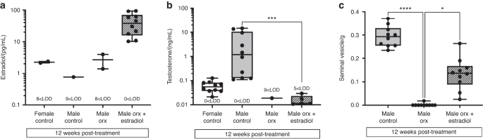
Circulating concentrations of serum E2 and T were quantified using high-sensitivity liquid chromatography-tandem mass spectrometry (LC–MS/MS) as described previously.42 The lower limit of quantification (LLOQ) of LC-MS/MS is 0.5 pg/mL for E2 and 5 pg/mL for T. Sex steroid concentrations below LLOQ were used as half of LLOQ (0.25 pg/mL for E2 and 2.5 pg/mL for T) in the statistical analyses. As a marker of bone formation, serum levels of procollagen type I N propeptide (P1NP, Immunodiagnostic Systems, Copenhagen, Denmark) were analyzed by ELISA and as a marker for bone resorption, terminal type I collagen fragments were measured in serum using an ELISA RatLaps kit (CTX-I, Immunodiagostic Systems).
DNA and RNA isolation, and real-time PCR
DNA was isolated from tissues using DNeasy Blood & Tissue Kit (Qiagen). Total mRNA from cortical bone was isolated using TriZol (Life Technologies, Thermo Fisher Scientific, Waltham, MA) and RNeasy Mini Kit (Qiagen). Total mRNA from soft tissues was isolated using RNeasy Mini Kit. The isolated mRNA was reversely transcribed into complementary DNA (cDNA) using the High-Capacity cDNA Reverse Transcription kit (Applied Biosystems, Thermo Fisher Scientific). Real-time PCR amplification was performed by Applied Biosystem StepOnePlus Real-Time PCR System (Thermo Fisher Scientific). The Assay-on-Demand primer and probe sets (Thermo Fisher Scientific) used in this study included Estrogen receptor alpha (Esr1: Mm00433147_m, targeting a cDNA region upstream of exon 6, thereby detecting both the WT and the mutated Esr1 expression) and 18S (4310893E), which was used for normalization. A customized assay (Thermo Fisher Scientific) was used to quantify the relative DNA abundance of the C451A-Esr1 gene: C451A F: GCCCAACCACACAGTCCATA, C451A R: TCTCCTCCTGATGTGTCTTGA, with FAM-MGB fluorescent dye to detect the C451A mutated DNA allele, and for normalization, an intronic region (Ctrl) upstream of the LoxP sites was quantified using the following primers with VIC-MGB fluorescent dye: Crtl F: CACATGACTGCTGGGCATTT, Ctrl R: AATGCACGTATGAGCACTGG. The relative RNA expression of WT Esr1 was measured by using PowerUpTM SYBRTM Green Master Mix (Applied Biosystems) and customized primers targeting the WT codon bases TGC at site 451 of Esr1 (Applied Biosystems, F: GGAAGCTCCTGTTTGCTCCT, R: GCAAAATGATGGATTTGAGGCA) normalized to the expression of Ppia3 (PrimerBank-MGH-PGA ID: 6679439a1, Applied Biosystems). The relative abundance of target sequences was normalized to either 18S, Ppia3 or Ctrl, and calculated using the ΔΔCt method.
Peripheral quantitative computed tomography
Peripheral quantitative computed tomography (pQCT) was performed with the pQCT XCT RESEARCH M (version 4.5B, Norland, UK) operating at a resolution of 70 μm as described before.43
Femur
Cortical bone parameters were analyzed in the mid-diaphyseal region at a distance proximal from the distal growth plate corresponding to 36% of the total length of the femur. The scan for trabecular bone parameters was positioned in the metaphysis of the femur at a distance proximal from the distal growth plate corresponding to 3% of the total length of the femur. The trabecular bone region was defined as the inner 45% of the total cross-sectional area.
Tibia
Cortical bone parameters were analyzed in the mid-diaphyseal region at a distance distal from the proximal growth plate corresponding to 30% of the total length of the tibia. The scan for trabecular bone parameters was positioned in the metaphysis of the tibia at a distance distal from the proximal growth plate corresponding to 2.6% of the total length of the tibia. The trabecular bone region was defined as the inner 45% of the total cross-sectional area.
High‐resolution microcomputed tomography
High-resolution microcomputed tomography (μCT) analysis was performed on the femur, tibia, and vertebra L5 using the Skyscan 1275 model (Bruker MicroCT, Billerica, Massachusetts, United States), with an X-ray tube voltage of 40 kV and a current of 200 μA. The angular rotation was set at 180°, with an angular increment of 0.40°. Voxel size was isotropically maintained at 7 μm.
Femur
Cortical parameters of femur were assessed in the diaphyseal region, initiating 5.2 mm from the distal growth plate, and extending longitudinally for 210 μm in the proximal direction. The trabecular bone analysis focused on the region proximal to the distal growth plate, within a defined volume of interest excluding cortical bone. The analysis of trabecular bone started 504 μm from the growth plate and extended a further longitudinal distance of 210 μm in the proximal direction.
Tibia
Cortical parameters of tibia were assessed in the diaphyseal region, initiating 5.2 mm from the proximal growth plate, and extending longitudinally for 210 μm in the distal direction. The trabecular bone analysis focused on the region distal to the proximal growth plate, within a defined volume of interest excluding cortical bone. The analysis of trabecular bone started 504 μm from the growth plate and extended a further longitudinal distance of 210 μm in the distal direction.
Vertebra
Analysis of cortical and trabecular parameters of L5 were initiated 7 µm caudal of the lower end of the pedicles, extending a further longitudinal distance of approximately 245 µm in the caudal direction. The region of interest for cortical and trabecular bone were manually drawn and analyzed using CTAn software.
Three-point bending
The three-point bending was performed with a span length of 5.5 mm and a loading speed of 0.155 mm/s was applied to the humerus using an Instron 3366 (Instron, Norwood, Massachusetts, United States). Biomechanical parameters were derived from the load deformation curves using Bluehill Universal software version 4.25 (Instron), and calculations were performed using Excel (Microsoft).
Dynamic histomorphometry
For dynamic histomorphometry, mice were administered intraperitoneal injections of the fluorochromes calcein and alizarin (Merck GmbH, Darmstadt, Germany) 9 days and 2 days prior to sacrifice, respectively. Following dissection, the femurs were fixed in 4% formaldehyde for 48 h, dehydrated using a graded ethanol series, and embedded in LR White Resin (London Resin Co, Ltd, UK). The femurs were sectioned transversely at the mid-diaphysis into 200-µm-thick slices. The slices were ground down to a single-cell layer of approximately 7 µm thickness using 800, 1 200, and 4 000 grit silicon carbide paper (Struers A/S, Rødovre, Denmark) with the aid of an automatic grinder (EXAKT® cutting and grinding equipment, EXAKT® Apparatebau GmbH & Co, Norderstedt, Germany). The dynamic cortical bone parameters were evaluated without any additional staining by using the Bioquant OSTEO software version 2023.v23.5.60 (Bioquant Image Analysis Corporation, Heidelberg, Germany) according to the American Society for Bone and Mineral Research guidelines.44
Primary osteoblast culture
Primary periosteal bone cells were isolated from calvariae of 3–5 days old global C451A mice and WT littermate controls by sequential enzymatic digestion.45 5–7 dissected calvariae per genotype were used in each experiment. The calvariae were washed in PBS after dissection and incubated in 5 mL 4 mmol/L EDTA in PBS at 37 °C, 600 r/min rotation table for 2 sequential 10-min digestions, followed by 7 sequential 10-min digestions of 5 mL 180 U/mL Collagenase type II (BioNordika, Worthington, Ohio, United States) in PBS. The first two collagenase fractions were discarded and the last five were pooled. Thereafter, the primary calvarial periosteal bone cells were cultured in complete α-MEM medium (Gibco, Thermo Fisher Scientific) supplemented with 10% heat-inactivated fetal bovine serum (Sigma), 2 mmol/L GlutaMAX (Gibco), 50 µg/mL gentamicin (Gibco), 100 U/mL penicillin and 100 µg/mL streptomycin (Gibco) for 3–5 days prior to the experiments. At the start of experiments, cells were detached with trypsin and re-seeded at approximately 18 000 cells/cm2 in osteogenic media (complete α-MEM medium supplemented with 4 mmol/L β-glycerophosphate, Sigma, and 0.28 mmol/L L-Ascorbic acid 2-phosphate sesquimagnesium salt hydrate, Sigma).
RNA isolation and real-time PCR
At the point of harvesting RNA (after 1, 4, and 7 days), cultured cells were lysed in RNeasy lysis (RLT) buffer with beta-mercaptoethanol (Qiagen). Total mRNA was isolated using RNeasy Micro Kit (Qiagen). cDNA reverse transcription and real-time PCR were performed and analyzed as described above. The Assay-on-Demand primer and probe sets (Thermo Fisher Scientific) used included alkaline phosphatase (Alpl: Mm00475834_m1), Sp7 transcription factor 7 (Sp7/Osterix/Osx: Mm04209856_m1), integrin binding sialoprotein (Ibsp: Mm00492555_m1), and 18S (4310893E), which was used for normalization.
Alkaline phosphatase (ALP) staining
At day 7, the cells were fixed by citrate buffered acetone and stained according to the commercial kit protocol (85L2-1KT, Sigma). In short, cells were fixed for 30 s and washed three times with distilled water (dH2O), followed by 20 min of ALP staining and three washes with dH2O. The photo for quantification was taken with ChemiDoc XRS+ (Bio-Rad, Hercules, California, United States) and analyzed by Image Lab 6.1.
All in vitro studies were repeated three times with similar results.
Statistical analysis
Statistical analysis and figure visualization were conducted using GraphPad Prism (version 10.0.3), while table analyses were carried out using Microsoft Excel (version 16.79.2). Values in the figures are displayed as individual values with mean (horizontal line) and SEM (vertical line), while tables present values as mean ± SD. For normally distributed continuous data, differences between two independent groups were assessed using Student’s t test (Excel). For data without normal distribution, the non-parametric two-tailed Mann–Whitney U test was applied to evaluate differences between two independent groups (GraphPad Prism). Two-way ANOVA was applied to examine the influence of two independent variables (GraphPad Prism), and Šidák´s multiple comparisons test was used to compare the difference between genotypes within each time point. In all conducted analyses, a difference was considered significant when P < 0.05.


Comments (0)