Cell lines and culture
The HCC cell lines (HepG2, Huh7, HepG2.2.15, MHCC97H), Treg cells, and HUVECs were acquired from the TCC cell bank (Shanghai, China). HepG2, Huh7, Treg cells, and HUVECs were grown in Dulbecco’s modified Eagle’s medium (DMEM, Wisent, 390-010-cl), and MHCC97H and HepG2.2.15 cells were cultured in Eagle’s minimum essential medium (EMEM, Wisent, 320-006-CL) supplemented with 10% fetal bovine serum (FBS, Wisent, 085–150) and penicillin-streptomycin solution (100 U/ml and 0.1 mg/ml) (Invitrogen, 15140-122, USA). The cells were grown in a humidified incubator at 37 ℃ and 5% CO2 concentration.
Plasmid construction and transfection
The amplified full-length HBx gene was inserted into the pcDNA3.1 plasmid (VT1001, YouBio, China) to constitute a HBx expression plasmid. The shRNA-WDR5 targeting sequence is 5′-CCAACCTTATTGTCTCAGGAT-3′, the shRNA-IGF-1 targeting sequence is 5′-GAAGCTGATAAACCAGAACTG-3′, and pLKO.1-puro empty vector (SHC001, Sigma-Aldrich, China) is used as a control. All plasmids transfected into cells by using Lipofectamine 3000 reagent (Invitrogen, L3000150, USA).
RNA extraction and qPCR assays
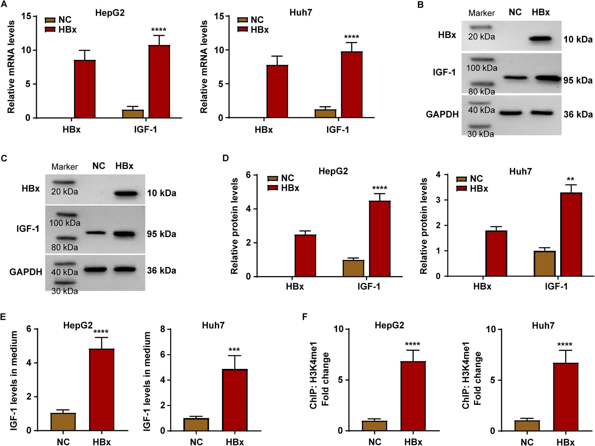
The RNA isolation kit (Promega) was used to separate total RNA from cells. Reverse transcription was conducted using a Reverse Transcription Kit (TOYOBO). qPCR was carried out utilizing the Mini Opticon Real-time PCR system (Bio-Rad) and SYBR Green (Invitrogen, USA). β-actin was used as an internal control. The 2−ΔΔCT method was used to compute the data. The following sets of primers were used: β-actin, (F) 5′-GCTATCCAGGCTGTGCTAT-3′ and (R) 5′-GATGGAGTTGAAGGTAGTTT-3′; HBx, (F) 5′-GCTGCTAGGCTGTACTGC-3′ and (R) 5′-TTAGGCAGAGGTGAAAAAG-3′; IGF-1, (F) 5′-CTCTTCAGTTCGTGTGTGGAGAC-3′ and (R) 5′-CAGCCTCCTTAGATCACAGCTC-3′; and WDR5, (F) 5′-AGTGCCTCAAGACTTTGCCAGC-3′ and (R) 5′-CGATGAGCGTCTTCAGGCACTG-3′.
Western blot
Cells were lysed on ice with pre-chilled RIPA lysate buffer (CW2333S, CWBIO, China) containing protease inhibitors (CW2200S, CWBIO, China), and cell lysates were collected and centrifuged for 10 min at 4 ℃ and 12,000 rpm, and the supernatant protein samples were collected. Protein concentration was determined using a BCA Protein Assay Kit (Thermo, QL227061, USA). Aliquots of protein samples were heat denatured in a water bath, separated using SDS-PAGE and electrotransferred onto a PVDF membrane (Sigma, 46978100, China). After blocking the membrane with 5% skimmed milk for 2 h at room temperature, anti-IGF-1 (CST, #3027), anti-HBx (CST, #14633), anti-WDR5 (CST, #13105), and anti-GAPDH (Abcam, ab8245) were incubated with the membrane at 4 °C overnight. Then, the membrane was incubated with HRP-conjugated secondary antibodies (CST, #7074, #7076) at room temperature for 1.5 h. Enhanced chemiluminescence (ECL) (Thermo, 32106) and a chemiluminescence scanner (Tanon) made protein bands visible. Bands density was measured using Quantity One software (Bio-Rad Laboratories).
ELISA assay
The culture medium was collected and the levels of IGF-1 and IL-10 in the medium were detected using ELISA reagents (Abcam, ab108873 and ab185986) according to the manufacturer’s product instructions.
Chromatin immunoprecipitation (ChIP) and ChIP-qPCR assay
ChIP experiments were performed using the SimpleChIP®Plus Sonication Chromatin IP Kit (CST, #56383) according to the manufacturer’s instructions. Briefly, 1×10⁷ cells were fixed with formaldehyde to crosslink DNA and proteins. Chromatin was fragmented by sonication. Ten micrograms of chromatin was incubated with 1 µl of antibody (HBx, H3K4me1 (Abcam, ab4729), and WDR5) overnight at 4 °C with shaking. Subsequently, 30 µl of Protein G Magnetic Beads were added and incubated for 2 h at 4 °C with shaking. DNA was isolated and purified from the mixture and finally subjected to qPCR.
Coculture of HCC and Treg cells
1×10⁵ HCC cells were inoculated into one well of a 24-well plate and incubated for 12 h for adherent growth. Subsequently, Transwell chamber (pore size 0.4 µm) (Corning Inc.) was placed into the well, and 3×10⁵ Treg cells were inoculated in the upper chamber. After 48 h of co-culture, subsequent assays were performed. For infiltration mimicry experiments, 5×10⁴ HUVECs were inoculated into the upper chamber of the Transwell and incubated for 12 h to allow the HUVECs to form single cell layer on the membrane. After removing the culture medium and washing the HUVECs layer with PBS, the upper chamber was then inoculated with Treg cells.
CCK-8 assay
Suspended cells from the upper chamber in the Transwell were collected with slow centrifugation (1000 rpm for 3 min), resuspended with 10% CCK-8 reagent (PF00004, PTG) (90% culture medium) and incubated in a cell culture incubator at 37 ℃ for 2 h. The absorbance was then measured at 450 nm using a microplate reader (BioTek Epoch2, Winooski).
Flow cytometry
Cells were collected and resuspended with 100 µl PBS. Subsequently, APC-CD4 antibody (Thermo,17–0042-82) and PE-FOXP3 antibody (Thermo,12–5773-82) were added to the cell suspension and incubated for 30 min at room temperature and in a dark environment. Subsequently, the assay was performed by flow cytometry (BD, Franklin Lakes, USA).
Statistical analysis
Data were analyzed using GraphPad Prism 7 (GraphPad, Chicago, USA) and SPSS 21 (SPSS, Chicago, USA) software. Experiment results with at least three independent replications are presented as mean ± standard deviation. Student’s t-test was used for comparison between the two groups. P-values less than 0.05 were considered statistically significant.


Comments (0)