Clinical cross-sectional study
The human study was approved by the Ethics Committee of West China Hospital, Sichuan University (Approval No. 2020–961) and registered with the Chinese Clinical Trial Registry (ChiCTR2100050683). The study adhered to the principles outlined in the Declaration of Helsinki and the Declaration of Tokyo. Informed written consent was obtained from patients or their closest relatives.
Participants
COPD patients aged 40–80 years, either in their stable stage (stable COPD, SCOPD) or exacerbation stage (ECOPD), who were admitted to West China Hospital between 1 May 2020 to 31 December 2021, were included in the study. COPD diagnosis followed the GOLD 2020 criteria. ECOPD was identified based on the International Classification of Diseases- 10 codes J44.0 or J44.1 for both inpatient and outpatient cases.
Exclusion criteria included: 1) A primary diagnosis other than COPD, such as pulmonary tuberculosis, bronchiectasis, pulmonary fibrosis, acute pulmonary embolism, acute pulmonary oedema, pneumothorax, or arrhythmia; 2) Complications potentially affecting blood eosinophil levels, such as asthma, leucocytosis, parasite infection, allergic disorders, eczema, eosinophilic pneumonia, solid tumors, severe immunodeficiency disease or autoimmune diseases; 3) Severe liver, heart or renal failures; 4) Requirement of mechanical ventilation (invasive or noninvasive); 5) Participation in any interventional trials within 30 days before enrolment.
Demographic characteristics (including gender, age, weight, height, and smoking history, etc.) and clinical information (including disease course, comorbidities, number of exacerbations and hospitalizations in the past 12 months, etc.) were recorded. Bacterial infection-related ECOPD was identified as the presence of cardinal symptoms including an increase in dyspnea, sputum volume, and sputum purulence, with or without positive pathogenic bacterial cultures in valid sputum or bronchoalveolar lavage fluid (BALF), increased serum levels of procalcitonin, C-reactive protein, or IL- 6, and infectious lesions visible in chest imaging.
Sample collection and reagents
Induced sputum and peripheral blood samples were collected from participants in a cross-sectional cohort. Eosinophil counts were determined from blood routine examination and sputum smear. For ECOPD patients, eosinophil counts were recorded before any antibiotic or systemic glucocorticoid use, if these treatments were required. Signature cytokines specific to different ILC subsets (including IFN-γ, IL- 1β, IL- 2, IL- 4, IL- 5, IL- 13, IL- 6, IL- 17 A, IL- 21, and IL- 22) in serum were measured by enzyme-linked immunosorbent assay (ELISA) (R&D systems, Emeryville, CA, USA).
Peripheral blood mononuclear cells were isolated by Ficoll (Biochrom) density gradient centrifugation within 3 h of sample collection. ILCs were evaluated by flow cytometry (BD, LSRFortessaTM, NJ, USA), and the results were analyzed by FlowJo software (Treestar, USA). The following antibodies were used: anti-human CD3 (300328, 1:100, Biolegend, CA, USA), anti-human CD14 (325622, 1:100, Biolegend, CA, USA), anti-human CD19 (302230, 1:100, Biolegend, CA, USA), anti-human CD56 (318322, 1:100, Biolegend, CA, USA), anti-human CD11c (565227, 1:100, Biolegend, CA, USA), anti-human FcεRI (334622, 1:100, Biolegend, CA, USA), anti-human CD123 (306016, 1:100, Biolegend, CA, USA), anti-human CD45 (11045942, 1:100, eBioscience, CA, USA), anti-human CD127 (25127842, 1:100, eBioscience, CA, USA), anti-human CRTH2 (12294942, 1:50, eBioscience, CA, USA), anti-human CD117 (NB100 - 77477 APCCY7, 1:100, NOVUS biologicals, Colorado, USA), anti-human ST2 (FAB5232 N- 100UG, 1:100, R and D system, USA), anti-human IL- 13 (501907, 1:100, Biolegend, CA, USA), anti-human RoRγt (17–6988- 82, 1:100, eBioscience, CA, USA), anti-human IL- 22 (17–7222- 82, 1:100, Invitrogen, CA, USA), anti-human IL- 5 (253764, 1:100, Biolegend, CA, USA), anti-human GATA3 (563349, 1:100, Biolegend, CA, USA), anti-human IFN-γ (561980, 1:100, BD Bioscience, CA, USA), anti-human IL- 17 (69–7179- 42, 1:100, eBioscience, CA, USA), anti-human T-bet (562467, 1:100, BD Bioscience, CA, USA). ILC1, ILC2, and ILC3 were gated as Lin−CD117−CD45+CD127+CRTH2−, Lin−CD117+/−CD45+CD127+CRTH2+, and Lin−CD117+CD45+CD127+CRTH2− respectively in viable cells. The expression level of cytokines and transcription factors were presented with geometric mean fluorescence intensity.
In vivo studyExperiment mice
Male C57BL/6 mice, aged 6–8 weeks and weighing approximately 20 g, were obtained from GemPharmatech Co. Ltd (Nanjing, Jiangsu, China). All mice were housed at the animal facility of West China Hospital of Sichuan University under controlled conditions (relative humidity of 50% ± 5% and room temperature of 21 ± 2℃ under a 12 h light/dark cycle), with free access to food and water. All experiments were carried out in accordance with the National Institute of Health Guide for the Care and Use of Laboratory Animals and were approved by the Animal Ethics Committee of West China Hospital of Sichuan University (Approval No. 2021023 A). Efforts were made to minimize animal numbers and discomfort. This animal study was reported in compliance with the guidelines of animal research in vivo experiments [21].
Drugs and reagents
Cigarettes were purchased from Longyan Tobacco Industry Co. Ltd. (Longyan, Fujian, China). Baicalin (purity ≥ 99.5%) and carboxymethylcellulose sodium were purchased from Macklin Biochemical Technology Co., Ltd. (Shanghai, China). LPS, paraformaldehyde, β-mercaptoethanol, and E.Z.N.A. Total RNA Kit II was purchased from Sigma-Aldrich Co (St. Louis, MO, USA). PBS was purchased from Mediatech, Inc. (Herndon, VA, USA). Acridine orange/propidium iodide was purchased from ScyTek Laboratories, Inc. (Logan, UT, USA). Wright-Giemsa Stain was purchased from Nanjing Jiancheng Bioengineering Institute (Nanjing, Jiangsu, China). The red blood cell lysate was purchased from Beyotime Biotechnology Co. Ltd. (Shanghai, China). Protease phosphate inhibitor and protease inhibitor were purchased from Prilai Gene Technology Co. Ltd. (Beijing, China). SDS-PAGE protein loading buffer, RIPA cracking liquid, and PMSF were purchased from Epizyme Biomedical Technology Co., Ltd (Shanghai, China). ELISA kits for IL- 22, IL- 33, IL- 5, and IL- 13 were supplied by R&D Systems Co., Ltd. (Minneapolis, MND, USA). iScript cDNA synthesis kit was purchased from Bio-Rad Laboratories (Hercules, CA, USA). FastStart Essential DNA Green Master was purchased from Roche Group (Basel, Switzerland). The primers were purchased from Qingke Biotechnology Co., LTD. (Beijing, China). Flow cytometry antibodies in this study included anti-mouse CD3ε (APC conjugated) (17,003,182, eBioscience, San Diego, CA, USA), anti-mouse CD11b (APC conjugated) (550,019, BD Biosciences, Franklin Lakes, NJ, USA), anti-human/mouse CD45R (B220) (APC conjugated) (17,045,282, eBioscience, San Diego, CA, USA), anti-mouse Ly- 6G (Gr- 1) (APC conjugated) (17,966,882, eBioscience, San Diego, CA, USA), anti-mouse Ly- 76(Ly- 6 C) (APC conjugated) (128,016, Biolegend, San Diego, CA, USA), rat anti-mouse CD127 (bv711 conjugated) (550,426, BD Biosciences, NJ, USA), mouse anti-mouse CD45.2 (APC-Cy7 conjugated) (565,490, 1:50, BD Biosciences, Franklin Lakes, NJ, USA), rat anti-mouse Ly- 6 A/E (Sca- 1) (bv421 conjugated) (565,490, Biolegend, San Diego, CA, USA), hamster anti-mouse KLRG1 (PE-Cy7 conjugated) (180,709, SouthernBiotech, Birmingham, AL, USA) and anti-mouse CD117(c-kit) (PE conjugated) (188009, SouthernBiotech, Birmingham, AL, USA). Reg3γ antibody (A2146) and Recombinant mouse IL- 22 (RP02942) were purchased from ABclonal Technology Co.,Ltd. (Wuhan, Hubei, China). Lung Dissociation Kit was purchased from Miltenyi Biotechnology Company (Bergisch Gladbach, North Rhine-Westphalia, Germany). Anti-mouse IL- 22 antibody was purchased from Bioss (bs- 2623R, Beijing, China), and anti-mouse GATA3 antibody was purchased from Abcepta (AP5606c, San Diego, CA, USA).
Mice interventions
Mice were fixed in specially-made nose-only exposure tubes and exposed to cigarette smoke (CS) (300 mg/m3 to 350 mg/m3) for 75 min, twice daily, five days per week, for a total of 12 weeks. Mice in the control group were exposed to fresh air. After CS exposure, an atomizing spray of lipopolysaccharide (LPS) (0.2 mg/mL) was administrated through endotracheal intubation after visualizing the glottis through the oral cavity to establish the ECOPD model.
To observe the effect of IL- 22, 50 μL of recombinant mouse IL- 22 (20 μg/mL) or normal saline was administrated through airway intubation 24 h after CS exposure [22, 23]. LPS was endotracheally sprayed to induce an acute airway inflammation 24 h post IL- 22 or normal saline treatment.
To evaluate the effect of baicalin in ECOPD mice, 200 μL baicalin dissolved in 3% carboxymethylcellulose sodium was administered intragastrically for consecutive five days to pre-modeled ECOPD mice. Mice were randomly divided into three baicalin treatment groups: high-dose group (200 mg/kg), medium-dose group (100 mg/kg), and low-dose group (50 mg/kg) [24, 25]. Normal saline was used as the control treatment.
Lung function measurement
Twenty-four hours after intervention, mice were anesthetized with sodium pentobarbital (50 mg/kg) via intraperitoneal injection and underwent tracheostomy. Lung function was assessed using a Buxco system (Buxco, Saint Paul, MN, USA) [26]. The system employs invasive plethysmography to measure respiratory mechanics. Following tracheostomy, mice were connected to the apparatus, and forced maneuvers were performed by applying controlled airway pressures. Parameters including forced expiratory volume at 20, 50, and 100 ms (FEV20, FEV50, FEV100), forced vital capacity (FVC), functional residual capacity (FRC), and total lung capacity (TLC) were recorded. These measurements reflect airflow limitation and lung hyperinflation, key pathophysiological features of COPD.
Mice sample preparation and processing
BALF was collected as previously described [26]. Briefly, the left bronchial tubes of mice were ligated, and the right lungs were lavaged three times with 0.5 mL of sterile pre-cooled PBS. Then, the BALF was centrifuged to pellet the cells, which were resuspended for cell count. The supernatant was kept at − 80 °C for cytokine analysis. After washing cell resuspensions with red blood cell lysate and analyzing cellular viability, cells in BALF were calculated. Cell counts were observed and counted in five randomly selected views per slice by two double-blind evaluators who were unaware of the experimental interventions.
After lavage, the right lung was isolated, stored in a cryopreserved tube, and kept in liquid nitrogen for protein and RNA extraction. The left lung was fixed in 10% formaldehyde for pathological examination. For lung tissue homogenate collection, freshly removed lung tissue (20 mg) was homogenized in tropomyosin receptor kinase lysate/β-mercaptoethanol (Sigma, St. Louis, Missouri, USA), and the supernatant was stored at − 80 °C for further analyzing.
Fresh lung tissue (20 mg) was immediately crushed and transferred to 2 mL enzyme-free EP tubes containing lysate (PMSF in the lysate was diluted with RIPA lysate at 1:100 volume). Protein phosphatase and protease inhibitor mixture were added. After grinding in the grinder (70 Hz, 70 s, twice), lung tissues were centrifuged and stored at − 80 ℃. Protein was extracted according to the manufacturer’s instructions (Prilai Gene Technology, Beijing, China), and protein concentration was measured using a BCA kit (Prilai Gene Technology, Beijing, China). For RNA extraction, crushed lung tissues were transferred into 2 mL enzyme-free EP tubes with pre-cooled β-mercaptoethanol and GTC lysis buffer. Total RNA was extracted according to the manufacturer’s instructions (R6812 - 02, OMEGA, St. Louis, MO, USA).
Single-cell suspensions were rapidly prepared using the lung dissociation kit (130–095–927, Miltenyi Biotec, Köln, Germany) from the remaining lung tissues, after the removal of trachea and connective tissue as much as possible. Collagenase and DNase were added to dissociate the lung tissue. Cells were isolated using the gentleMACS™ Dissociator (130–093–235, Miltenyi Biotec, Köln, Germany) and filtered to obtain a single-cell suspension. To increase the ILCs yield for flow cytometry, the above procedures were processed for pooled lung segments from 5 mice in each group. Percoll gradient fractionation was used to further separate lymphocytes. After lysing red blood cells with ammonium chloride solution, cells were centrifuged and resuspended in pre-cooled PMI- 1640 medium (10% FCS, 200 U/mL cyan-streptomycin dual antibodies) to a final concentration of 1.5 × 10⁷ cells/mL.
Flow cytometric analysis
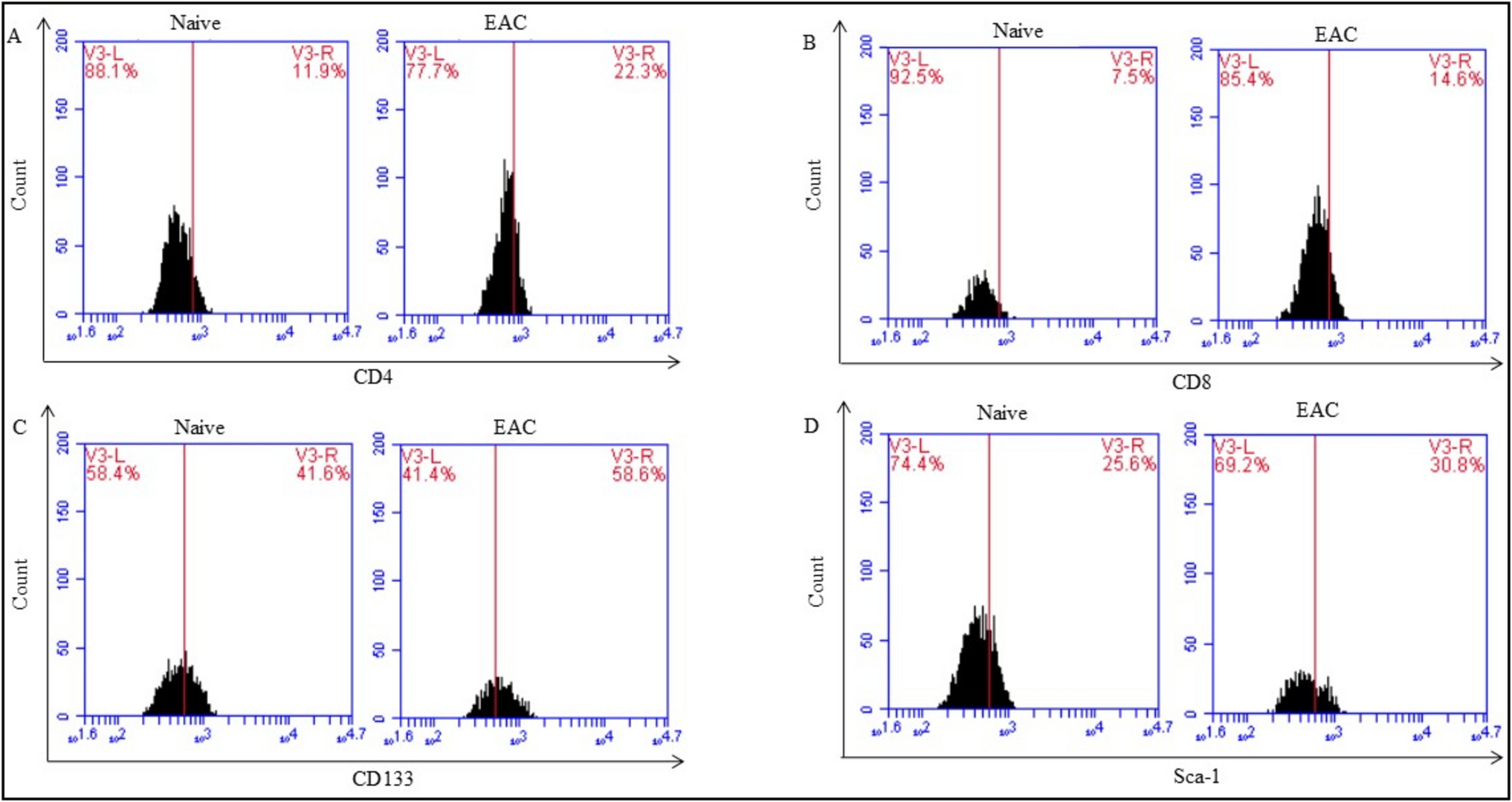
Cells were stained with flow cytometry antibodies to sort or analyze by fluorescence-activated cell sorting. Lung tissue ILCs were identified as Lin−CD45+CD127+ cells. ILC1, ILC2, ILC3 were further identified as Lin−CD45+CD127+CD117–Sca- 1+ cells, Lin−CD45+CD127+CD117+Sca- 1+KLRG1+ cells, and Lin−CD45+CD127+CD117+Sca- 1– cells. Eosinophils in BALF were identified as SSChiLin−CD45+Siglec-F+ cells. Flow cytometry was also performed using BD LSRFortessaTM (Franklin Lakes, NJ, USA), and the results were analyzed by FlowJo software (Treestar, Ashland, OR, USA).
Histopathological assessment of mice lung tissues
The left lungs fixed with 10% formaldehyde were prepared for paraffin embedding, sectioning, and hematoxylin–eosin staining. Five different fields per section were examined under optical microscopy. Inflammatory lesions were scored on a numeric scale for alveolar septal, perivascular, and peribronchiolar infiltrates [18]. The total inflammation score was the sum of these subscales. Scoring was conducted by two independent, blinded researchers.
Measurement of cytokines, Reg3γ, and GATA3
IL- 5, IL- 13, IL- 33, and IL- 22 levels in lung homogenate and BALF were measured using the ELISA method according to the manufacturer’s instructions (R&D systems, Emeryville, CA, USA). IL- 22 and Reg3γ in lung tissue were tested by western blotting with fluorescent-based anti-rabbit (ZB- 2301, 1:10,000, ZSGB-BIO, Beijing, China) or anti-mouse (ZB- 2305, 1:10,000, ZSGB-BIO, Beijing, China) IgG secondary antibodies. Reg3γ, IL- 22, and GATA3 in lung tissue were detected by immunohistochemistry. Chemiluminescence (Bio-Rad, Hercules, CA, USA) was used to visualize protein expression, and band intensities were analyzed by ImageJ software (NIH, Bethesda, MD, USA). The mRNA levels of Il33, Il22, and Reg3γ in lung tissues were detected by qPCR. Gapdh was used as the housekeeping gene according to the manufacturer’s instructions (Qingke Biotechnology, Beijing, China). The sequences were shown in Table 1.
Liquid chromatography-tandem mass spectrometry (LC–MS/MS) proteomics analysis of mice lung tissues
Mice lung tissue cells were lysed using SDT buffer, followed by proteins extraction, and quantification, and digestion by trypsin [27]. The digest peptides were desalted and separated using IntelliFlow technology on a reversed-phase analytical column (Thermo Scientific Acclaim PepMap100, 100 μm × 2 cm, nanoViper C18) maintained at 45 °C. Chromatographic separation was performed using a 120-min linear gradient from 5 to 40% mobile phase B (mobile phase A: 0.1% formic acid in water; mobile phase B: 0.1% formic acid in 84% acetonitrile) at a constant flow rate of 300 nL/min. The auto-sampler temperature was maintained at 4 °C with an injection volume of 2 μL.
LC–MS/MS analysis was conducted on a Q Exactive HF mass spectrometer (Thermo Scientific, Waltham, MA, USA) coupled to an Easy nLC 1200 system (Thermo Fisher Scientific, San Josse, CA) in positive ion mode. Data were acquired in data-dependent top 10 mode with a scan range of 300 − 1800 m/z and an automatic gain control target of 3 × 10⁶. The ion transfer tube temperature was set to 275 °C with a spray voltage of 2.1 kV.
MaxQuant (v.1.6.6) was used to analyze raw MS files against the Swissprot_mouse_database with peptide and protein FDR ≤ 0.01. Protein modifications included carbamidomethyl on Cys (fixed), acetylation on protein N-terminal, and oxidation on Met (variable), allowing up to 5 variable modifications. Identified proteins were filtered to exclude decoy hits, contaminants, or site-specific identifications before further analysis.
Protein data were processed and visualized using RStudio (Boston, MA, USA). Functional annotations and pathway analysis were performed using the clusterProfiler package (v.3.10.1) for Gene Ontology (GO) and Kyoto Encyclopedia of Genes and Genomes (KEGG). Protein–protein interaction networks were constructed with STRING (v.12.0) using a minimum interaction score threshold of > 0.4, and visualized using the QGraph package (v.1.9.2).
Transcriptomics sequencing of mice lung tissues
RNA sequencing libraries were prepared using the NEBNext Ultra RNA Library Prep Kit for Illumina. mRNA was purified with poly-T oligo magnetic beads and quantified with NanoDrop and Agilent 2100 bioanalyzer. First-strand cDNA was synthesized using random hexamers and M-MuLV Reverse Transcriptase, followed by second-strand cDNA synthesis. The resulting double-stranded DNA was circularized and amplified with phi29 to form DNA nanoballs (DNBs), each with over 300 copies of the original molecules. The library was loaded onto a nanoarray, and single-end 50-base reads were generated on the BGIseq500 platform. Quality was validated using the Agilent Technologies 2100 bioanalyzer.
Sequencing data were analyzed using DESeq2 to identify differentially expressed genes (DEGs), with correction for multiple testing using the Benjamini & Hochberg method (P ≤ 0.05). Enrichment analysis for DEGs was performed with the hypergeometric test. Cluster analysis and functional enrichment were conducted using Pheatmap and Kmeans in R, and ClusterProfiler for biological pathways. The top 20 enriched pathways were visualized in a bubble diagram, where point size indicated the number of annotated genes and color depth represented enrichment significance.
Immunofluorescence assessment of ILC3 s in mice lung tissues
IL- 22-producing ILC3 s (CD3–RORγt+IL- 22+ cells) were visualized with immunofluorescence histology in the paraffin lung tissues sections according to previous published protocols [28, 29]. The following antibodies were used: IL- 22 antibody (orb8397, Biorbyt, Cambridge, UK), CD3 antibody (orb11308, Biorbyt, Cambridge, UK), RORγt antibody (PA5 - 23,148, Thermo, MA, USA). All micrographs were acquired on a microscope (BA210 T, Motic).
In vitro studyHuman blood ILC3 isolation and stimulation
After the depletion of Lin+ and CRTH2+ cells from human blood by using the Human ILC2 isolation kit (No.130–114–825, Miltenyi Biotec, Köln, Germany), ILC3 s were separated from ILC1 s using the anti-human antibodies of CD127 and CD117 through flow cytometry. The purity of ILC3 s (> 90%) was confirmed by flow cytometry after sorting. From 100 mL of human blood, approximately 1 × 10⁴ cells could be yielded. Isolated ILC3 s were cultured in 200 μL RPMI 1640 with 2% human serum, isolated from the same human donor, for 1 h, and then stimulated with 40 μM baicalin. After 8 h, cells were harvested and the supernatants were stored at − 80 °C after centrifugation.
IL- 22 detection in the supernatant of ILC3 s
IL- 22 levels in the supernatant of ILC3 s were detected before or after baicalin treatment by using the Human IL- 22 DuoSet ELISA kit (DY782, R&D systems, Emeryville, CA, USA).
Statistical analysis
All the experiments were repeated thrice and the data presented was collective data of three individual experiments. Western blotting images provided were representative of three experiments. Continuous variables were expressed as mean ± standard deviation (SD) and ANOVA was used for comparison between groups for normal distribution data. P < 0.05 was considered statistically significant. GraphPad Prism 9.0 statistical software (GraphPad Software Inc., USA) was used for data statistical analysis.


Comments (0)