Animals
The Sprague Dawley (SD) male rats, aged 6 weeks, were acquired from Beijing Spafu Biotechnology Co., LTD (Beijing, China). The rats were fed in a Specific Pathogen Free (SPF) animal laboratory at 21–23 °C and 60%–65% humidity for 1 week, during which they ate and drank freely. The experiment began one week after the rats had acclimated. All animal experiments in this study were approved by the Animal Ethics Committee of Affiliated Hospital of Guizhou Medical University. The dead rats in the experiment were properly disposed in accordance with the Operating Procedures for Disposal of Laboratory Animal Carcasses of Guizhou Medical University. Animal carcasses were sealed in a special biosafety bag and stored in a temporary refrigerator at −20 ℃ for centralized freezing storage, which finally were incinerated by a professional company for harmless treatment.
Chronic constriction injury of the sciatic nerve (CCI) rat model
The 40 rats were randomly divided into 4 groups: control group, Sham group, CCI group and CCI + PRF group. After the rats were anesthetized by intraperitoneal injection, the left sciatic nerve in the middle thigh of the rats was exposed by blunt muscle stripping. The nerves were then isolated from the adhesive tissue adjacent to the sciatic nerve, following with ligating at four sites 1 mm apart above the trigeminal branch of the sciatic nerve. The tightness of the ligature was adjusted to an appropriate level where slight twitching of the surrounding muscles was able to be observed. The same procedure was performed on the Sham group of rats, but only the left sciatic nerve was exposed with no ligating. To investigate the effect of PRF on NP, PRF treatment was performed as previously described at the sciatic nerve ligation site in CCI rats at the day after surgery [7]. In brief, PRF treatment lasted for 300 s at a pulse frequency of 2 Hz, a temperature of 42 °C and an output voltage of 45 V. During PRF treatment, the right thigh of the rats exhibited a slight twitch. During CCI modeling, 3 out of 40 rats died. There were no deaths in the Control group and the Sham group, 2 deaths in the CCI group and 1 death in the CCI + PRF group.
Afterwards, to explore the effect of MG53 knockdown on NP, 40 rats were randomly divided into CCI group, CCI + PRF group, CCI + PRF + AAV-sh-NC group and CCI + PRF + AAV-sh-MG53 group. The latter two groups were, respectively, treated with intrathecal injection of AAV-sh-NC or AAV-sh-MG53 at 6 weeks before CCI modeling. The rats were treated with molding and PRF according to the above methods. In addition, in order to investigate the role of upregulation of MG53 in NP, 30 rats were randomly divided into 3 groups for CCI modeling. Two out of three groups, respectively, received intrathecal injection of AAV-NC or AAV-MG53 before modeling. In order to clarify the mechanism of MG53 regulating microglial activation, another 30 rats were randomly divided into 3 groups and CCI modeling was performed as above. Two groups out of them were, respectively, treated with CCI after intrathecal injection of AAV-MG53 or AAV-MG53 + tunicamycin. During this period, nine rats died, one each from the CCI + PRF group, the CCI + PRF + AAV-sh-MG53 group, the CCI + AAV-NC group, the CCI + AAV-MG53 group and the CCI + AAV-MG53 + tunicamycin group, as well as two each from CCI + PRF + AAV-sh-NC group and CCI + AAV-MG53 group. Euthanasia of the rats was performed as previously [24]. Briefly, 14 days after surgery, the rats were killed by cervical dislocation companied with inhalation of isoflurane (0.41 mL/min). Spinal cord tissues from the euthanized rats were isolated for further testing. The instrument of PRF treatment was RFE4 series 4 channels radiofrequency (RF) Lesion Generator (Beijing Neo Science, Beijing, China), including RF electrode CD2201(OD = 0.4 mm, L = 50 mm) and RF cannula 240,100 (22G-50 mm, −4 mm, straight).
Mechanical withdrawal threshold (MWT) test
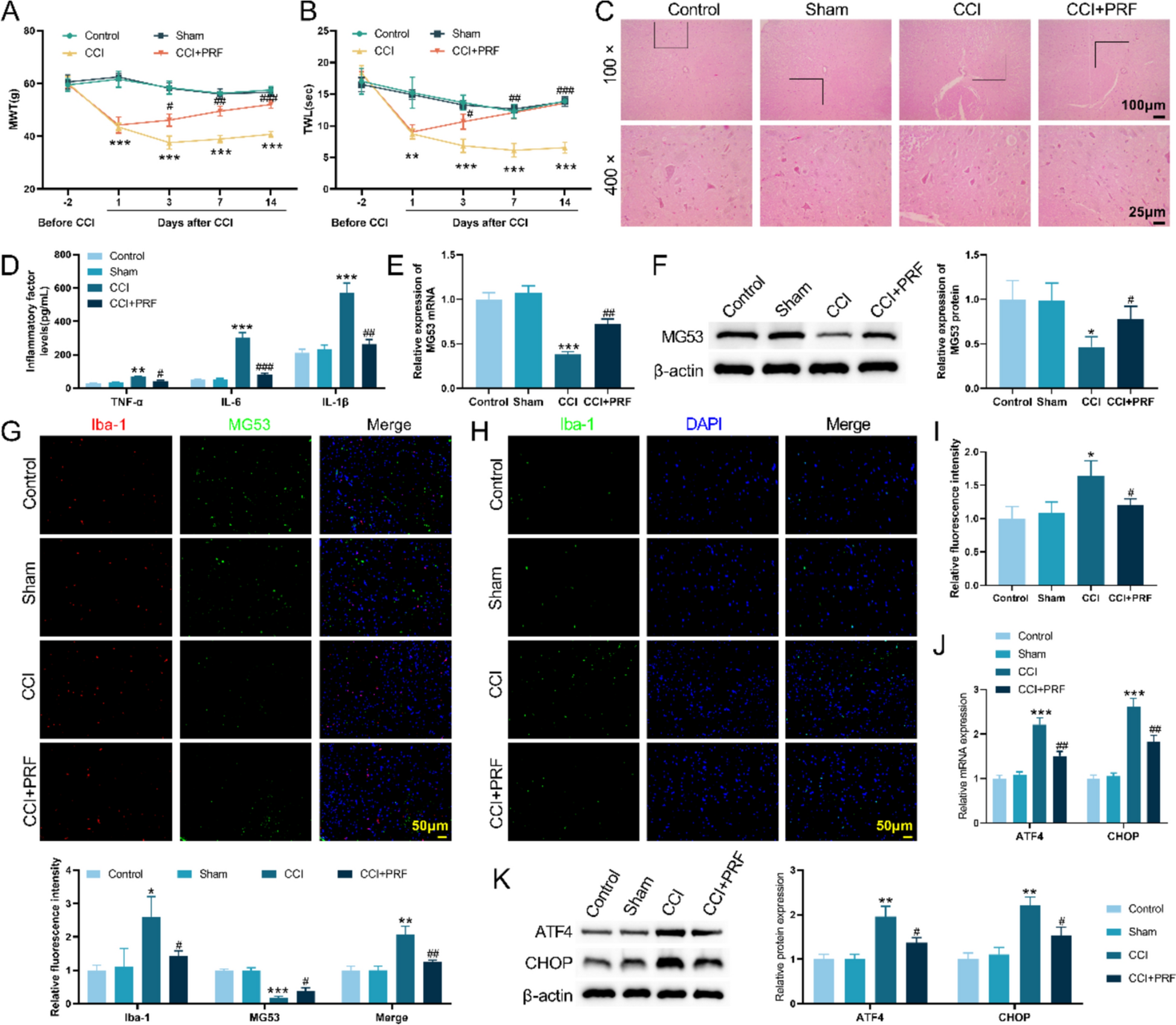
MWT tests were performed at 2 days before CCI surgery and 1, 3, 7, and 14 days after surgery. The plantar mechanical pain test system was used to stimulate the center of the plantar on both sides of the rats with 16 different intensity stimuli, 10 times for each intensity, according to the intensity from small to large. The intensity of the stimulus, in which more than 5 times paw withdrawal responses were observed in rats, was identified as the threshold for the response of rats to mechanical stimuli.
Thermal withdrawal latency (TWL) test
TWL tests were also performed at 2 days before CCI surgery and 1, 3, 7, and 14 days after surgery. The procedure was as follows: the rats were placed in a glass box containing a smart hot-plate pain meter, in which the rats were able to move their whole body freely. After the rats stood for 10 min, the left and right heels of the rats were illuminated with a strong light of the same intensity. The time from the onset of light to the withdrawal of the rat's feet was subsequently recorded and measured every 5 min for a total of three measurements. The average of the three results was taken to evaluate the thermal hyperalgesia of the rats.
Cell culture
Rat primary microglia (Cybercon Biotechnology Co., LTD, Shanghai, China) were cultured in primary astrocyte medium containing 5% fetal bovine serum, 1% penicillin streptomycin and 1% primary astrocyte culture additive. The conventional culture conditions of the cells were 37 °C and 5% CO2.
Cell transfection
Primary rat microglia were cultured until the number of cells had increased by about 70%–80%, following with the removing of previous medium. After washing the cells twice with PBS, the cells were cultured overnight in a new serum-free and antibiotic-free medium. After dilution with serum-free Dulbecco's modified eagle medium (DMEM), primary rat microglia were transfected with si-MG53-1, si-MG53-2, si-MG53-3 and si-NC plasmid (2.5 μg) (Genepharma, Suzhou, Jiangsu, China) applying Lipofectamine 2000 (4 μL) (Thermo Fisher Scientific Inc., Waltham, MA, USA). After an additional 6 h of incubation, the supernatant was aspirated and the medium was replaced. Subsequently, the cells continued to be cultured for 48 h. Microglia were divided into Control group, si-NC group and si-MG53 group.
Hematoxylin–eosin staining (HE)
Spinal cord tissue of rats fixed with 4% polyformaldehyde was dehydrated and paraffin embedded. Afterwards, the intact tissue was cut into wax sheets with a thickness of 4 µm, which were then applied and dried. Subsequently, the sections were stained with hematoxylin dye (Beyotime, Shanghai, China) for 8 min, following with staining with eosin dye (Beyotime, Shanghai, China) for 5 min. The pathological injury of rat spinal cord was observed by optical microscope after sealing the stained tissue sections.
Enzyme-linked immunosorbent assay (ELISA)
The expression levels of TNF-α, IL-6 and IL-1β were detected by ELISA kits (Elabscience, Wuhan, Hubei, China). Under the condition of adding protease inhibitors, the spinal cord tissue was fully ground and ultrasonically broken on ice. The treated tissue was then centrifuged and the supernatant is placed on ice for use. The cells of logarithmic growth stage were collected and washed 3 times with cold PBS. Then, the cells were broken by repeated freezing and thawing after suspension. The cell extract was centrifuged at 2–8 °C for 10 min to obtain the supernatant for use. At the same, the extract of tissue or cell was analyzed according to the instructions of the ELISA kit. Finally, enzyme-labeled analyzer (Thermo Fisher Scientific, Waltham, MA, USA) was used to measure the absorbance of each hole in sequence. ELISA kits for rat TNF-α, IL-6 and IL-1β were purchased from Wuhan Ilerite Biological Co., LTD.
Western blotting (WB)
The total protein extracted was quantified for protein concentration utilizing the bicinchoninic acid (BCA) kit (Biosharp, Guangzhou, Guangdong, China). The protein samples underwent electrophoresis on sodium dodecyl sulfate (SDS) polyacrylamide gel. Subsequently, the proteins were transferred from the gel to polyvinylidene fluoride (PVDF) membrane and blocked with 5% BSA for a duration of 2 h at ambient temperature. The membrane was then subjected to incubation with primary antibodies for MG53 (1:1,000, ab307593; Abcam, Cambridge, MA, USA), ATF4 (1:1,000, ab270980; Abcam, Cambridge, MA, USA), CHOP (1:1,000, A20987; ABclonal Technology, Wuhan, Hubei, China) and β-actin (1:1,000, ab8226; Abcam, Cambridge, MA, USA) overnight at 4 °C. After washing three times with TBST buffer, the membrane was incubated with horseradish peroxidase (HRP)-labeled goat anti-rabbit IgG (1:5000, Ab0101; Abways Technology, Shanghai, China) and HRP-labeled goat anti-mouse IgG (1:5000, Ab0102; Abways Technology, Shanghai, China) secondary antibody for 2 h at 37 °C. The relative expression of the target protein was analyzed by Image J v1.8.0 software.
Quantitative real-time PCR (qRT-PCR)
Total RNA was extracted by adding TRIzol reagent (Invitrogen, Thermo Fisher Scientific Inc., Waltham, MA, USA) in each group, and the extracted total RNA was reversely transcribed into cDNA according to the instructions of PrimeScript RT reagent Kit (Takara, Tokyo, Japan), and then the cDNA was amplified. The primers were designed by Primer-BLAST software and synthesized by Thermo Fisher Scientific (Waltham, MA, USA), and the primer sequences and product lengths were shown in Table 1. The relative expression of MG53, ATF4 and CHOP mRNAs was calculated using the 2−ΔΔCT method with GAPDH as an internal reference.
Homologous double label immunofluorescence (IF) analysis
After deparaffinization and rehydration, tissue slides were treated with 100 μL endogenous peroxidase blockers for 10 min. Then, tissue slides were incubated with monoclonal primary antibody against Iba-1 (1:200, Ab178846; Abcam, Cambridge, MA, USA) overnight at 4 °C. Subsequently, HRP-labeled goat anti-rabbit IgG (1:5000, Ab0101; Abways Technology, Shanghai, China) secondary antibody was applied to the sections at room temperature for 50 min, following with tyramide signal amplification (TSA) stain. After antigen repair of the sections again, another polyclonal primary antibody against MG53 (1:200, ab307593; Abcam, Cambridge, MA, USA) was added to the sections overnight at 4 °C. Afterwards, the sections were incubated again with HRP-labeled goat anti-rabbit IgG secondary antibody in the dark at room temperature for 50 min. Next, the slides were counterstained with 4',6-diamidino-2-phenylindole (DAPI) (Servicebio Biotechnology, Wuhan, Hubei, China) and then were visualized using microscope.
Statistical analysis
The data from this chapter of the study were statistically analyzed using SPSS 23.0 and the results are presented as mean ± standard deviation (mean ± SD). Statistical methods were one-way ANOVA, Chi-square test, or non-parametric test depending on the data characteristics of the different comparison groups. Differences were considered statistically significant if P < 0.05.


Comments (0)