
Remember me


Wild-type (WT) C57BL/6NTac (Taconic Farms) male and female 12–24-week-old mice were used in most experiments. Fos2A-iCreER (TRAP2) (RRID:IMSR_JAX:030323, Jackson Laboratory) mice were crossed with R26AI14/+ (AI14) (RRID:IMSR_JAX:007914, Jackson Laboratory) mice to generate double-transgenic TRAP2×Ai14 mice and maintained on a C57BL/6 Jackson background. Double-transgenic TRAP2×Ai14 or single-transgenic TRAP2 male and female 12–32-week-old mice were used in designated experiments. Male and female 12–32-week-old Ndnftm1.1(cre)Rudy/J (NDNF-Cre) (RRID:IMSR_JAX:030757, Jackson Laboratory) mice were used in designated experiments. All mice used in this study were group-housed with free access to food and water and maintained on a 12-h light–dark cycle. The temperature set point was 72 °F with humidity between 30% and 70%. Mice were single-housed for 1 week before behavioral experiments except following a surgical procedure, in which case they were single-housed for 2 weeks before the beginning of a behavioral experiment. Littermates were assigned to different experimental groups to prevent confounding effects of cage. All experiments were performed during the light phase of the cycle. All studies were approved by the Chancellor’s Animal Research Committee at UCLA.
Memory linking taskAll mice were handled and habituated to transportation and the behavioral room before initiating a behavioral experiment. Single-housed mice were first handled for three consecutive days in their colony room. Mice were handled by allowing them to freely rest or explore on the palm of the experimenter for 1 min on the first day, 1.5 min on the second day and 2 min on the third day. Then, mice were transported to the behavioral room every day for the next five consecutive days. Four cages (one mouse per cage) were transported at a time on a wheeled cart and covered with a black plastic bag to prevent mice from seeing their surroundings. Once inside the behavioral room, each mouse was handled for 2 min. All handling and habituation were performed at the same time of day (between 10 a.m. and 12 p.m.) and gloves were changed between each mouse.
Then, 1 day following the end of the habituation sessions, mice underwent a context linking experiment16,17,18. Mice were first exposed to context A for 10 min and, 5 h or 7 days later, were exposed to context B for 10 min, as detailed below. Then, 2 days later, mice were reexposed to context B, received a 2-s foot shock (0.75 mA) 10 s upon entering the context and remained in the context for an additional 30 s. During the next three consecutive days, mice were exposed to context A, a novel context C or context B and their freezing levels were recorded using an automated scoring system (Med Associates).
Each context consisted of a small chamber with particular visual, tactile and odor cues (Med Associates contextual fear condition system). Context 1 consisted of a smooth white plastic floor, a white plastic insert with round walls and a black insert ceiling and was cleaned with a 5% solution of Simple Green all-purpose cleaner. Context 2 consisted of a smooth white plastic floor topped with a roughed transparent floor, black triangle enclosure and one black–white checkered wall and was cleaned with a 5% solution of Windex glass cleaner. Context 3 consisted of horizontal metal rods (shocker) and a square enclosure with metal walls and was cleaned with 70% ethanol. All contexts were illuminated with white visible light. Contexts 1 and 2 were used for context A described in our experiments and were counterbalanced to be either the ‘linked context’ or a novel context. Context 3 was used as context B where mice received a mild foot shock.
Miniscope recordingsAll mice were handled and habituated to the behavioral room and the weight of the miniscope before starting a recording session. Then, 3 days after implantation of the baseplate, as described below, mice were handled in the colony room for 2 mins per day for five consecutive days. Then, 2 days later, mice were transported to the behavioral room as described above and handled for 2 min per day for five consecutive days. During the last 3 days of habituation, a V4 UCLA miniscope was attached to the baseplate and mice were allowed to freely move in their home cage for 2 min. On the last day of habituation, a 5-min recoding was performed in the home cage. Then, 2 days later, mice were allowed to explore context A for 10 min and, 7 days or 5 h later, explored context A, B or C while calcium activity was recorded in all sessions. In experiments where vmPFC was chemogenetically inhibited, as described below, CNO (5 mg kg−1) was injected 30 min before exposure to context B or C.
Three-chamber social testMice with hSyn–hM4Di–mCherry, CamKII–hM4Di–mCherry or hSyn–Cherry in the vmPFC were tested on a classic three-chamber spontaneous social preference assay52. All mice were injected with CNO (5 mg kg−1) 30 min before being introduced into the three-chamber apparatus. Mice were first introduced in the central chamber and allowed to explore it for 5 min without access to the adjacent chambers. After this, mice gained access to the adjacent chambers and were allowed to explore the entire apparatus for 10 min. One of the outer chambers contained a conspecific inside a wire cup. The opposite outer chamber contained an object inside a wire cup. The object and conspecific chambers were counterbalanced across mice. Mouse behavior was recorded and the time spent on each chamber was manually scored during the 10-min exploration.
Social transmission of food preferenceObserver mice were tested for their relative preference for two new odor-laced foods after interacting with a demonstrator mouse. First, all mice were habituated to eating ground food in a bowl for 1 week in their home cage and were habituated to daily transport to the behavioral room. Demonstrator mice were then food deprived for 24 h and subsequently fed cinnamon-laced food (2% w/w) for 1 h. These mice were then introduced to the home cage of observer mice inside a wire cup and allowed to interact for 10 min. Observer mice were then food deprived for 24 h. The following day, observer mice were given a choice between cinnamon-laced food (2% w/w) or cocoa-laced food (1% w/w) in their home cage for 1 h. Total food consumption was determined by weighing each bowl before and after the trial. Innate preference for these two flavors was tested in naive mice.
ChemogeneticsMice were injected with CNO in saline (5 mg kg−1; Tocris, 4936) 30 min before being introduced to a context.
OptogeneticsFollowing recovery from surgery, mice were handled and habituated as described above but an optic fiber cable was connected to the optic fiber during habituation. On the first day of recordings, calcium activity was first recorded for 5 min while mice were in their home cage. Immediately after, mice were exposed to a novel context (A) for 10 min and calcium activity was recorded. Then, 7 days later, calcium activity was again recorded in the home cage for 5 min followed by optogenetic stimulation of vmPFC–MEC terminals.
Optogenetic inhibition was achieved by delivering 535-nm laser light (500 ms, 0.5 Hz, 10–15 mA) for 2 min to half the mice while in their home cage. Immediately following light stimulation, mice were introduced to a novel context (B) and activity was recorded for 5 min. Recording was then stopped and laser light was again delivered for 1 min using the same pulse parameters. Activity was recorded for an additional 5 min before mice were returned to their home cage. Then, 10 days later, the procedure described above was repeated with new contexts (C and D) and light was delivered to the other half of mice.
Optogenetic activation was achieved by by delivering 653-nm laser light (10 ms, 20 Hz, 10–15 mA) for 10 min to half of the mice while they explored context A, 7 days after having visited the same context. Calcium activity in the dCA1 was recorded as described above. Then, 10 days later the procedure described above was repeated with a new context (B) and light was delivered to the other half of mice. This procedure was similar for the 5-h experiment but all mice visited two different pairs of contexts with light on or off.
Stereotaxic surgeryAnesthesia was induced with 5% isoflurane in oxygen in an anesthesia chamber before mice were placed on a stereotaxic alignment system (Kopf Model 1900). Anesthesia was maintained using 1.5% isoflurane in oxygen. Petrolatum eye ointment was applied in both eyes (Puralube vet ointment) to prevent them from drying. Body temperature was maintained using a heating pad. Aseptic techniques were used throughout the entire surgery. Carprofen (5 mg kg−1) and dexamethasone (0.2 mg kg−1) were subcutaneously injected at the end of each surgery and for two (virus surgery) or seven (miniscope surgery) additional days. Following surgery, mice were allowed to recover over a heating pad in their home cage. Amoxicillin was provided in water for 2 weeks following the surgery.
For intracranial injection of viral vectors, a midline incision was made in the scalp and a small craniotomy was performed over the desired brain region using an air-driven dental drill (Henry Schein). A glass pipette was used in a nanoliter injector (World Precision Instruments) with a Micro4 Controller (World Precision Instruments) to inject viral vectors. All coordinates for injections were relative to Bregma unless otherwise specified. For vmPFC viral injections, +1.8 mm anterior–posterior (AP), ±0.35 mm medial–lateral (ML) and −2.1 mm dorsal–ventral (DV). For dHPC, −2.1 mm AP, ±2 mm ML and −1.65 DV. For MEC, −4.85 mm AP, ±3.4 mm ML and −3.5 mm DV. For LEC, −3.65 mm AP, ±4 mm ML and −4.25 mm DV. Viral vectors were injected at a rate of 50 nl min−1. After injection, the scalp was closed using 9-mm wound clips (Clay Adams) that were removed after 8 days.
For in vivo imaging of calcium activity, a GRIN lens was implanted over the target region 30 min following viral injection. To this end, the skull was scorched and a 1.2-mm-diameter screw was implanted over the posterior parietal cortex on the opposite side of the lens to improve adherence of dental cement. A circular craniotomy (diameter: 1 mm) was then carefully performed over the target region. Part of the neocortex was carefully aspirated using a blunt hypodermic needle (27 G) while artificial cerebrospinal fluid was applied to prevent drying. Once bleeding stopped, a 1-mm GRIN lens was slowly lowered to the target position. For vmPFC, the lens was implanted at +1.8 mm AP, −0.5 mm ML and −1.9 mm DV. For dHPC, the lens was implanted at −2.1 mm AP, −2 mm MLand −1.35 mm DV (from the top of the craniotomy). The GRIN lens was secured to the skull using cyanoacrylate glue and dental cement (Ortho-Jet, LANG). Kwik-Sil (World Precision Instruments) was used to cover the GRIN lens. Then, 2 weeks later, a baseplate was cemented over the GRIN lens using the V4 miniscope to determine the position that ensured the best focal plane.
For experiments involving in vivo calcium imaging and optogenetics, surgeries were performed in two phases. First, all viruses were injected as described above. Then, 1 week later, both the GRIN lens and the optic fiber were implanted on their respective target areas. The optic fiber was implanted over the MEC at an angle to ensure sufficient space between the miniscope and optic fiber (−4.85 mm AP ±3.4 mm ML and −3 mm DV; 16° angle on AP axis towards posterior).
Viral vectorsThe viral vectors described below were injected using the coordinates and methodology described above.
For vmPFC, the vectors consisted of 200 nl of pAAV-hSyn-mCherry (Addgene, 114472-AAV5), 200 nl of pAAV-hSyn-hM4D(Gi)-mCherry (Addgene, 50475-AAV5), 400 nl of pAAV.Syn.GCaMP6f.WPRE.SV40 (Addgene, 100837-AAV1), 400 nl of pAAV.CAG.Flex.GCaMP6f.WPRE.SV40 (Addgene, 100835-AAV1), 250 nl of pAAV-hSyn-DIO-hM4D(Gi)-mCherry (Addgene, 44362-AAV8), 250 nl of AAV8-hSyn-DIO-mCherry (Addgene, 50459-AAV8), 200 nl of pAAV-CaMKIIa-hM4D(Gi)-mCherry (Addgene, 50477-AAV5), 200 nl of pOTTC1484-pAAV SYN1 HA-hM4D(Gi) (Addgene, 121538-AAV5), 200 nl of pAAV-hSyn1-SIO-eOPN3-mScarlet-WPRE (Addgene, 125713-AAV5), 250 nl of pENN.AAV.hSyn.Cre.WPRE.hGH (Addgene, 105553-AAV1) and 250 nl of pAAV-Syn-ChrimsonR-tdT (Addgene, 59171-AAV5).
For dHPC, the vectors consisted of 400 nl of pAAV.Syn.GCaMP6f.WPRE.SV40 (Addgene, 100837-AAV1), 300 nl of pAAV-EF1a-Cre (Addgene, 55636-AAVrg) and 300 nl of AAV8-hSyn-DIO-mCherry (Addgene, 50459-AAV8).
For MEC, the vectors consisted of 350 nl of pAAV-EF1a-Cre (Addgene, 55636-AAVrg), 400 nl of pAAV-hSyn-DIO-hM4D(Gi)-mCherry (Addgene, 44362-AAV8), 400 nl of AAV8-hSyn-DIO-mCherry (Addgene, 50459-AAV8) and 400 nl of` pAAV-Syn-FLEX-rc[ChrimsonR-tdTomato] (Addgene, 62723-AAV5).
For LEC, the vector consisted of 350 nl of pAAV-EF1a-Cre (Addgene, 55636-AAVrg).
ImmunohistochemistryFirst, 90 min after behavior, mice were transcardially perfused with PBS followed by 4% paraformaldehyde (PFA) in 0.1 M phosphate buffer. Brains were collected, incubated overnight in 4% PFA and then transferred to PBS for storage at 4 °C. Brains were sliced at 50 µm using a vibratome (Leica VT100S) or incubated in a 30% sucrose solution to be sliced at 50 µm using a cryostat (Leica CM1850).
Free-floating brain slices were incubated with a blocking solution containing 0.3% Triton X-100 in PBS and 10% normal goat serum (Vector Laboratories, S-1000) at room temperature for 1 h to minimize nonspecific antibody binding in the subsequent steps. Primary and secondary antibodies were diluted in the same blocking solution. Free-floating sections were incubated with the primary antibody overnight at 4 °C with constant shaking, repeatedly washed in PBS and then incubated with the secondary antibody for 2 h at room temperature with constant shaking. Slices where then incubated with DAPI (Invitrogen; 1:1,000) for 20 min and repeatedly washed in PBS before being mounted on glass slides (VWR Superfrost Plus) using DAPI Fluoromount-G mounting media (Southern Biotech).
Primary antibodies were as follows: rabbit anti-c-Fos (Cell Signaling, 9F6, 2250; 1:700), chicken anti-RFP (Synaptic Systems, 409006; 1:700), mouse anti-GAD67, clone 1G10.2. (EMD Millipore, MAB5406; 1:1,000), rabbit anti-cholera toxin B antibody (Abcam, ab34992; 1:1,000), mouse anti-PV (Sigma-Aldrich, P3088; 1:1,000), guinea pig anti-VIP (Synaptic Systems, 443005; 1:100), rat anti-SOM (EMD Millipore, MAB354; 1:100), guinea pig anti-Calbindin D28k (Synaptic Systems, 214004; 1:700) and goat anti-Reelin polyclonal antibody (Invitrogen, PA5-47537; 1:1,000).
Secondary antibodies were as follows: goat anti-rabbit Alexa Fluor 647 (Invitrogen, A-21244; 1:1,000), goat anti-rabbit Alexa Fluor 488 (Invitrogen, A-11008; 1:1,000), goat anti-chicken Alexa Fluor 594 (Invitrogen, A-11042; 1:1,000), goat anti-mouse Alexa Fluor 488 (Invitrogen, A-11001; 1:1,000), goat anti-guinea pig Alexa Fluor 568 (Invitrogen, A-11075; 1:1,000), goat anti-rat Alexa Fluor 594 (Invitrogen, A-11007; 1:1,000) and donkey anti-goat Alexa Fluor 647 (Invitrogen, A-21447; 1:1,000).
RNAscopeBrains were collected 60 min after behavior and fast-frozen in optimal cutting temperature compound by dry ice without PFA fixation. Frozen brains were sliced at 15 µm using a Leica Cryostat. In situ hybridization was performed using the RNAscope fluorescent multiplex reagent (ACD, 323120) according to the instructions from the manufacturer. Probe-Mm-Ndnf (ACD, 447471) and Probe-Mm-Fos (316921) were used for mRNA labeling.
Confocal microscopyImages were acquired using a Nikon A1 laser scanning confocal microscope and analysis was performed using NIS-Elements AR Analysis software (Nikon; version 4.40.00).
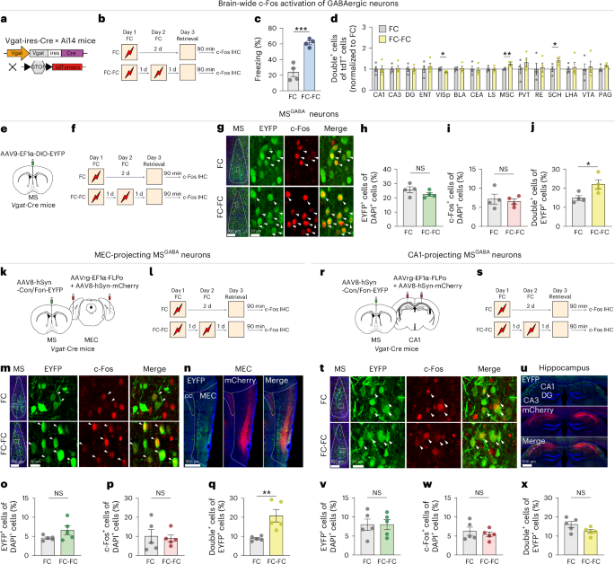
Slices from TRAP2×Ai14 mice (Fig. 2e) were imaged at ×10 magnification and 6–8 slices per mouse were used to automatically determine the number of tdTomato-positive cells in the vmPFC.
Slices from TRAP2×Ai14 mice (Fig. 3h) were imaged using at ×20 magnification with a z step of 2 µm for a total of 27 steps per slice. A maximum z projection was generated for each field of view and a total of four slices per mouse were used to determine the number of DAPI-positive, c-Fos-positive and RFP-positive cells in the HPC. In this case, DAPI-positive cells were automatically counted but c-Fos-positive, RFP-positive and double-positive cells were obtained by averaging the manual counts of 2–3 experimenters blind to the experimental conditions. Chance levels were calculated as (c-Fos/DAPI) × (TRAP/DAPI).
Slices from WT mice (Fig. 7b) were imaged at ×20 magnification with a z step of 2 µm for a total of 27 steps per slice. A total of four slices per mouse was used to determine the number of c-Fos-positive, PV-positive, SOM-positive, VIP-positive and GAD67-positive cells in the dCA1. Counts were obtained by averaging the manual counts of 2–3 experimenters blind to the experimental conditions. DAPI was automatically counted. MEC DAPI and c-Fos were also automatically counted.
For RNAscope (Fig. 7f), brain slices were imaged at ×20 magnification and 2–3 slices per mouse were used to manually count NDNF-positive and C-Fos-positive cells in the SLM. Final counts were obtained by averaging the scores of three experimenters blind to the experimental conditions.
Miniscope analysisOne-photon calcium imagining was performed using the UCLA V4 miniscope (https://github.com/Aharoni-Lab/Miniscope-v4)17. Specifically, during recordings, digital imaging data were sent from the complementary metal–oxide–semiconductor (CMOS) imaging sensor (Aptina, MT9V032) to custom data acquisition electronics and a USB host controller (Cypress, CYUSB3013) over a lightweight, highly flexible coaxial cable. Images were acquired at 30 frames per second, using a display resolution of 600 × 600 pixels (1 pixel = 1–2 µm) and saved into uncompressed mp4v files. The analysis pipeline was written in MATLAB R2020b using the ConcatMiniscope pipeline53. The algorithm first runs the NoRMCorre algorithm for motion correction (rigid registration) of individual sessions from a single animal54, followed by the alignment and normalization of brightness across sessions and concatenation into a single long video. Alignment was performed aligned using a semiautomatic alignment tool based on the ‘imregtform’ function (MATLAB R2020b, image-processing toolbox) followed by manual checking of landmarks (usually blood vessels). Individual neurons were identified and extracted using the CNMF-E algorithm55. During motion correction, videos were 2× spatially downsampled using the default built-in NoRMCorre protocol. During CNMF-E initialization, videos were further 2× spatially downsampled and 5× temporally downsampled. The quality of neuron extraction was then verified using a MATLAB R2020b custom-made neuron deletion GUI. We excluded the detected putative neurons exhibiting ROI morphology or calcium trace abnormalities or incoherencies between the calcium trace peaks and the expected correspondent fluorescence increases in the video.
For quantification of neuronal activity across sessions, after the detection of individual neurons, CNMF-E projects their raw activity as a relative brightness of each ROI and compiles them into the variable neuron.C_raw. To quantify the activity of the same neuron across multiple sessions, we first separated the neuron.C_raw from each session and deconvolved the calcium signal form each neuron using an empirical method based on multiple one-dimensional convolutions of the raw signal with gaussian kernels. According to the assumption that true calcium transients are represented by steep increases in fluorescence followed by slow exponential decay, we devised a fast method to separate steep increases in brightness that are related to actual calcium transients from the background noise using a nonparametric analysis. Briefly, we convolved the raw data separately with two Gaussian kernels with s.d. of 50 and 100 ms. Then, for each convolution, we performed a 3× decimation (each data point comprised ~100 ms) and calculated the first derivative of the data. The distribution of first derivatives represents the distribution of steepness of calcium increases within 100-ms time windows, well within the timeframe of the calcium indicator. For each convolution with each of the two kernels, we calculated a threshold as the third quartile plus 1.5× the interquartile range (~3 s.d. above the mean in a normal distribution). We only considered actual calcium transients the data points with steepness that were higher than both thresholds, which were deemed as 1, while the remaining data points were deemed as 0. As an additional quality-control step, putative noisy neurons were automatically detected and then manually removed. Neurons were deemed noisy and sorted out for further inspection if their peak-to-noise ratio was lower than 20 or if the distribution of the noise showed a difference between the first and third quartiles higher than 200%. The number of calcium events (putative firing rates) per second for each neuron was calculated as the number of 1 values per time interval and their average on each session was determined by the total number of 1 values divided by the number of data points. The top 10% of active cells (Fig. 6) were identified by sorting the average number of calcium events per second on a given session from highest to lowest.
For overlap quantification, for the determination of the percentage of overlapping ensembles between two sessions, the 10-min recordings of each session were individually analyzed, as described above (without concatenation). Recordings from both sessions in the same mouse were first aligned using the spatial footprints (neuron.A, output from CNMF-E) of each one of the detected cells for individual sessions. The centroid distance and spatial correlation were calculated for all cell pairs. Cell pairs from different sessions were considered to match if their spatial correlation was at least 0.8 and their centroid distance was 5 pixels or less. Overlapping percentages between two sessions were calculated as the number of matched cells over the average of the total number of detected cells in each one of the two sessions. Overlapping index = matched cells from session 1 and session 2 (number of overlapping cells)/((number of session 1 cells + number of session 2 cells)/2).
The probability in Extended Data Fig. 7a–c was calculated by determining the probability of a subset of neurons from one session (for example, top 10% of Ctx A) to be present in the subset of neurons from a given percentile of activity in Ctx B (for example, top 20%)17. This was defined as PA10,B20 = NA10,B20/U, where NA10,B20 is the real number of neurons present on both percentiles and U is the universe of all cells detected in the concatenated video. The probability values were normalized by chance defined as PA10 × PB20 (0.1 × 0.2).
For statistical analyses of overlap significance in top 10% active neurons (Extended Data Fig. 7d), to evaluate whether the observed overlap of high-firing neurons between sessions exceeded chance levels, we performed a one-sided permutation test. For each subject, the top 10% most active neurons in Ctx A, HC1 and Ctx B were identified on the basis of descending firing rates. The real overlap between Ctx A and Ctx B or HC1 and Ctx B was computed as the proportion of top 10% neurons shared across the two sessions, excluding neurons present in the three sessions (that is, CtxA∩HC1∩CtxB). To establish a null distribution, we generated 1,000 shuffled datasets per subject by randomly permuting neuron identities in Ctx B while keeping the firing rate ranks in Ctx A and HC1 fixed. For each shuffle, we recalculated the overlap proportion. The P value was computed as the proportion of shuffled overlaps that were greater than or equal to the real (observed) overlap, reflecting the probability of obtaining such overlap by chance under the null hypothesis of no structured reactivation across sessions. Group-level P values were derived from aggregating real and shuffled overlap distributions across subjects.
Whole-brain clearingBrains were cleared according to the Adipo-clear protocol56, itself an adaption of the iDISCO protocol57. Animals were transcardially perfused as described above. Brains were then left overnight in 4% PFA at 4 °C. Hemisections of samples were taken by sagitally cutting the sample 2 mm to the right of the midline before being treated and immunostained21. Briefly, samples were dehydrated in a series of methanol washes before being washed in a dichloromethane solution. Samples were then bleached, rehydrated and incubated with primary antibody (rabbit anti-c-Fos; Synaptic Systems, 226008) at 1:500 for 11 days on a nutating orbital shaker at 37 °C. Following 2 days of washes, samples were then incubated with anti-rabbit Alexa Fluor 647 at 1:2,000 for 8 days at 37 °C. Samples were then washed for 3 days, dehydrated, washed in dichloromethane and finally cleared in dibenzyl ether.
Whole-brain imagingBrains were imaged using a light-sheet microscope (Ultramicroscope II, LaVision Biotec) that contained a ×2 (numerical aperture: 0.5) objective lens (MVPLAPO 2x), a dipping cap with a 6-mm working distance and a scientific CMOS camera (Andor Neo). Inspector Microscope v285 controller software was used to acquire images. All images were acquired at ×0.8 optical zoom. Samples were imaged with a step-size of 3 µm with a contrast adaptive algorithm for the 640-nm signal channel (set at 20 acquisitions per plane). The 488-nm channel was acquired without horizontal scanning.
Whole-brain analysisFor model training, Light-sheet images of fluorescently labeled cell bodies were used to train a new model for c-Fos-positive detection, as described and implemented in the TrailMap pipeline58. Initial model weights were derived from a model previously trained on c-Fos-positive cells and further refined for this dataset. Three sessions of training with 20 steps per epoch and 100 epochs per session were performed. Each session included 8–12 hand-labeled image cubes (120 × 120 × 120 pixels). Training cubes were selected from different subjects across a variety of brain regions. Model performance was tested on new training cubes and compared to expert scoring.
For whole-brain c-Fos-positive quantification, image segmentation, registration and quantification were performed using the DeepCOUNT pipeline59. Briefly, the 488-nm autofluorescence channel was scaled and registered to the Gubra Lab light-sheet fluorescence microscopy atlas average template using annotations from the Allen CCF60,61. The same transformation was applied to the 640-nm channel. Transformed 640-nm images were processed using c-Fos-positive detection model in TrailMap. Model output probability maps were processed in MATLAB R2020b using the extended-maxima transform function and the connected component’s function. Count values per region are reported as the number of c-Fos-positive cells per volume of brain region in pixels.
Statistics and reproducibilityData collection and manual analysis were performed by experimenters blind to the experimental conditions. Sample sizes were based on prior studies from the laboratory and the field of study. Mice were pseudorandomly assigned to each group in all experiments. Control and experimental groups were matched by age and sex. Littermates were assigned to different groups whenever possible. All statistical analyses were performed using GraphPad Prism (version 10.2.3; GraphPad Software). In all figures, n designates the number of neurons and N designates the number of mice. Statistical significance was assessed using a Friedman test, multiple paired t-test with Holm–Šídák correction, one-way analysis of variance (ANOVA), two-way repeated measures (RM) ANOVA, Wilcoxon matched-pairs test, Mann–Whitney test or paired or unpaired Student’s t-test where appropriate, followed by the post hoc test indicated in figures. Normality was tested by Shapiro–Wilk and Kolmogorov–Smirnov tests. The level of significance was set at α < 0.05. Mice were excluded on the basis of an outlier test (Grubbs’ test 95% confidence) performed by GraphPad Prism software. A minimum of two experimental cohorts was used for Figs. 1e,f, 2b,c, 3c–h, 5a–l and 6a–f and Extended Data Figs. 1a,b, 6a–c and 7a–d. Representative histological images were repeated independently in different mice with similar results in Figs. 1c,d, 3b,i, 5d and 7e,m,r and Extended Data Figs. 4e, 5a–c, 9c and 10c,d.
Reporting summaryFurther information on research design is available in the Nature Portfolio Reporting Summary linked to this article.
Comments (0)