There were two important findings in the present study. First, on the basis of statin treatment, TXL treatment further increased the minimum FCT and decreased the maximum lipid arc of the OCT-assessed lesions relative to placebo treatment in patients with ACS. Second, TXL treatment attenuated angina severity and improved the SAQ scores and exhibited good safety in these patients. To the best of our knowledge, this is the first clinical study to demonstrate the beneficial effect of the traditional Chinese medicine TXL capsule on the stability of coronary plaques assessed by OCT.
Pathological studies over recent decades have demonstrated that vulnerable plaques are characterized by a large plaque burden, a large lipid-rich core and a thin fibrous cap.5 Among them, fibrous cap thickness is the main determinant of plaque vulnerability. Early studies before the advent of statin therapy showed that the fibrous cap thickness of thin-cap fibroatheroma (TCFA) was defined as < 65 μm,6 but this cutoff value of 65 μm was originally derived from pathological studies in human specimens. A minimum FCT of < 120 μm and a lipid arc of > 90° were defined as the inclusion criteria of the HUYGENS study,12 and the baseline FCT was ~100–110 μm in the PACMAN-AMI trial.10 In the present study, almost all recruited patients were receiving statin therapy at baseline, which may increase FCT by 30–40 μm,10,11 and our pilot study showed that the proportion of coronary lesions with a minimum FCT < 65 μm was very low. In addition, a fibrous cap thickness of <100 μm was defined as a major criterion in diagnosing vulnerable plaque by an early expert consensus.9 Based on previous studies6,10,11,12,24,25 and the baseline measurements of FCT in our patients, we defined the minimum FCT < 100 μm and the maximum lipid arc > 90° as the inclusion criteria for our study population.
Despite tremendous achievements in the prevention and therapy of ACS, considerable residual risks of cardiovascular events remain in these patients.26 Previous studies reported that TXL may exert an effective anti-atherosclerotic effect beyond that of statin therapy. The CAPITAL trial showed that TXL capsule treatment for 24 months significantly blocked the progression of carotid IMT and plaque area in asymptomatic patients in whom more than one-third of subjects received statin therapy.21 The CTS-AMI trial22 demonstrated that combined treatment with TXL and statins significantly lowered the 30-day incidence of MACE in patients with acute myocardial infarction, presenting definite clinical benefits. Wu et al. found that TXL administration in combination with guideline-directed medical therapy reduced the frequency of acute episodes and improved the symptoms of angina pectoris.27 In the current study, TXL capsule administration was effective in alleviating the severity of angina pectoris and improving the SAQ scores in patients with ACS, consistent with previous reports.
To verify whether TXL exerts an anti-atherosclerotic effect by stabilizing vulnerable plaques, we measured the minimum FCT of high-risk coronary plaques and the changes from baseline to 12 months and found that on the basis of statin therapy, TXL treatment significantly increased the FCT of OCT-assessed plaques from 62.5 μm at baseline to 115 μm at the 12-month follow-up. The mean increase in the minimum FCT was 61.2 μm in the TXL group and 33.7 μm in the placebo group after 12 months of treatment, with an intergroup difference in FCT of 35 μm. Previous studies in patients with ACS found a 20 μm to 30 μm difference in the thinnest FCT between ruptured and nonruptured plaques.7 Notably, the intergroup difference of 35 μm in FCT in the current study exceeded the critical threshold, suggesting that TXL treatment may change high-risk plaques into low-risk ones. Correlative analysis in the present study showed that the increase in FCT correlated with the improvement of angina symptoms, suggesting that the symptomatic relief in our patients was partially induced by coronary plaque stabilization. Additionally, the TXL group showed a significant reduction in maximum lipid arc and plaque length relative to the placebo group, indicating that TXL reduced coronary plaque lipid content and plaque burden. These results were consistent with previous findings demonstrating that the lipid arc and plaque length are independent predictors of plaque vulnerability.28,29,30 Our previous studies in a rabbit model of vulnerable plaques16,17 showed that TXL thickened vulnerable plaque fibrous caps, reduced intraplaque macrophages, and lowered rupture incidence. In the current study, TXL treatment stabilized thin-cap and lipid-rich plaques by increasing FCT, reducing lipid arc, and shortening plaque length. These animal and clinical studies substantiated the anti-atherosclerotic effects of TXL therapy. Although TXL treatment tended to lower MACE, no significant difference was found compared with the placebo group, which may be related to the limited sample size and short follow-up period in the current study.
The mechanism underlying the beneficial effects of TXL treatment on atherosclerosis is not completely understood. Using high-performance liquid chromatography (HPLC) method, many high content components have been identified from TXL, including ginsenoside Rg1, Rg3, paeoniflorin, acanthopanax saponins, and jujube saponin A. Animal experiments have confirmed a lipid-lowering effect of TXL treatment.17 In the present study, the serum levels of TC, LDL-C and triglycerides were significantly lower in the TXL group than in the placebo group after 12 months of treatment, although no difference existed in the administration of statin, ezetimibe and PCSK9 inhibitor between the two groups. Considering the intolerance of Chinese patients to high-intensity statins, the Chinese guidelines for the management of dyslipidemia in adults published in 201623 and 202331 recommended moderate-intensity statins in patients with dyslipidemia. Thus, more than 90% of the recruited patients in this study received moderate-intensity statins, and ezetimibe and PCSK9 inhibitor were not commonly used due to low accessibility to these drugs at the initiation of the study. However, after adjusting for on-treatment LDL-C changes, the beneficial effect of TXL on FCT remained statistically significant. In addition, subgroup analyses revealed no heterogeneity in the effect of TXL on increasing FCT, regardless of statin use and baseline LDL-C levels. These results suggested that the mechanisms underlying the therapeutic efficacy of TXL were independent of lipid-lowering effects and may involve a distinct and direct pharmacological pathway. Although the anti-inflammatory effect of TXL was found in experimental studies,19,32 hs-CRP, a well-recognized systemic inflammatory biomarker, did not differ significantly between the TXL and placebo groups in the present study, which may be related to the relatively low baseline hs-CRP levels in our enrolled patients. In addition, the anti-inflammatory effect of TXL remains to be verified by more specific inflammatory biomarkers (e.g., IL-6 or TNF-α) in future studies. Another possible mechanism underlying the stabilizing effect of TXL on FCT may involve improved endothelial function by upregulating the expression of endothelial nitric oxide synthase,33 although this mechanism requires further verification in clinical patients.
An important finding of this study was that no significant difference was found in the overall incidence of adverse events caused by TXL or placebo administration between the two groups of patients. The incidence of drug discontinuation caused by adverse events in the TXL group and placebo group was 0.9% and 1.8%, respectively, which was not significantly different. These results indicated that TXL was safe for long-term treatment.
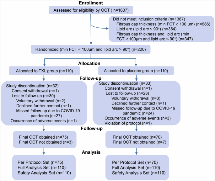
Our study contains several limitations. First, owing to the impact of the COVID-19 pandemic, some patients were lost to follow-up, and the possibility of selection bias could not be ruled out, which might diminish the impact of our results. However, there was no significant difference in baseline characteristics between patients who completed the study and those lost to follow-up (supplementary Table 8). Furthermore, sensitivity analysis demonstrated that our primary results were robust under different assumptions about the missing data. Although we cannot completely rule out selection bias, these analyses increased our confidence in the internal validity of the trial’s conclusions. Second, although there was a trend toward reduced composite cardiovascular events in the TXL group compared with the placebo group, our study was underpowered to detect such a difference in secondary endpoints. Further large sample and long follow-up studies are needed to assess the therapeutic effect of TXL on cardiovascular events. Third, only Chinese patients were enrolled in this study, which may limit the immediate generalizability of our findings to other ethnic groups. Future studies in diverse populations are warranted to confirm the beneficial effect of TXL on plaque stabilization globally.
In conclusion, on the basis of statin therapy, TXL treatment increased the minimum fibrous cap thickness and decreased the maximum lipid arc of coronary thin-cap lipid-rich plaques in patients with ACS. Furthermore, TXL treatment attenuated the severity of angina pectoris and improved the SAQ scores with a good safety profile. Our results provide evidence that on top of statin therapy, treatment with TXL further stabilized coronary atherosclerotic plaques in patients with ACS, which may explain the beneficial effects of TXL on cardiovascular events observed in clinical trials. Larger-scale, longer-term randomized controlled trials with hard clinical endpoints are warranted to further validate the effects of TXL on plaque progression and stability and explore its underlying mechanisms.
Materials and methods (see supplementary materials I for details)Design and eligibility
The TXL-CAP study was an investigator-initiated, multicenter, randomized and double-blind clinical trial that was conducted in 17 medical centers in China. The protocol of the trial has been published previously.34
The trial was approved by the Ethics Committee on Scientific Research of Qilu Hospital of Shandong University [No. 2021(069)] (Supplementary Materials II). The study was performed in accordance with the principles of the Declaration of Helsinki and registered at http://www.chictr.org.cn with the number ChiCTR1900025842.
Subjects underwent a comprehensive baseline screening for eligibility. The inclusion criteria were34: (1) patients who were 18 to 80 years old; (2) patients meeting the diagnostic criteria of ACS by 2015 European Society of Cardiology (ESC) Guidelines35; and (3) the OCT-assessed lesion had a fibrous cap thickness < 100 μm and lipid arc > 90° as determined by OCT technique that was not treated by PCI. If there were multiple lesions that met this criterion, the plaque with the thinnest fibrous cap was chosen as the OCT-assessed lesion, which could be one of the following34: (1) stenotic lesions with a diameter stenosis < 70% without the need for stent implantation; (2) stenotic lesions with diameter stenosis ≥ 70% were stented, and plaques in other coronary vessels that did not require PCI were selected as OCT-assessed lesions; (3) stenotic lesions with diameter stenosis ≥ 70% were stented, and lesions in the same coronary artery that were located at least 5 mm away from the edge of the stent and did not require PCI were selected as OCT-assessed lesions. Thus, the OCT-assessed lesions were divided into three categories according to the aforementioned definitions.
Exclusion criteria included left main coronary disease and/or right coronary artery orifice stenosis; medical history of type 1 diabetes; familial hypercholesterolemia; requiring warfarin for anticoagulation; Takayasu arteritis; severe liver and/or kidney dysfunction; severe chronic obstructive pulmonary disease or respiratory failure; and gastrointestinal, respiratory or other organ bleeding within the previous month.34 All patients or their legal authorized representatives signed written informed consent before randomization.
Randomization and blinding
Patients were randomized to the TXL group or the placebo group at a 1:1 ratio. The information of an eligible patient was entered into a central randomization system, which generated a double-blind random number for enrolled patients. Patients who met the criteria first completed the baseline assessment. The investigators then logged into a randomization and trial supply management (RTSM) system to generate random patient numbers and the corresponding drug numbers in the order of enrolling time. All participants in the trial were blinded to the allocation of treatment. The details are presented in the study protocol in Supplementary Materials I.
ProceduresDrug administration and follow-up
After randomization, patients in the TXL group were treated with an oral dose of TXL capsule (4 capsules, 1.04 g) three times daily for 12 months. The placebo group received a placebo capsule that was completely the same as a TXL capsule. The details of the study drugs, including TXL capsule and placebo, are described in Supplementary Materials III. During the entire study period, other proprietary Chinese medicines that contain the same ingredients as TXL capsules were prohibited. All patients received treatment recommended by the ESC guidelines for ACS, including dual antiplatelet agents and statins. The intensity of statin administration adhered to the 2016 Chinese Guidelines for the Management of Dyslipidemia, which recommended a moderate intensity of statin therapy.23 Patients were followed up for 12 months.
OCT image acquisition and analysis
Patients underwent OCT examination after coronary angiography at the beginning and the end of 12 months of the trial. An OCT system was used, and images were acquired as previously reported.36 All coronary plaques imaged by OCT were divided into three types: lipid plaques, fibrous plaques, and calcified plaques. Lipid plaques manifested as poor signal regions with diffuse borders and exhibited both enhanced backscattering and attenuation. To ensure an accurate measurement, the entire plaque was first scanned by an OCT catheter along its longaxis and imaged at 1-mm intervals along the longaxis to determine the cross-sectional image with the thinnest FCT and the largest lipid arc, which were used for measurement of FCT and lipid arc. The lipid plaque length was measured in the longitudinal imaging view. All measurements were repeated 3 times, and the values were averaged. Fibrous plaques manifested as homogeneous hyperintense regions, and calcified plaques manifested as well-demarcated hypointense regions interspersed with hyperintense foci.37,38 In addition, plaque calcification arc and length, macrophage infiltration, cholesterol crystal and microchannels depicted by OCT were recorded. Macrophage infiltration was defined as hyperintense punctate areas—either discrete or confluent—that exhibited higher signal intensity than the background speckle noise. Cholesterol crystals were defined as linear, highly back-scattering structures within the plaque. Microchannels were identified as small lacunar structures within plaques, with a diameter ranging from 50 μm to 300 μm, and were required to be visible on no fewer than three consecutive imaging frames. A lipid plaque occupying ≥ 2 quadrants with an FCT < 65 μm was defined as TCFA.39
To verify the reproducibility of OCT measurements, two key variables, FCT and lipid arc, were measured twice in 60 randomly selected patients by two independent experts. Interobserver variability was evaluated between the two experts, and intraobserver variability was evaluated by one expert at different times. Bland‒Altman plots were used to analyze the interobserver and intraobserver variability, and ICCs were calculated.
Laboratory assessment
Fasting blood samples were obtained from all recruited patients. The serum levels of lipids, blood glucose, cTnI and hs-CRP, as well as liver and renal functions, were measured at baseline and during follow-up.
Study outcomes
The primary endpoint was the difference in the minimum FCT of the OCT-assessed coronary lesions between the TXL and placebo groups at the end of the 12-month follow-up. Secondary endpoints included the changes in the minimum FCT of the OCT-assessed lesion in the TXL and placebo groups from baseline to 12 months; the changes in maximum lipid arc and length of the OCT-assessed plaque from baseline to 12 months; the percentage of lipid, fibrous and calcified plaques at the end of 12 months of follow-up; the incidence of composite cardiovascular events at 12 months; severity of angina pectoris according to the criteria of the Canadian Cardiovascular Society40; SAQ score41; and serum hs-CRP levels.34
The baseline severity of angina pectoris in enrolled patients was assessed before discharge. Clinical events were recorded by the researchers and submitted to the Clinical Endpoint Committee (CEC), which was independently reviewed and determined by two members of the Committee, and disputed endpoint events were reassessed by all members of the Committee.
Adverse events related to drug treatment were adjudicated and evaluated by CEC blinded to treatment allocation, regardless of whether they were causally related to the study drug.
Data analysis
The sample size of the TXL-CAP trial was estimated based on a previous report.42 In the current study, the standard deviation (SD) was assumed to be 0.07 mm for both groups of patients. To detect a mean difference of 0.03 mm between the TXL group and the placebo group with 80% statistical power, a sample size of 87 subjects per group is required when a two-sided Type I error rate (α) is set at 0.05. Assuming a 20% dropout rate, a total of 220 subjects (110 in TXL and 110 in placebo) are needed to obtain final sample sizes of 87 in each group. The sample size calculation software used was PASS 13. Details of the statistical analysis plan (SAP) are available in Supplementary Materials IV.
Continuous variables were expressed as the mean ± SD. Nonnormally distributed data were summarized as medians (Q1-Q3). Categorical variables are described as numbers (%). Normally distributed continuous variables were analyzed using the two-sample t test, and nonnormally distributed continuous variables were assessed via the Wilcoxon rank-sum test. Categorical data were compared using the chi-square test or Fisher’s exact test.
Efficacy analysis was performed on the full analysis set (FAS). Supportive analysis was also conducted to supplement the primary findings based on the PPS population. Safety analysis, including adverse events, toxicity and laboratory measurements, was performed in the Safety Analysis Set.
We conducted an initial analysis of the primary outcome using the Wilcoxon rank sum test due to the nonnormal distribution of the data. To account for missing data, the last observation carried forward (LOCF) technique was used. We analyzed the change difference of the minimum FCT with a linear mixed-effects model, which included the fixed effects of treatment group and baseline FCT value, with a random intercept for study center to account for variability between sites. Furthermore, as a sensitivity analysis, we included the change in LDL-C from baseline to the 12-month follow-up time point as a covariate in the model. To test the robustness of our findings against missing data due to loss to follow-up, we performed multiple imputation under the assumption of MAR and a series of scenarios for MNAR using delta-adjusted multiple imputation.
For safety variables, we applied descriptive statistics for all patients who were randomized and received at least one dose of the study drug. All treatment-emergent adverse events were recorded, and the rates of adverse events were summarized.
Statistical analyses adhered to the prespecified plan (Supplementary Materials IV), and all statistical analyses were performed using SAS software (version 9.4). Two-sided P < 0.05 denoted statistical significance.

Comments (0)