
Remember me

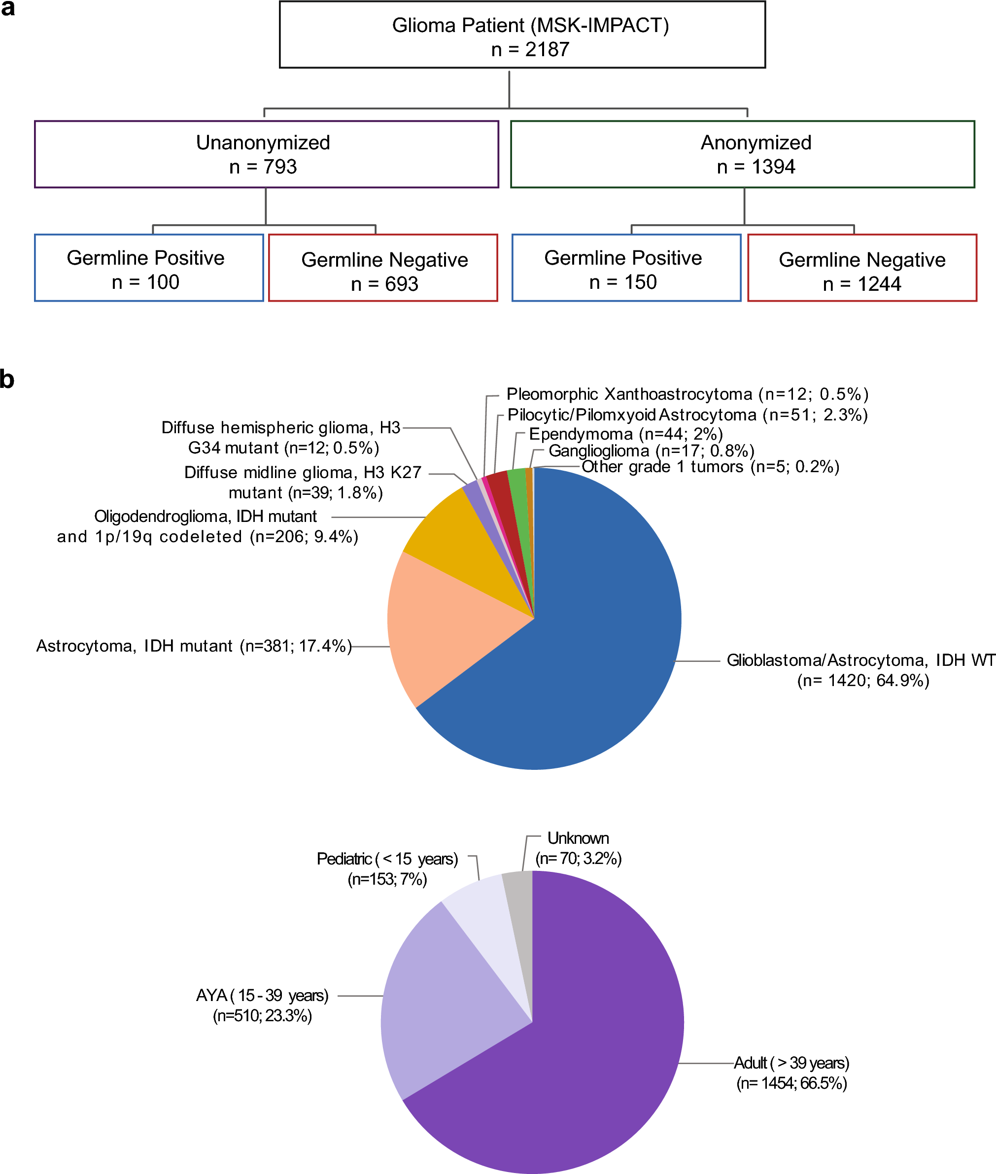


iPSC lines were utilized following IRB review and approval through MGB/BWH IRB (#2015P001676). Previously reported iPSC lines were used, which were generated from cryopreserved peripheral blood mononuclear cell (PBMC) samples from autopsied participants from the ROS and MAP cohorts using the Sendai reprogramming method [46]. ROS and MAP were both reviewed by an Institutional Review Board of Rush University Medical Center [8]. Participants enrolled without known dementia and agreed to annual clinical evaluation and brain donation. All signed informed and repository consents and an Anatomic Gift Act. Cognitive impairment and dementia, global cognition, and several measures of AD pathology methods were previously described [9,10,11,12,13, 68, 75, 94].
iPSCs undergo a rigorous quality control procedure that includes a sterility check, mycoplasma testing, karyotyping, and pluripotency assays performed by the New York Stem Cell Foundation (NYSCF) [46]. iPSCs were maintained using StemFlex Medium (Thermo Fisher Scientific). All cell lines were routinely tested for mycoplasma using a PCR kit (MP0035-1KT) and STR profiling to prevent potential contamination or alteration of the cell lines. iPSC cell lines harboring two homozygous familial Alzheimer’s disease mutations (APPSWE/PSEN1M146V; APPSWE/PSEN1M146V) and their isogenic WT control (Coriell Institute, catalog ID: AG07889) were obtained from NYSCF and are previously described [44, 57].
CRISPR/Cas9 editing to generate BAG3 WT, HET, and KO iPSCsThe following lines were chosen for CRISPR/Cas9 mutagenesis of BAG3: BR33 and BR24, two iPSC lines derived from non-cognitively impaired individuals (male and female, respectively) from the ROSMAP cohorts [46]. gRNA ‘GCAGCGATTCCGAACTGAGG’ was used to target exon 2 within the BAG3 sequence, which is conserved within relevant BAG3 transcript isoforms. The Zhang Lab CRISPR Design website (crispr.mit.edu) was used to generate guide RNAs (gRNAs) with minimal off-target effects. gRNAs were cloned into the pXPR-003 vector. hiPSCs were electroporated with the sgRNA-encoding plasmid and Cas9, and cells were isolated for monoclonal selection and Sanger sequenced to determine BAG3 mutations. For each targeted iPSC line, one unedited/wild-type clone and two mutant clones (HET and KO) were chosen for downstream analyses. IDT CRISPR Cas9 guide RNA design checker was used to check for any potential off-target effects.
Differentiation of iPSCs to induced neurons (iNs)iPSC-derived neurons (iNs) were differentiated following a previously published paper with minor modifications [46, 66, 101]. iPSCs were plated at a density of 95 k cells/cm2 on plates coated with growth factor reduced Matrigel 1 day prior to virus transduction (Corning #354230). Then, iPSCs were transduced with two lentiviruses—pTet-O-NGN2-puro (Addgene plasmid #52047, a gift from Marius Wernig) and FUdeltaGW-rtTA (Addgene plasmid #19780, a gift from Konrad Hochedlinger). The cells were then replated at 200 k cells/cm2 using StemFlex Medium (Thermo Fisher Scientific) and ROCK inhibitor (10 µM) (day 0). The media were changed to KSR media (day 1), 1:1 of KSR and N2B media (day 2), and N2B media (day 3). On day 4, cells were dissociated using accutase and plated at 50 k cells/cm2 using iN D4 media (NBM media + 1:50 B27 + BDNF, GDNF, CNTF (10 ng/mL, Peprotech). Doxycycline (2 μg/ml, Sigma) was added from day 1 to the end of the differentiation, and puromycin (5 µg/ml, Gibco) was added from day 2 to the end of the differentiation. On day 3, B27 supplement (1:100, Life Technologies) was added. From day 4 to the end of the differentiation, cells were cultured in iN day 4 media and fed every 2–3 days [57].
Induced neuron protocol media:
KSR media: Knockout DMEM, 15% KOSR, 1 × MEM-NEAA, 55 µM beta-mercaptoethanol, 1 × GlutaMAX (Life Technologies).
N2B media: DMEM/F12, 1 × GlutaMAX (Life Technologies), 1 × N2 supplement B (Stemcell Technologies), 0.3% dextrose (D-(+)-glucose, Sigma).
NBM media: Neurobasal medium, 0.5 × MEM-NEAA, 1 × GlutaMAX (Life Technologies), 0.3% dextrose (D-(+)-glucose, Sigma).
Cell culture treatmentsFor cell cultures receiving pharmacological treatment, cells were fed with fresh iN media containing the following compounds where applicable:
Bortezomib (Cat# 179324-69-7): Day 18 iNs were treated with 5 nM bortezomib as previously described [33]. The vehicle for this inhibitor was DMSO.
Chloroquine (Cat# 5648): Day 20 iNs were treated with 40 µM chloroquine. The vehicle for this treatment was water.
Cycloheximide (CHX; Cat# C4859): Day 19 iNs were treated with 20 µM CHX for 48 h with or without 10 nM Bortezomib.
Western blottingCells were lysed with RIPA lysis buffer (Thermo Fisher Scientific #89900) with the protease inhibitor (Complete TM mini-protease inhibitor, Roche) and phosphatase inhibitor (phosphoSTOP, Roche) added freshly before lysis. Cells were lysed for 30 min on ice before transferring lysates to microcentrifuge tubes. Cell debris was pelleted by centrifugation (15,000 × g) for 15 min at 4 °C. Supernatant (cell lysate) was collected and stored at – 20 °C until use. Protein concentration in cell lysate samples was determined with the Pierce BCA Protein Assay kit when applicable (ThermoFisher, #23225). Cell lysates were prepared with 4X LI-COR loading buffer (Fisher Scientific, #NC9779096) and 2.5% β-mercaptoethanol, centrifuged, and incubated at 95 °C for 5 min. Samples were resolved using Novex NuPAGE™ 4–12% Bis–Tris gels (ThermoFisher, #NP0336BOX) and NuPAGE™ 1X MOPS-SDS or MES-SDS running buffer (ThermoFisher, #NP0001). Gel electrophoresis was run at 200 V for 50 min. SeeBlue Plus2 (ThermoFisher, #LC5925) pre-stained protein standard was used for evaluation of molecular weight. The gel was extracted and transferred to a nitrocellulose membrane by incubation with 20% methanol Tris–glycine transfer buffer at 400 mA for 2 h. The transferred blot was blocked with Odyssey blocking buffer (LI-COR, #927-50100) for 1 h at room temperature with agitation and incubated with primary antibody (diluted in blocking buffer) overnight at 4 °C with agitation. Blots were incubated with LI-COR secondary antibody diluted 1:10,000 in TBST for 1 h at room temperature with agitation. Blots were washed twice (10 min per wash) with TBST and stored in 1X TBS until imaging. Blots were imaged on a LI-COR Odyssey machine and quantified using ImageStudio software.
Autophagic fluxOn Day 20 of iN differentiation, neurons were fed with media containing 40 µM chloroquine for 24 h along with vehicle control (water). After 24 h, samples were harvested as described above. LC3B has two bands, LC3B-I (top band) and LC3B-II (bottom band). Autophagic flux was calculated by subtracting the amount of LC3B-II in the vehicle-treated controls from the quantified amount of LC3B-II in the chloroquine-treated cells following normalization to GAPDH.
Revert™ 700 total protein stainWhen normalizing to total protein, western blots were stained with REVERT 700 total protein stain per manufacturer guidelines (Cat# 926-11011). Briefly, before blocking or antibody exposure, blots were covered in REVERT Staining Solution for 5 min, washed with Wash Solution two times, and imaged on the LI-COR Odyssey machine. Following imaging, blots were stripped with destaining solution and then proceeded to blocking. The proceeding protocol is then performed as described above.
AHA labeling and protein turnover measurementsOn day 20 of iN cell culture, cells were switched to methionine starvation media (DMEM no methionine no cysteine (Gibco 21–013-024) with 1 × B27 supplement, 0.22 µM sodium pyruvate, 5 µg/mL NAC, 10 ng/mL CNTF, 10 ng/mL BDNF, 10 ng/mL GDNF, 2 µg/mL doxycycline) overnight at 37 °C. On day 21, starvation media was supplemented with 500 µM L-Azidohomoalanine (AHA; Vector Laboratories, CCT-1066) for 24 h to label newly synthesized proteins. After 24 h, iN media was fully changed to complete NBM after DPBS wash. AHA-labeled proteins were chased out in NBM for 7 days. The translation (100%) control for each genotype was lysed immediately after AHA 24-h incubation. Lysates were then prepared as normal for western blotting, as described above. To measure protein turnover, cell lysates were incubated with 5 mM iodoacetamide (Sigma, I6125) for 30 min at room temperature. Lysates were then incubated with 100 µM DBCO-PEG4-Biotin (Tocris, 7480) for 30 min at room temperature. Western blots were performed as described above. For the rabbit anti-Biotin antibody, it was used at a 1:2000 dilution. The amount of Biotin signal per sample was normalized to REVERT total protein stain and then the 100% translation control to yield the % of Biotin remaining, an indicator of protein turnover during the 7-day chase period.
Antibody
Source
Identifier
Rabbit anti-BAG3
Proteintech
Cat# 10599-1-AP
Mouse anti-phospho-tau (Ser202, Thr205)
Thermo Fisher Scientific
Cat# MN1020
Mouse anti-GAPDH
Proteintech
Cat# 60004-1-Ig
Rabbit anti-TAU
Cell Signaling Technologies
Cat# 46687S
Rabbit anti-pTau-214
Cell Signaling Technologies
Cat# 77348
Rabbit anti-pTau-404
Thermo Fisher Scientific
Cat# 44-758G
Mouse anti-TAU
Thermo Fisher Scientific
Cat# AHB0042
Rabbit anti-LC3B
Novus
Cat# NB100-2220
Rabbit anti-PSMB5
Bethyl
Cat# A303-847A-M
Mouse anti-ubiquitin
Millipore
Cat# Mab1510-I-100UG
Mouse anti-CAPZA1
Proteintech
Cat# 66066-1-Ig
Rabbit anti-CFL1
Cell Signaling Technologies
Cat# 5175
Rabbit anti-beta-actin
Abcam
Cat# ab8227
Rabbit anti-alpha-synuclein
Abcam
Cat# ab138501
Rabbit anti-biotin
Abcam
Cat# ab53494
Proteasome activity assayInduced neurons were washed with ice-cold DPBS on Day 21, followed by homogenization in 25 mM HEPES–KOH (pH 7.5), 5 mM MgCl2, 10% glycerol, 0.1% NP-40, 1 mM dithiothreitol (DTT), 1 mM adenosine triphosphate (ATP), 0.1 mM phenylmethylsulfonyl fluoride (PMSF), and 1 mM NaF, followed by sonication in a water bath sonicator (B2500A-MT, VWR) for 20 times for 10 s each, with 30 s pauses between pulses. Protein lysates were centrifuged at 10,000 × g for 10 min at 4 °C. Samples normalized for protein concentration (5∼10 μg total protein) were loaded into a black-walled 96-well plate. Proteasome activity was monitored using an Infinite 200 PRO plate reader (Tecan) with the i-control microplate reader software (Tecan). The reaction was started by adding 20 μM Suc-LLVY-amc (for chymotrypsin-like activity) in 50 mM Tris, 40 mM KCl, 5 mM MgCl2, 0.05 mg/mL Bovine Serum Albumin (BSA), 1 mM ATP, 1 mM DTT, and 1 mM NaF at the zero-time point. For background detection, 2 μM epoxomicin was added to the reactions with Suc-LLVY-amc. The activity was assayed by measuring the fluorescence intensity for 2 h at 37 °C in 1-min intervals (excitation 380 nm; emission 460 nm) [33].
Stable isotope labeling by amino acids in cell culture (SILAC) using iNsSILAC labelingSILAC was employed to investigate protein dynamics during neuronal differentiation. Cells were cultured in media supplemented with heavy isotope-labeled amino acids (Lys + 8, CNLM29-H-1 and Arg + 10 CNLM539-H1 from Cambridge Isotopes) starting at day 14 of differentiation (designated D0 for SILAC labeling). The heavy-labeled media contained 15N. Following the onset of heavy isotope labeling (after D0 harvest), cells were harvested at various time points: D1, D2, D7, D14, and D21, corresponding to 1-, 2-, 7-, 14-, and 21-day post-labeling, respectively. Four distinct lines: BR26, BR57, BR58, and BR98 were used in parallel for all time points. Cell lysates from each line at every time point were prepared and subjected to quantitative mass spectrometry-based proteomic analysis to assess changes in protein expression and turnover during differentiation [89].
Mass spectrometry sample preparationFor the MS analysis, proteins from the cell lysate were precipitated using chloroform/methanol protein precipitation followed by denaturation, reduction, alkylation, enzymatic digestion, peptide cleanup, and high-pH reversed-phase fractionation. Methanol and chloroform were added in a 4:1:3 ratio (sample:methanol:chloroform), followed by vortexing and centrifugation at high speed to separate phases. The aqueous layer was discarded, and the protein pellet was washed with methanol and briefly air-dried. The pellet was then resuspended in 100 µL of 6 M guanidine hydrochloride and incubated at 95 °C for 10 min to ensure complete solubilization. Disulfide bonds were reduced by adding 0.5 µL of 1 M dithiothreitol (DTT), followed by incubation at 56 °C for 20 min. After cooling to room temperature, 3 µL of 500 mM iodoacetamide (IAA) was added to alkylate the reduced cysteine residues. Samples were incubated in the dark at room temperature for 20 min. Excess IAA was quenched by adding 5 µL of 1 M DTT and incubating for 15 min at room temperature. Then, samples were digested with mass spectrometry grade trypsin/Lys-c mix (Promega, Cat# V5073) overnight at 37 °C. The reaction was subsequently stopped by acidification with 1% formic acid (FA) and desalted using HyperSep C18 Cartridges (Thermo Scientific, Cat# 60108-302) per manufacturer’s instructions. Eluates were dried using a vacuum concentrator and stored at − 80 °C.
Dried peptides were reconstituted in 300 µL of 0.1% triethylamine (TEA) in water and fractionated using the Pierce™ High-pH Reversed-Phase Peptide Fractionation Kit (Thermo Scientific, Cat# 84868). Fractionation was performed according to the manufacturer’s protocol, with stepwise elution using 5%, 10%, 15%, and 50% acetonitrile (ACN) in 0.1% TEA. Each of the four resulting fractions was collected, dried under vacuum, and stored at − 80 °C until LC–MS/MS analysis. Each dried high-pH fraction was further desalted using Pierce™ C18 Spin Columns (Thermo Scientific, Cat# 89852) to remove residual TEA and salts. Final eluates were dried using a vacuum concentrator and stored at − 80 °C until LC–MS/MS analysis [73].
Liquid chromatography–mass spectrometry (LC–MS/MS)Dried peptide samples were resuspended in 20 µL of Buffer A (94.875% H₂O, 5% acetonitrile [ACN], and 0.125% formic acid [FA]). A total of 3 µg of peptide was injected via autosampler and loaded onto a PepMap™ 100 C18 nanoViper™ trap column (75 µm × 2 cm, Thermo Fisher Scientific) using an UltiMate™ 3000 HPLC system. Peptides were then separated on an analytical PepMap™ RSLC C18 column (Thermo Fisher Scientific) coupled to a stainless-steel emitter tip and analyzed using a Nanospray Flex™ ion source operating at 2,000 V.
Chromatographic separation was performed over a 2.5-h gradient using Buffer A (94.875% H₂O, 5% ACN, 0.125% FA) and Buffer B (99.875% ACN, 0.125% FA). The elution gradient consisted of 2–8% Buffer B over 6 min, 8–24% over 64 min, 24–36% over 20 min, 36–55% over 10 min, 55–95% over 10 min, followed by 10 min at 95%, and finally re-equilibration to 2% Buffer B over 30 min. Peptides were analyzed on an Orbitrap Fusion mass spectrometer (Thermo Fisher Scientific) using data-dependent acquisition. MS1 survey scans were acquired in the Orbitrap at a resolution of 60,000, over a scan range of 300–1500 m/z. MS parameters were: ion transfer tube temperature, 300 °C; Easy-IC internal mass calibration; default charge state, 2; automatic gain control (AGC) target, 2 × 10⁵; maximum injection time, 50 ms; microscans, 1; and S-lens RF level, 60. Data were collected in positive ion mode with centroid format. MIPS was enabled. Charge states from 2 to 6 were selected for fragmentation; unassigned precursors were excluded. Dynamic exclusion was enabled with a repeat count of 1, exclusion duration of 30 s (high) and 45 s (low), and a ± 10 ppm mass tolerance.
For MS/MS (MS2) acquisition, the top 20 most intense precursor ions were selected for fragmentation. Precursor isolation was performed with a 1.6 m/z window using the quadrupole. Higher energy collisional dissociation (HCD) was employed with a normalized collision energy (NCE) of 30%. Fragment ions were detected in the ion trap using a rapid scan rate, with a maximum injection time of 75 ms and an AGC target of 10,000. Q value was set to 0.25, and ion accumulation used all available parallelizable time [30].
Mass spectrometry data analysis and quantificationProtein identification, quantification, and analysis were performed with Integrated Proteomics Pipeline—IP2 (Integrated Proteomics Applications, Inc., San Diego, CA. http://www.integratedproteomics.com/) using ProLuCID, DTASelect2, Census, and QuantCompare. Spectrum raw files were extracted into MS1, MS2 files using RawExtract 1.9.9 (http://fields.scripps.edu/downloads.php). The tandem mass spectra were searched against the UniProt human protein database (downloaded on 06-03-2022), matched to sequences using the ProLuCID/SEQUEST algorithm (ProLuCID version 3.1) with 20 ppm peptide mass tolerance for precursor ions and 600 ppm for fragment ions. ProLuCID searches included all fully and half-tryptic peptide candidates that fell within the mass tolerance window and had no miscleavages. Carbamidomethylation (+ 57.02146 Da) of cysteine was considered as a static modification. Peptide/spectrum matches (PSMs) were assessed in DTASelect2 using the cross-correlation score (XCorr) and normalized difference in cross-correlation scores (DeltaCN). Peptide probabilities and false-discovery rates (FDR) were calculated based on a target/decoy database containing the reversed sequences of all the proteins appended to the target. For each brain region per biological replicates, proteins identified had an FDR of ≤ 1% at the protein level. Each protein identified was required to have a minimum of one peptide of minimal length of six amino acid residues.
For the SILAC experiments, each dataset was searched twice, once against light (14N) and then against heavy (8.0142 K and 10.0083 R) protein databases. After the results from ProLuCID were filtered using DTASelect2, ion chromatograms were generated using “Census”. Census allows users to filter peptide ratio measurements based on a correlation threshold because the correlation coefficient (values between zero and one) represents the quality of the correlation between the unlabeled and labeled chromatograms and can be used to filter out poor quality measurements. This correlation coefficient also considers the retention time of the identified spectra. Peptide ratio measurements used for further analysis exceeded a profile score of 0.8. Fractional abundance was calculated from the “light” and “heavy” peptide chromatograms, and for each respective protein, the 14N and 15N chromatograms for the respective peptides were averaged. The protein remaining was then calculated from those “light” and “heavy” averages to finally compare across DIVs. Final protein ratios were generated with QuantCompare, which uses Log twofold change on the biological replicates. The statistical significance of the differential expression for all proteins was assessed using a two-tailed one-sample Student’s t-test on their corresponding peptide quantification ratios between both conditions. The obtained p values were FDR-adjusted for multiple hypothesis testing using the Benjamini–Hochberg correction. Proteins with FDR-adjusted p values < 0.05 and measured in at least two biological replicates in both conditions were considered for further analyses [89]. The percentage of “light” protein remaining at an individual harvest day was calculated by dividing the light/heavy ratio at the day of interest by the corresponding value at harvest D1. Proteins that displayed light/heavy ratios > 1 were excluded from analysis. Proteins measured in all four genetic backgrounds in at least 2 differentiations at harvest D7 and D14 were used for principal component analysis (PCA). PCA was performed in R studio using the factoextra library. Dimension coordinates for individual lines and proteins were exported and visualized in GraphPad Prism 10.
TMT-MS proteomics and data analysis for iNs and human brain extractsSample processingiNs were plated in 6-well plates (1 million and 500 k cells per well, respectively) and differentiated to day 21. Media was removed from the wells and cells were washed two times with ice-cold DPBS. Samples were processed as previously described [20, 49]. Cells were lysed in 250 μL of urea lysis buffer (8 M urea, 100 mM NaHPO4, pH 8.5) with the protease inhibitor (Complete TM mini-protease inhibitor, Roche) and phosphatase inhibitor (phosphoSTOP, Roche) added freshly before the lysis. All homogenization was performed using a Bullet Blender (Next Advance) according to manufacturer protocols. Briefly, each tissue piece was added to urea lysis buffer in a 1.5 mL Rino tube (Next Advance) harboring 750 mg stainless-steel beads (0.9–2 mm in diameter) and blended twice for 5 min intervals in the cold room (4 °C). Protein supernatants were transferred to 1.5 mL Eppendorf tubes and sonicated (Sonic Dismembrator, Fisher Scientific) 3 times for 5 s with 15-s intervals of rest at 30% amplitude to disrupt nucleic acids and subsequently vortexed. Protein concentration was determined with BCA assay, and samples were frozen in aliquots at − 80 °C. Protein homogenates (50 μg) were treated with 1 mM dithiothreitol (DTT) at 25 °C for 30 min, followed by 5 mM iodoacetamide (IAA) at 25 °C for 30 min in the dark. The protein mixture was digested overnight with 1:100 (w/w) lysyl endopeptidase (Wako) at room temperature. The samples were then diluted with 50 mM NH4HCO3 to a final concentration of less than 2 M urea and further digested overnight with 1:50 (w/w) trypsin (Promega) at 25 °C. Resulting peptides were desalted with a Sep-Pak C18 column (Waters) and dried under vacuum.
Tandem mass tag (TMT) labelingPeptides were reconstituted in 100 μl of 100 mM triethyl ammonium bicarbonate (TEAB), and labeling was performed using TMTPro isobaric tags (Thermofisher Scientific, A44520) as previously described [31, 70]. Briefly, the TMT labeling reagents were equilibrated to room temperature, and anhydrous ACN (200 mL) was added to each reagent channel. Each channel was gently vortexed for 5 min, and then 20 mL from each TMT channel was transferred to the peptide solutions and allowed to incubate for 1 h at room temperature. The reaction was quenched with 5% (v/v) hydroxylamine (5 mL) (Pierce). All 16 channels were then combined and dried by SpeedVac (LabConco) to approximately 100 mL and diluted with 1 mL of 0.1% (v/v) TFA, then acidified to a final concentration of 1% (v/v) FA and 0.1% (v/v) TFA. Peptides were desalted with a 60 mg HLB plate (Waters). The eluates were then dried to completeness. High-pH fractionation was performed essentially as described with slight modification [71]. Dried samples were resuspended in high-pH loading buffer (0.07% v/v NH4OH, 0.045% v/v FA, 2% v/v ACN) and loaded onto a Water’s BEH (2.1 x 150 mm with 1.7 µm beads). A Thermo Vanquish UPLC system was used to carry out the fractionation. Solvent A consisted of 0.0175% (v/v) NH4OH, 0.01125% (v/v) FA, and 2% (v/v) ACN; solvent B consisted of 0.0175% (v/v) NH4OH, 0.01125% (v/v) FA, and 90% (v/v) ACN. The sample elution was performed over a 25 min gradient with a flow rate of 0.6 mL/min with a gradient from 0 to 50% B. A total of 96 individual equal volume fractions were collected across the gradient and dried to completeness using vacuum centrifugation.
Data processing protocolAll raw files were searched using Thermo’s Proteome Discoverer suite (version 2.4.1.15) with Sequest HT. The spectra were searched against a human Uniprot database downloaded August 2020 (86,395 target sequences). Search parameters included a 10 ppm precursor mass window, a 0.05 Da product mass window, dynamic modifications for methionine (+ 15.995 Da), deamidated asparagine and glutamine (+ 0.984 Da), phosphorylated serine, threonine, and tyrosine (+ 79.966 Da), and static modifications for carbamidomethyl cysteines (+ 57.021 Da) and N-terminal and Lysine-tagged TMT (+ 304.207 Da). Percolator was used to filter PSMs to 0.1%. Peptides were grouped using strict parsimony, and only razor and unique peptides were used for protein level quantitation. Reporter ions were quantified from MS2 scans using an integration tolerance of 20 ppm with the most confident centroid setting. Abundance data were log2 transformed, and batch effects were regressed using ComBat [51]. Differential expression was calculated using the DEP package in R [100].
Gene set variation analyses (GSVA)BR107 iNs were either differentiated to day 20 and treated for 24 h with 40 µM chloroquine or differentiated to day 18 and treated for 72 h with 5 nM bortezomib, as previously described [4, 33]. Briefly, iNs were harvested and processed for TMT-MS proteomics, as described above. The top 100 downregulated (q < 0.05) proteins with autophagy inhibition or proteasome inhibition were used as the “chloroquine enrichment score” or “bortezomib enrichment score,” respectively, using GSVA. These GSVA enrichment scores were then correlated to BAG3 protein abundance across 53 ROSMAP lines [33].
TBS-soluble brain extract preparation and iN treatmentHuman brain tissue applied to 10 iN genetic backgrounds was obtained from Albany Medical Center [34]. Tris-buffered saline (TBS: 20 mM Tris–HCl, 150 mM NaCl, pH 7.4) brain extracts were prepared using a method previously described [34, 78]. Briefly, the tissue was dissected to isolate the gray matter, which was homogenized in ice-cold TBS at a ratio of 1:4 tissue weight to buffer volume within a Dounce homogenizer. This suspension was then subjected to ultra-centrifugation at 1.75 × 10⁵g in a TL100 centrifuge (Beckman Coulter) to pellet cellular debris. The supernatant was placed in a 2-kDa dialysis cassette (Slide-A-Lyzer, Thermo Fisher) and dialyzed 1:104 (volume:volume) in artificial cerebrospinal fluid (aCSF: 124 mM NaCl, 2.5 mM KCl, 2.0 mM MgSO4, 1.25 mM KH2PO4, 26 mM NaHCO3, 10 mM glucose, 4 mM sucrose, 2.5 mM CaCl2). Following dialysis, the TBS brain extracts were aliquoted and stored at − 80 ℃. To treat iNs, the TBS brain extracts were ‘buffer-exchanged’ into culture media by placing the extract in a 3-kDa centrifugation filter (Amicon Ultra, EMD Millipore) and performing a 90-min centrifugation at 3000 × g in an RC-5C centrifuge (Sorvall, Thermo Fisher). Following centrifugation, the retentate was reconstituted to 1X volume in BrainPhys Media with SM-1 neuronal supplement (StemCell Technologies). The protein concentration within the reconstituted extracts was determined by performing a bicinchoninic acid (BCA) assay (Thermo Fisher) on an 8-step, twofold dilution series of reconstituted extract and media to determine the proportion of protein contributed by the brain extract. The reconstituted extract was then back diluted in BrainPhys media to a concentration of 1 mg/mL brain extract protein. Day 18 iNs were treated with this human AD brain extract (BE) by diluting the 1 mg/mL BE 1:8 in iN culture media. Treatments were carried out for 72 h at 37 °C with the media changed and fresh 1:8 AD BE added every 24 h. After the final treatment, cells were washed three times with ice-cold DPBS and harvested in RIPA for western blotting, as described.
TBS-soluble human brain extract preparation for proteomic analysisFrozen angular gyrus tissue was thawed, and meninges were peeled off and discarded. Aqueous TBS extracts were prepared according to the “soaking” method as previously described [32, 81], with some modifications. The tissue was chopped on a McIlwain tissue chopper set to 0.05 mm width, then soaked for 30 min in a 5-ml Eppendorf Protein LoBind tube with nutation at 4 °C in five volumes TBS (20 mM Tris, 150 mM NaCl, pH 7.4) supplemented with 5 μg/ml leupeptin, 5 μg/ml aprotinin, 2 μg/ml pepstatin, 120 μg/ml 4-benzenesulfonyl fluoride hydrochloride, and 5 mM NaF. The suspension was centrifuged at 2,000 g in a swinging-bucket rotor for 10 min at 4 °C. The supernatants were then centrifuged in 1.5-ml LoBind tubes at 20,000 g for 110 min at 4 °C in a tabletop centrifuge. The supernatants were pooled, re-aliquoted, and frozen at – 80 °C as the TBS-soluble fraction for further analysis. The pellets were frozen at – 80 °C for subsequent extraction.
To create the SDS and urea extracts, a pellet from the TBS extract was thawed on ice and reweighed. The pellet was resuspended in five volumes TBS with 2% SDS. The samples were sonicated at 35% power three times for 10 s. They were then centrifuged again at 20,000 g for 110 min at 4 °C in a tabletop centrifuge as above. The supernatants were re-aliquoted and frozen at – 80 °C as the SDS extract. The pellets were resuspended in another five volumes of 8 M urea followed by sonication, centrifugation, and aliquoting as for the SDS extracts.
Aβ immunodepletion of AD brain extract and iN treatmentTreatment of iNs with Aβ-immunodepleted AD brain extracts was performed as previously described [92], with some modifications. AD soaking extracts were immunodepleted by adding anti-Aβ rabbit polyclonal antibody S97 or mock-immunodepleted with pre-immune rabbit purified IgG (45 µg/ml), along with 50 µl/ml protein A sepharose beads. Binding and depletion were carried out overnight three times, followed by a clearance step with protein A beads but no antibody. Following immunodepletion and prior to addition to iN cultures, extracts were buffer exchanged into iN growth medium using PD MiniTrap columns by the spin method (Cytiva) followed by aliquoting and storage at – 80 °C. Efficiency of immunodepletion and nonspecific loss during buffer exchange was monitored by ELISA. Buffer exchanged extracts were added at a v:v ratio of 1:3 to day 21 iNs (BR24) at a density of 5,000 cells per well of a 96-well plate for 72 h without media exchange, along with a media-only control. Neurite length was monitored using the IncuCyte Zoom system. Following treatment, RNA was harvested using an Invitrogen PureLink RNA Mini Kit. Immunodepletion, mock-immunodepletion, iN treatment, and RNA harvesting were performed for three AD brain extracts in two experimental batches and submitted for RNA sequencing.
RNAseqPurity and concentration of RNA extractions were assessed using a NanoDrop spectrophotometer. Samples were sent to Genewiz (Azenta) for library preparation and sequencing. Adaptors were removed using the R package cutadapt, followed by quality control with fastqc. Alignment was accomplished with tophat, followed by gene expression matrices generated by cufflinks and htseq-count. Last, differentially expressed genes were accomplished by DEseq2 using a general linear model.
ELISAs48-h conditioned media was collected immediately prior to harvest. Extracellular proteins were measured following manufacturer instructions for the Aβ Peptide Panel 1 (6E10) Kit (MesoScale Discoveries, Cat# K15200E-2). Samples were normalized to total protein concentration as determined by the Pierce BCA Protein Assay kit (ThermoFisher, #23225).
Data visualizationSchematics were generated using Biorender. Graphs and heat maps were generated using R Studio or GraphPad Prism 10.
Quantification and statistical analysisInformation regarding statistical analyses can be found in the figure legends. All statistical tests were performed using GraphPad Prism 10 or R Studio. All data are shown as mean ± SD or SEM. Where appropriate, paired statistical tests were employed.
Comments (0)