Blood Collection
Platelets, leukocytes and red blood cells (RBCs) used in this study were obtained from healthy adult volunteers. Blood was collected into polypropylene tubes containing 0.109 M sodium citrate as an anticoagulant. Samples were processed and isolated in Department of laboratory medicine at West China Hospital, Sichuan University. All procedures were approved by the Ethics Committee of West China Hospital, Sichuan University, and informed consent was obtained from all volunteers.
Preparation of Platelets, Leukocytes, and Whole Blood
Platelet-rich plasma (PRP) was prepared by centrifugation at 200 g for 5 min. The resulting pellet was washed and resuspended in phosphate buffer saline (PBS). For activation, platelets were treated with thrombin (10602400001, Roche, 1 U/mL), collagen (FD034945, Sysmex, 100 µg/mL), or thrombin receptor agonist peptide (TRAP) (CLP0512, Solarbio, 50 µM) for 15 min at 37 °C (P-sel positive platelet was around 70–80% as evidenced by flow cytometry). Peripheral blood mononuclear cells (PBMCs) and neutrophils were isolated from whole blood using Polymorphprep™ (Serumwerk, Germany) through density gradient centrifugation at 500 × g for 30 min at room temperature with slow deceleration. After centrifugation, the layer above Polymorphprep™ contained only peripheral blood mononuclear cells (PBMCs), while the lower layer contained polymorphonuclear leukocytes (mainly neutrophils). Both PBMCs and neutrophils had a purity greater than 90%. Platelets were removed by centrifugation at 200 g for 5 min for whole blood preparation. All procedures were at room temperature.
Co-culture Experiments with Activated Platelets
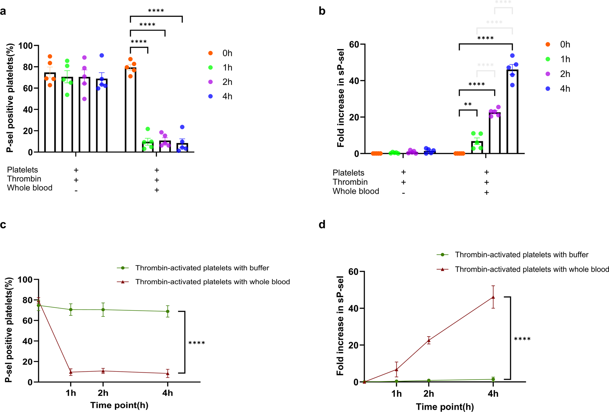
Activated platelets were co-cultured with whole blood, PBMCs, or neutrophils at 37 °C for 4 h. In some experiments, before co-culturing with activated platelets, neutrophils were pre-treated with neutrophil elastase (NE) inhibitor Sivelestat (HY-17443, MedChemExpress, 1 mM), myeloperoxidase (MPO) inhibitor ABAH (475944-1GM, EMD Millipore, 2 mM), Latrunculin B (LatB) (L5288, Merck Millipore, 5 µM), lipopolysaccharide (LPS) (L2880, Sigma-Aldrich, 100 µg/mL), or Pam3CSK4 (tlrl-pms, InvivoGen, 25 µg/mL) for 1 h. Samples were taken at four time points: 0, 1, 2, and 4 h of co-culture. After centrifugation, the supernatant was collected, and the cell pellets were fixed with 4% paraformaldehyde.
Detection of P-sel on the Platelet Surface
Flow Cytometry Detection of P-sel on Platelets
The cell pellets were blocked with 2% Bovine Serum Albumin (BSA) and then stained with CD42b (303934, Brilliant Violet 510™ anti-human CD42b, Biolegend), CD62P antibodies (304905, PE-anti-human CD62P, Biolegend) and CD45 (304025, PerCP-anti-human CD45, Biolegend) for 1 h at room temperature, protected from light. After washing with PBS buffer, the cells were analyzed using a flow cytometer (BD FACSCanto II, BD Biosciences). At least 50,000 events were analyzed for each sample. Data were analyzed using FlowJo software (FlowJo, LLC) to determine the percentage of CD62P positive platelet (%) and the number of platelet–leukocyte aggregates (PLAs, CD62P positive/PerCP-anti-human CD45 positive).
Chemiluminescence Immunoassay Detection of P-sel on Platelets
The expression of platelet P-sel was detected in real-time using a homogeneous chemiluminescence immunoassay instrument (HSCL-5000, Poclight Biotech, China). After sampling at the corresponding time points, the samples were centrifuged and washed with PBS to obtain the cell pellets. Homogeneous chemiluminescence immunoassay is a two-site (“sandwich”) chemiluminescence immunoassay based on chemiluminescence (CL) resonance energy transfer (CRET) technology. The acridinium ester (AE) emits a strong CL signal in the presence of H₂O₂. Graphene oxide (GO) serves as an efficient quencher, creating a “signal off” state via CRET between GO and AE-labelled DNA3. Upon sample application, DNA1-coated CD61 antibody, DNA2-coated CD61 antibody, and AE-labelled DNA3 form a sandwich complex, detaching DNA3 from GO and inhibiting CRET. This produces a CD61-CL signal, mimicking quantitative platelet count detection. For P-sel expression on platelets, the same principle is applied using DNA1-coated CD62P antibody and DNA2-coated CD41 antibody. The sandwich complex forms with CD62P-positive platelets, detaching AE-labelled DNA3 from GO and generating a CD62P-CL signal, mimicking quantitative P-sel positive platelet count detection. The CL intensity ratio of CD62P to CD61 is used to represent the P-sel intensity. Due to methodological limitations, chemiluminescence immunoassay could not be used to analyze whole blood samples.
Detection of sP-sel and NE
sP-sel and NE in the supernatants were determined using ELISA kits (Boster Bio, China) according to the manufacturer’s instructions. The absorbance was measured at a wavelength of 450 nm using a microplate reader.
Statistics
Data were analyzed using GraphPad Prism 9.5. The normality of the data was assessed using the Shapiro-Wilk test. For normally distributed data, results are presented as mean ± SEM. A two-way ANOVA was conducted to evaluate differences among groups across various time points (0, 1, 2, and 4 h). Tukey’s multiple comparisons test was used to analyze differences between groups at each time point, as well as changes from 0 h to 4 h measurements within each group. Spearman correlation analysis was performed to evaluate the relationship between PLAs and the fold increase of sP-sel. Statistical significance was defined as P < 0.05.


Comments (0)