Preparation of P. gingivalis and Inactivated B. subtilis R0179
P. gingivalis ATCC 33277 was cultured following established methods [26]. The B. subtilis R0179 strain was grown in Luria-Bertani (LB) broth [27]. The bacteria were harvested by centrifuging at 4000 rpm for 10 min and rinsed three times with phosphate-buffered saline (PBS). B. subtilis R0179 bacterial suspension was mixed with sterile glycerol (Beijing Solarbio Science & Technology Co., Ltd., Beijing, China) and inactivated by autoclaving at 120 °C for 15 min under high pressure, followed by centrifugation to collect the heat-inactivated bacterial cells.
Culture of Gingival Epithelial Cells
Human gingival epithelial cells were purchased from iCell and subsequently reconstituted in a suitable amount of complete culture medium, specifically iCell primary keratinocyte basal medium supplemented with primary keratinocyte culture supplement (iCell Bioscience Inc., Shanghai, China).
Construction of the Gingival Epithelial Cells Invasion Model
1 × 10⁶ gingival epithelial cells were plated into 6-well plates (Corning Incorporated, Corning, New York, USA) and maintained in antibiotic-free complete medium for overnight incubation to allow cell attachment. P. gingivalis in the logarithmic growth phase was collected by centrifugation, washed three times with PBS, and resuspended in an antibiotic-free complete medium. P. gingivalis was then co-cultured with gingival epithelial cells for 2 h at different multiplicity of infection (MOI), while control cells were left unstimulated. After 2 h, the medium was discarded, and cells were gently washed three times with sterile PBS. Fresh medium containing 0.5 mg/mL gentamicin (Beijing Solarbio Science & Technology Co., Ltd., Beijing, China) and 0.1 mg/mL metronidazole (Beijing Solarbio Science & Technology Co., Ltd., Beijing, China) was then added. In certain groups, B. subtilis R0179 or 1 µM agonists of TLR2 (MedChemExpress, New Jersey, USA) or TLR4 (MedChemExpress, New Jersey, USA) were introduced for further treatment, and incubation continued for 10 h. The total stimulation time was the sum of both incubation phases.
Transmission Electron Microscopy Observation
Gingival epithelial cells were plated in a 6-well plate and stimulated with P. gingivalis (MOI = 50:1) for 2 h. The cells were washed twice with sterile PBS, fixed with pre-cooled 2.5% glutaraldehyde (Beijing Solarbio Science & Technology Co., Ltd., Beijing, China) for 1 h, and scraped off. The collected cells were centrifuged, and fresh 2.5% glutaraldehyde was added, followed by fixation at 4 °C. The pellet was treated with 1% osmium tetroxide (Condice Chemical Co., Ltd., Hubei, China) for 2 h, washed with PBS, and sequentially dehydrated in a graded ethanol series. The samples were subjected to ultrathin sectioning, followed by double staining with uranyl acetate and lead citrate (both from Condice Chemical Co., Ltd., Hubei, China), and were finally observed under a transmission electron microscope (Japan Electron Optics Laboratory Ltd., Tokyo, Japan).
Quantitative PCR (qPCR) Analysis
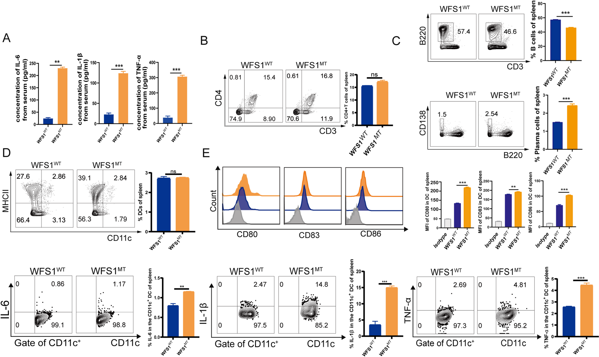
Total RNA was extracted from the co-cultured gingival epithelial cells using TRIzol reagent (Invitrogen Corporation, California, USA), followed by cDNA synthesis with a reverse transcription kit (Takara Bio Inc., Shiga, Japan). qPCR was conducted to analyze the expression of TLR1, TLR2, TLR4, TLR5, TLR6, TNF-α, IL-1β, IL-6, and GAPDH genes. The gene-specific primers used are listed in Table S1. The gene expression levels were normalized to the GAPDH housekeeping gene and analyzed using the ΔΔCt method.
Western Blot Analysis
Proteins were isolated from the co-cultured cells by RIPA lysis buffer (Huaxingbio Co., Ltd., Beijing, China), and their concentrations were quantified via the BCA assay. Equivalent protein amounts were resolved by SDS-PAGE (Huaxingbio Co., Ltd., Beijing, China) and subsequently transferred onto PVDF membranes (Merck KGaA, Darmstadt, Germany). The membranes were blocked and incubated overnight at 4 °C with primary antibodies against TLR1, TLR2, TLR4, TLR5, TLR6, NF-κB, p-NF-κB, and GAPDH. The membranes were finally detected using an enhanced chemiluminescence (ECL) detection system (Bio-Rad Laboratories, Inc., California, USA). Detailed antibody information is provided in Table S2.
Antibody Array Assay
The cell culture was centrifuged at 3,000 rpm for 30 min at 4 °C, and the resulting supernatant was collected and filtered through a 0.22 μm membrane. Equilibrate the glass slide chip (RayBiotech Co., Ltd., Guangdong, China) at 37 °C for 30 min and dry for 2 h. Prepare standards and negative controls as per the RayBiotech manual. Block the chip with 100 µL blocking buffer per well for 1 h, then introduce 90 µL of standard or sample and incubate overnight at 4 °C. Wash 5 times with wash buffer I and 2 times with wash buffer II. Incubate with 80 µL detection antibody for 2 h, then with 80 µL Cy3-streptavidin (RayBiotech Co., Ltd., Guangdong, China) for 1 h. Wash and send the chip to RayBiotech for signal acquisition using the InnoScan 300 scanner. Analyze data with QAH-INF-1 software (RayBiotech Co., Ltd., Guangdong, China).
Mouse Model
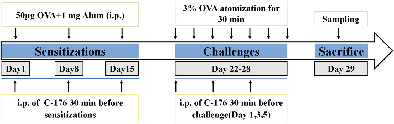
Application of inactivated B. subtilis R0179 in a mouse model of periodontitis. Six-week-old male C57BL/6 mice was obtained and housed under specific pathogen-free (SPF) conditions. The animals were randomly assigned into five groups (n = 8 mice per group): Control group (C), P. gingivalis-infected group (C + Pg), P. gingivalis + B. subtilis R0179-infected group (C + Pg + B. subtilis), P. gingivalis + B. subtilis infection + TLR2 agonist (TLR2-agonist), and P. gingivalis + B. subtilis infection + TLR4 agonist (TLR4-agonist). For infection, P. gingivalis ATCC 33277 or B. subtilis R0179 was suspended in 2% carboxymethyl cellulose to a concentration of 109 CFU/mL. The mice received 10 µL of the bacterial suspension via gingival sulcus application every three days for four weeks. TLR2-agonist and TLR4-agonist were applied simultaneously with bacterial infection, each at a concentration of 1 mM, with 2 µL administered into the gingival sulcus each time.
Hematoxylin and Eosin (H&E) Staining
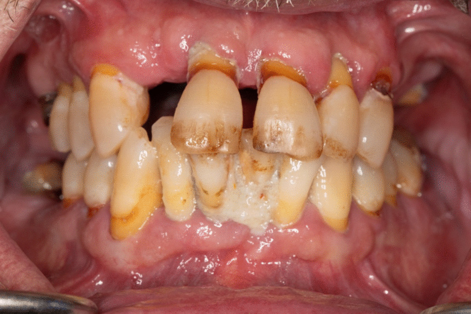
The gingival tissues of mice from the control group, C + Pg group, and C + Pg + B. subtilis group were used for H&E staining. The gingival tissues were collected, fixed, embedded, and sectioned. Tissue sections were stained with hematoxylin (Beijing Solarbio Science & Technology Co., Ltd., Beijing, China) for 3 min, rinsed with distilled water, and subsequently counterstained with eosin (Beijing Solarbio Science & Technology Co., Ltd., Beijing, China) for 30 s. After two additional rinses in distilled water, the samples were dehydrated through a graded ethanol series, cleared with xylene, and mounted using neutral resin and a coverslip.
Immunohistochemical (IHC) Staining
The gingival tissues of mice from the control group, C + Pg group, C + Pg + B. subtilis group, TLR2-agonist group, and TLR4-agonist group were used for IHC staining. Gingival tissues were harvested, fixed in polyformaldehyde, and embedded in paraffin. Sections were deparaffinized, rehydrated, and treated with antigen retrieval solution. After blocking peroxidase and nonspecific binding, the sections were incubated overnight with primary antibodies against TLR2, TLR4, p-NF-κB, IL-6, IL-1β, and TNF-α (Detailed antibody information is provided in Table S3). Biotinylated secondary antibodies (Proteintech Co., Ltd., Hubei, China) were applied, and visualization was done using a DAB substrate. Counterstaining with hematoxylin (Beijing Solarbio Science & Technology Co., Ltd., Beijing, China) was performed, and slides were examined under a light microscope.
Statistical Analysis
Statistical analysis for multiple comparisons was performed using GraphPad Prism 8.0 (San Diego, California, USA). Statistical comparisons were performed using one-way ANOVA, followed by Tukey’s post hoc test. Differences with P-values less than 0.05 were deemed statistically significant.


Comments (0)