Melmed S et al. Clinical biology of the pituitary adenoma. Endocr Rev. 2022;43(6):1003–1037. https://doi.org/10.1210/endrev/bnac010
Bashari WA, Gillett D, MacFarlane J, Scoffings D, Gurnell M, Pituitary Imaging,. Pituit. Jan. 2022;677–721. https://doi.org/10.1016/B978-0-323-99899-4.00022-6.
Tritos NA, Miller KK. Diagnosis and management of pituitary adenomas. JAMA. Apr. 2023;329(16):1386. https://doi.org/10.1001/jama.2023.5444.
Petersenn S et al. Dec., Diagnosis and management of prolactin-secreting pituitary adenomas: a pituitary society international consensus statement. Nat Rev Endocrinol. 2023;19(12):722–740. https://doi.org/10.1038/s41574-023-00886-5
Fleseriu M, Langlois F, Lim DST, Varlamov EV, Melmed S. Acromegaly: pathogenesis, diagnosis, and management. Lancet Diabetes Endocrinol. 2022;10(11):804–826. https://doi.org/10.1016/S2213-8587(22)00244-3
Fleseriu M, Varlamov EV, Hinojosa-Amaya JM, Langlois F, Melmed S. An individualized approach to the management of Cushing disease. Nat Rev Endocrinol. 2023;19(10):581–599. https://doi.org/10.1038/s41574-023-00868-7
Fleseriu M et al. Dec., Consensus on diagnosis and management of Cushing’s disease: a guideline update. Lancet Diabetes Endocrinol. 2021;9(12):847–875. https://doi.org/10.1016/S2213-8587(21)00235-7
MacFarlane J et al. Advances in the Imaging of Pituitary Tumors. 2020. W.B. Saunders. https://doi.org/10.1016/j.ecl.2020.06.002
Bashari WA et al. Modern imaging of pituitary adenomas. Apr 01 2019 Bailliere Tindall Ltd https://doi.org/10.1016/j.beem.2019.05.002
Bi WL, Laws ER. Metabolic imaging in the detection of growth hormone-secreting pituitary adenomas. World Neurosurg. Sep. 2014;82(3):329–30. https://doi.org/10.1016/j.wneu.2014.03.014.
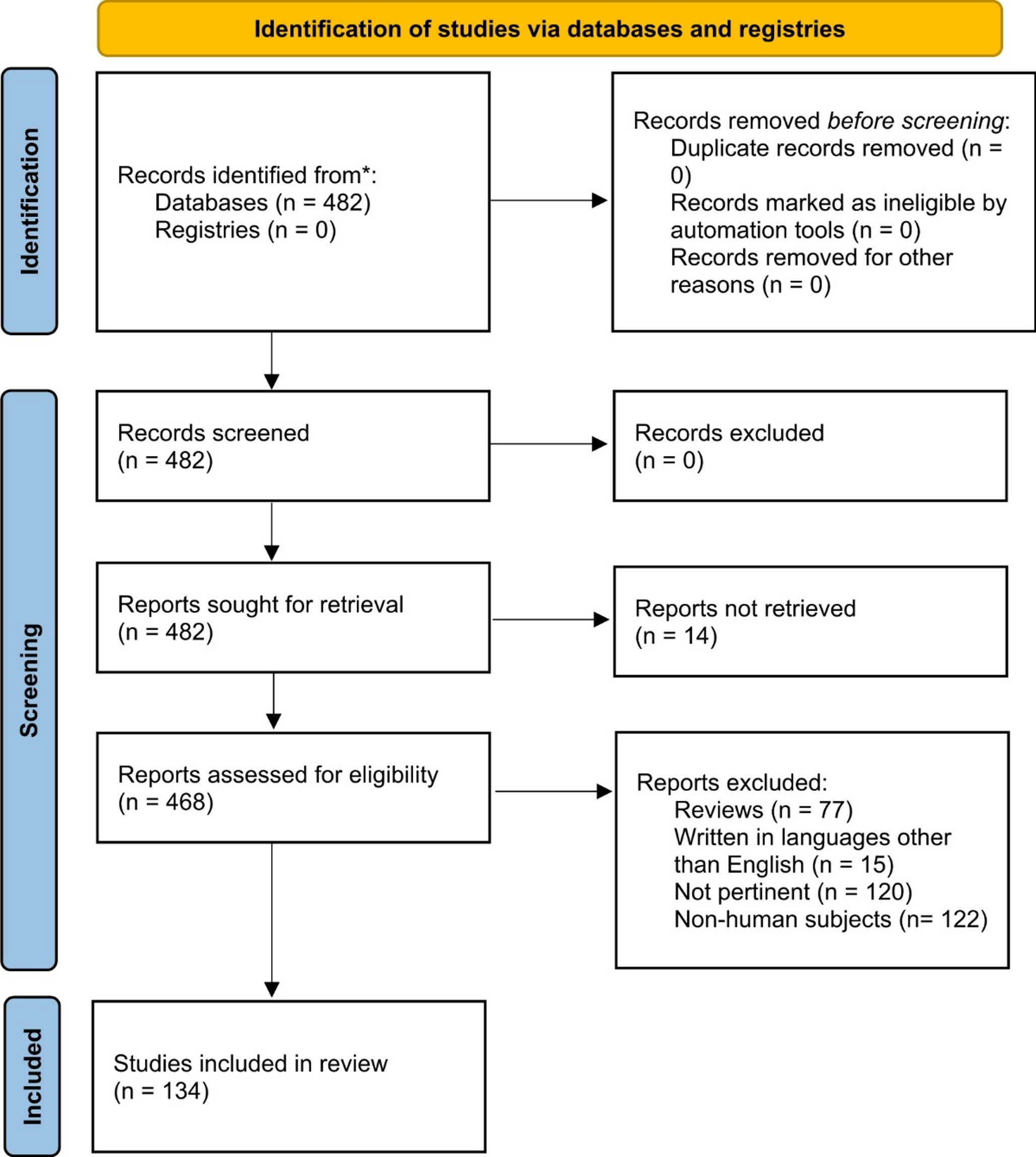
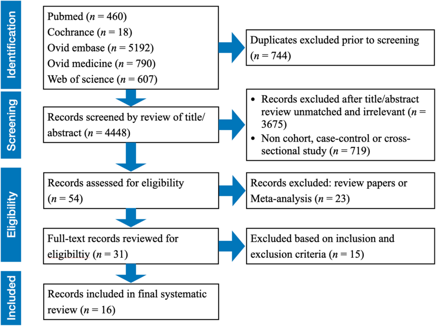
Page MJ et al. Mar., The PRISMA 2020 statement: an updated guideline for reporting systematic reviews. BMJ. 2021;n71. https://doi.org/10.1136/bmj.n71
Ikeda H, Ohhashi G. Demonstration of high coincidence of pituitary adenoma in patients with ruptured Rathke’s cleft cyst: Results of a prospective study. Clin Neurol Neurosurg. 2015;139:144–151. https://doi.org/10.1016/j.clineuro.2015.09.018
Senanayake R, et al. New types of localization methods for adrenocorticotropic hormone-dependent cushing’s syndrome. Jan 01. 2021. https://doi.org/10.1016/j.beem.2021.101513. Bailliere Tindall Ltd.
Article
Google Scholar
Rodriguez-Barcelo S, Gutierrez-Cardo A, Dominguez-Paez M, Medina-Imbroda J, Romero-Moreno L, Arraez-Sanchez M. Clinical usefulness of coregistered 11 C-methionine positron emission tomography/3-T magnetic resonance imaging at the follow-up of acromegaly. 2014. Elsevier Inc. https://doi.org/10.1016/j.wneu.2013.11.011
Haberbosch L et al. Feb., Dual Role for l-[Methyl-11 C]-Methionine PET in Acromegaly: Confirming Remission and Detecting Recurrence. J Nucl Med. 2024;65(2):327–328. https://doi.org/10.2967/jnumed.123.266446
Gillett D, et al. Development of a bespoke Phantom to optimize molecular PET imaging of pituitary tumors. EJNMMI Phys. Dec. 2023;10(1). https://doi.org/10.1186/s40658-023-00552-9.
Bendor-Samuel OM, Pal A, Cudlip S, Anderson G, Salgia S, Makaya T. Pituitary gigantism: A rare learning opportunity. Arch Dis Child Educ Pract Ed. 2020;105(2):111–116. https://doi.org/10.1136/archdischild-2018-316282
Haberbosch L et al. Mar., Real-world experience with 11 C-methionine positron emission tomography in the management of acromegaly. Eur J Endocrinol. 2024;190(4):307–313. https://doi.org/10.1093/ejendo/lvae028
Bashari WA et al. Jun., PET-guided repeat transsphenoidal surgery for previously deemed unresectable lateral disease in acromegaly. Neurosurg Focus. 2020;48(6):E8. https://doi.org/10.3171/2020.3.FOCUS2052
Zhang F, et al. The combination of 13 N-ammonia and 11 C-methionine in differentiation of residual/recurrent pituitary adenoma from the pituitary gland remnant after trans-sphenoidal Adenomectomy. BMC Cancer. Dec. 2021;21(1). https://doi.org/10.1186/s12885-021-08574-1.
Koulouri O et al. Successful treatment of residual pituitary adenoma in persistent acromegaly following 1 localisation by 11 C-methionine PET-CT co-registered with MRI 2 3.
Kontogeorgos G, Thodou E. Double adenomas of the pituitary: an imaging, pathological, and clinical diagnostic challenge. 2019. Springer. https://doi.org/10.1007/s42000-019-00126-4
Bashari WA et al. Jul., Using Molecular Imaging to Enhance Decision Making in the Management of Pituitary Adenomas. J Nucl Med. 2021;62:57S-62S. https://doi.org/10.2967/jnumed.120.251546
Taku N, Koulouri O, Scoffings D, Gurnell M, Burnet N. The use of 11 carbon methionine positron emission tomography (PET) imaging to enhance radiotherapy planning in the treatment of a giant, invasive pituitary adenoma. BJR|case Rep. Jun. 2017;3(2):20160098. https://doi.org/10.1259/bjrcr.20160098.
Kontogeorgos G. New horizons in diagnosis and management of endocrine tumors. Cancer Biomarkers. 2014;14:2–3. https://doi.org/10.3233/CBM-130353.
Article
CAS
Google Scholar
Wang Z, et al. Clinical application of combination [11 C]C-methionine and [13 N]N-ammonia PET/CT in recurrent functional pituitary adenomas with negative MRI or [18F]F-FDG PET/CT. BMC Endocr Disord. Dec. 2024;24(1). https://doi.org/10.1186/s12902-024-01543-2.
Quinn M, Bashari W, Smith D, Gurnell M, Agha A. A remarkable case of thyrotoxicosis initially caused by graves’ disease followed by a probable TSHoma-a case report. BMC Endocr Disord. Aug. 2020;20(1). https://doi.org/10.1186/s12902-020-00611-7.
Gillett D, et al. Localization of TSH-secreting pituitary adenoma using 11 C-methionine image subtraction. EJNMMI Res. 2022;12(1). https://doi.org/10.1186/s13550-022-00899-7.
Koulouri O, et al. Localisation of an occult thyrotropinoma with 11 C-methionine PET-CT before and after somatostatin analogue therapy. Dec 01. 2016. https://doi.org/10.1016/S2213-8587(16)30311-4. Lancet Publishing Group.
Article
Google Scholar
Feng Z, et al. Utility of 11 C-methionine and 18F-FDG PET/CT in patients with functioning pituitary adenomas. Clin Nucl Med. 2016;41(3):e130–4. https://doi.org/10.1097/RLU.0000000000001085.
Article
PubMed
Google Scholar
Bakker LEH et al. Aug., Implementation of functional imaging using 11 C-methionine PET-CT co-registered with MRI for advanced surgical planning and decision making in prolactinoma surgery. Pituitary. 2022;25(4):587–601. https://doi.org/10.1007/s11102-022-01230-2
Bashari WA, et al. 11 C-methionine PET aids localization of microprolactinomas in patients with intolerance or resistance to dopamine agonist therapy. Pituitary. Aug. 2022;25(4):573–86. https://doi.org/10.1007/s11102-022-01229-9.
Sagan KP, Andrysiak-Mamos E, Sagan L, Nowacki P, Małkowski B, Syrenicz A. Cushing’s Syndrome in a Patient With Rathke’s Cleft Cyst and ACTH Cell Hyperplasia Detected by 11 C-Methionine PET Imaging—A Case Presentation. Front Endocrinol (Lausanne). Jul. 2020;11. https://doi.org/10.3389/fendo.2020.00460.
Ikeda H, Abe T, Watanabe K. Usefulness of composite methionine-positron emission tomography/3.0-tesla magnetic resonance imaging to detect the localization and extent of early-stage Cushing adenoma. J Neurosurg. 2010;112(4):750–755. https://doi.org/10.3171/2009.7.JNS09285
Furnica RM, et al. Diagnostic value of 11 C-Methionine PET-CT imaging in persistent or recurrent Cushing disease after surgery. J Clin Endocrinol Metab Jan. 2025. https://doi.org/10.1210/clinem/dgaf047.
Article
Google Scholar
Lurquin F, et al. Ectopic sphenoidal ACTH-secreting adenoma revealed by 11 C Methionine PET scan: case report. BMC Endocr Disord. Dec. 2023;23(1). https://doi.org/10.1186/s12902-023-01298-2.
Koulouri O et al. Oct., A role for 11 C-methionine PET imaging in ACTH-dependent Cushing’s syndrome, in European Journal of Endocrinology, BioScientifica Ltd., 2015, pp. M107–M120. https://doi.org/10.1530/EJE-15-0616
Berkmann S et al. Dec., Selective resection of cushing microadenoma guided by preoperative hybrid 18-fluoroethyl-L-tyrosine and 11-C-methionine PET/MRI, Pituitary, vol. 24, no. 6, pp. 878–886, 2021, https://doi.org/10.1007/s11102-021-01160-5
Stanly A et al. Dec., Utility of F18-FDG PET/CT in the Evaluation of Pituitary Uptake, World J Nucl Med, vol. 23, no. 04, pp. 234–241, 2024, https://doi.org/10.1055/s-0044-1787967
Wang H et al. Mar., PET/MRI in the diagnosis of hormone-producing pituitary microadenoma: A prospective pilot study, Journal of Nuclear Medicine, vol. 59, no. 3, pp. 523–528, 2018, https://doi.org/10.2967/jnumed.117.191916
Giustina A, Gola M, Doga M, Rosei EA. Primary Lymphoma of the Pituitary: An Emerging Clinical Entity, J Clin Endocrinol Metab, vol. 86, no. 10, pp. 4567–4575, Oct. 2001, https://doi.org/10.1210/jcem.86.10.7909
Tsukamoto T, Miki Y. Imaging of pituitary tumors: an update with the 5th WHO Classifications—part 1. Pituitary neuroendocrine tumor (PitNET)/pituitary adenoma, Aug. 01, 2023, Springer. https://doi.org/10.1007/s11604-023-01400-7
Ryu SI, Tafti BA, Skirboll SL. Pituitary Adenomas Can Appear as Hypermetabolic Lesions in 18 F-FDG PET Imaging, Journal of Neuroimaging, vol. 20, no. 4, pp. 393–396, Oct. 2010, https://doi.org/10.1111/j.1552-6569.2008.00347.x
Hyun SH, Choi JY, Lee KH, Choe YS, Kim BT. Incidental focal 18F-FDG uptake in the pituitary gland: Clinical significance and differential diagnostic criteria, Journal of Nuclear Medicine, vol. 52, no. 4, pp. 547–550, Apr. 2011, https://doi.org/10.2967/jnumed.110.083733
Goulart CR, et al. Newly diagnosed Sellar tumors in patients with cancer: A diagnostic challenge and management dilemma. World Neurosurg. Oct. 2017;106:254–65. https://doi.org/10.1016/j.wneu.2017.06.139.
Hoang JK et al. Jul., Management of Incidental Pituitary Findings on CT, MRI, and 18F-Fluorodeoxyglucose PET: A White Paper of the ACR Incidental Findings Committee, Journal of the American College of Radiology, vol. 15, no. 7, pp. 966–972, 2018, https://doi.org/10.1016/j.jacr.2018.03.037
Maffei P, et al. A very rare case of nonfunctioning pituitary adenoma incidentally disclosed at 18F-FDG PET/CT. Clin Nucl Med. May 2012;37(5):e100–1. https://doi.org/10.1097/RLU.0b013e3182485217.
Oldfield EH, Merrill MJ. Apoplexy of pituitary adenomas: The perfect storm, J Neurosurg, vol. 122, no. 6, pp. 1444–1449, Jun. 2015, https://doi.org/10.3171/2014.10.JNS141720
Jeong SY et al. Dec., Incidental pituitary uptake on whole-body 18F-FDG PET/CT: A multicentre study, Eur J Nucl Med Mol Imaging, vol. 37, no. 12, pp. 2334–2343, 2010, https://doi.org/10.1007/s00259-010-1571-5
Gemmel F, Balink H, Collins J, Oomen P. Occult prolactinoma diagnosed by FDG PET/CT, 2010. [Online]. Available: www.nuclearmed.com.
Skoura E, Datseris IE, Xekouki P, Tolis G, Stratakis CA. SPECT and 18 F-FDG PET/CT imaging of multiple paragangliomas and a growth hormoneyproducing pituitary adenoma as phenotypes from a novel succinate dehydrogenase subunit D mutation, 2013. [Online]. Available: www.nuclearmed.com.
Alzahrani AS, Farhat R, Al-Arifi A, Al-Kahtani N, Kanaan I, Abouzied M. The diagnostic value of fused positron emission tomography/computed tomography in the localization of adrenocorticotropin-secreting pituitary adenoma in cushing’s disease. Pituitary. 2009;12(4):309–14. https://doi.org/10.1007/s11102-009-0180-4.
Article
CAS
PubMed
Google Scholar
Li X, et al. Case report and literature review: ectopic Thyrotropin-Secreting pituitary adenoma in the suprasellar region. Front Endocrinol (Lausanne). Mar. 2021;12. https://doi.org/10.3389/fendo.2021.619161.
Huang Y, Wen X, Liang X, Xu L. Mixed thyrotropin-secreting pituitary neuroendocrine tumor coexisting with Graves’ disease: a case report, Front Med (Lausanne), vol. 11, Sep. 2024, https://doi.org/10.3389/fmed.2024.1436400
Zhu C, et al. Central hyperthyroidism due to an ectopic TSH-secreting pituitary tumor: a case report and literature review. Front Endocrinol (Lausanne). 2024;15. https://doi.org/10.3389/fendo.2024.1301260.
Kim S, et al. Ectopic Thyroid-Stimulating Hormone–Secreting pituitary adenoma of the nasopharynx diagnosed by gallium 68 DOTATATE positron emission tomography/computed tomography. World Neurosurg. May 2019;125:400–4. https://doi.org/10.1016/j.wneu.2019.02.022.
Ishizaki U, Abe K, Takako K, Masui K, Sakai S, FDG PET/CT Findings of Ectopic Pituitary Adenoma. Feb.,, Clin Nucl Med, vol. 45, no. 2, pp. 151–153, 2020, https://doi.org/10.1097/RLU.0000000000002887
Ding L, et al. Differentiation of suprasellar meningiomas from non-functioning pituitary macroadenomas by 18F-FDG and 13 N-Ammonia PET/CT. BMC Cancer. Jun. 2020;20(1). https://doi.org/10.1186/s12885-020-06852-y.
Beyhan E, Fenercioğlu ÖE, Karagöz Y, Ergül N, Çermik TF. Mild68Ga PSMA-11 Uptake in Incidental Pituitary Adenoma, Mol Imaging Radionucl Ther, vol. 31, no. 3, pp. 244–245, Oct. 2022, https://doi.org/10.4274/mirt.galenos.2021.97752
Lemelin A, et al. Pheochromocytoma, paragangliomas, and pituitary adenoma: an unusual association in a patient with an SDHD mutation. Case report. Medicine. Jul. 2019;98:e16594. https://doi.org/10.1097/MD.0000000000016594.
Chai A, Soon AYQ, Manish B, Tan JL. Ectopic sphenoid sinus pituitary adenoma masquerading as metastatic head and neck cancer. BMJ Case Rep. Mar. 2021;14(3). https://doi.org/10.1136/bcr-2020-240411.
Andrioli M, Pecori Giraldi F, De Martin M, Cattaneo A, Carzaniga C, Cavagnini F. Differential diagnosis of ACTH-dependent hypercortisolism: imaging versus laboratory. Pituitary. 2009;12(4):294–6. https://doi.org/10.1007/s11102-009-0174-2.
Article
PubMed
Google Scholar
Sarkar S, Rajaratnam S, Chacko G, Mani S, Hesargatta AS, Chacko AG. Pure endoscopic transsphenoidal surgery for functional pituitary adenomas: outcomes with cushing’s disease. Acta Neurochir (Wien). Jan. 2016;158(1):77–86. https://doi.org/10.1007/s00701-015-2638-7.
Ammini A, et al. Etiology and clinical profile of patients with cushing’s syndrome: A single center experience. Indian J Endocrinol Metab. Jan. 2014;18(1):99–105. https://doi.org/10.4103/2230-8210.126586.
Chittiboina P, Montgomery BK, Millo C, Herscovitch P, Lonser RR. High-resolution 18F-fluorodeoxyglucose positron emission tomography and magnetic resonance imaging for pituitary adenoma detection in Cushing disease, J Neurosurg, vol. 122, no. 4, pp. 791–797, Apr. 2015, https://doi.org/10.3171/2014.10.JNS14911
Kong Z, Wang Y, Ma W, Cheng X. FDG-PET/CT in the detection of pituitary stalk ACTH-secreting adenoma, Jun. 01, 2020, Springer. https://doi.org/10.1007/s00259-019-04655-3
Patt HP, Lele V, Lila A, Bandgar T, Shah N. Utility of hCRH-stimulated (18) F-FDG PET-CT scan in localisation of pituitary microadenoma in cushing’s disease. Blackwell Publishing. 2014. https://doi.org/10.1111/1754-9485.12165.
Article
Google Scholar
Boyle J et al. Jul., CRH stimulation improves 18F-FDG-PET detection of pituitary adenomas in Cushing’s disease, Endocrine, vol. 65, no. 1, pp. 155–165, 2019, https://doi.org/10.1007/s12020-019-01944-7
Lu J, et al. Corticotropin releasing hormone can selectively stimulate glucose uptake in corticotropinoma via glucose transporter 1. Mol Cell Endocrinol. Jul. 2018;470:105–14. https://doi.org/10.1016/j.mce.2017.10.003.
Kim K, et al. Dexamethasone suppression for 18F-FDG PET/CT to localize ACTH-secreting pituitary tumors. Cancer Imaging. Dec. 2023;23(1). https://doi.org/10.1186/s40644-023-00600-8.
Thearle MS, Freda PU, Bruce JN, Isaacson SR, Lee Y, Fine RL. Temozolomide (Temodar®) and capecitabine (Xeloda®) treatment of an aggressive corticotroph pituitary tumor, Pituitary, vol. 14, no. 4, pp. 418–424, Dec. 2011, https://doi.org/10.1007/s11102-009-0211-1
Hintz E, Tomlin J, Chengazi V, Vates G. Positron emission tomography-Computed tomography coregistration for diagnosis and intraoperative localization in recurrent Nelson syndrome. J Neurol Surg Rep. May 2013;74(01):033–6. https://doi.org/10.1055/s-0033-1346974.
Xu H, Zhang M, Zhai G, Zhang M, Ning G, Li B. The role of integrated 18F-FDG PET/CT in identification of ectopic ACTH secretion tumors. Endocrine. 2009;36(3):385–91. https://doi.org/10.1007/s12020-009-9247-2.
Article
CAS
PubMed
Google Scholar
Hou Y, Zhu Z, Jin X, Wang R, Xing B. Combined 18 F-FDG PET/CT and 99m Tc 3PRGD2 SPECT/CT imaging in a case of pituitary metastases, 2013. [Online]. Available: www.nuclearmed.com.
Cheung IHW, Mahboobani NR, Poon WL. Case report of large solitary skull base metastasis from renal cell carcinoma as initial clinical presentation: Radiological findings and differentials, Radiol Case Rep, vol. 19, no. 11, pp. 5376–5379, Nov. 2024, https://doi.org/10.1016/j.radcr.2024.07.169
Ng S, et al. Pituitary metastasis of malignant melanoma misdiagnosed as pituitary adenoma: A case report and systematic review of the literature. Neurochirurgie. Nov. 2020;66(5):383–90. https://doi.org/10.1016/j.neuchi.2020.06.129.
Starc MT, Rosenblum MK, Meyers PA, Hatzoglou V. Rare presentation of ewing sarcoma metastasis to the Sella and suprasellar cistern. Clin Imaging. Jan. 2017;41:73–7. https://doi.org/10.1016/j.clinimag.2016.10.017.
Sahel O, Benameur Y, Nabih S, Biyi A, Doudouh A. A rare case of pituitary metastasis from breast cancer detected on fluorodeoxyglucose positron emission tomography/computed tomography that presented as insipid diabetes, World J Nucl Med, vol. 20, no. 02, pp. 211–214, Apr. 2021, https://doi.org/10.4103/wjnm.wjnm_88_20
Agarwal KK, Sharma P, Singla S, Suman S, Bal KCC, Kumar R. A rare case of Non–Small cell lung Cancer metastasizing to the pituitary gland. Clin Nucl Med. May 2014;39(5):e318–9. https://doi.org/10.1097/RLU.0b013e31828da679.
Lamorie-Foote K, et al. Melanoma metastasis to a nonfunctioning pituitary macroadenoma: illustrative case. J Neurosurgery: Case Lessons. Jun. 2021;1(23). https://doi.org/10.3171/CASE2167.
Donovan LE, Arnal AV, Wang SH, Odia Y. Widely metastatic atypical pituitary adenoma with mTOR pathway STK11(F298L) mutation treated with everolimus therapy, CNS Oncol, vol. 5, no. 4, pp. 203–209, Oct. 2016, https://doi.org/10.2217/cns-2016-0011
Hagen C, Schroeder HD, Hansen S, Hagen C, Andersen M. Temozolomide treatment of a pituitary carcinoma and two pituitary macroadenomas resistant to conventional therapy. Eur J Endocrinol. 2009;161(4):631–7. https://doi.org/10.1530/EJE-09-0389.
Article
CAS
PubMed
Google Scholar
Kamiya-Matsuoka C et al. Aug., Radiotherapy with concurrent temozolomide for the management of extraneural metastases in pituitary carcinoma, Pituitary, vol. 19, no. 4, pp. 415–421, 2016, https://doi.org/10.1007/s11102-016-0721-6
Kwok MM, Virk JS, Michael M, McKinley M, Magarey MJR. Cervical Nodal Metastatic Pituitary Carcinoma: A Case Report, Ear Nose Throat J, vol. 101, no. 2, pp. 110–113, Feb. 2022, https://doi.org/10.1177/0145561320944649
Ortiz LD, et al. Anti-VEGF therapy in pituitary carcinoma. Pituitary. Sep. 2012;15(3):445–9. https://doi.org/10.1007/s11102-011-0346-8.
Flores L, Karger AG et al. doi:


Comments (0)