Reagents
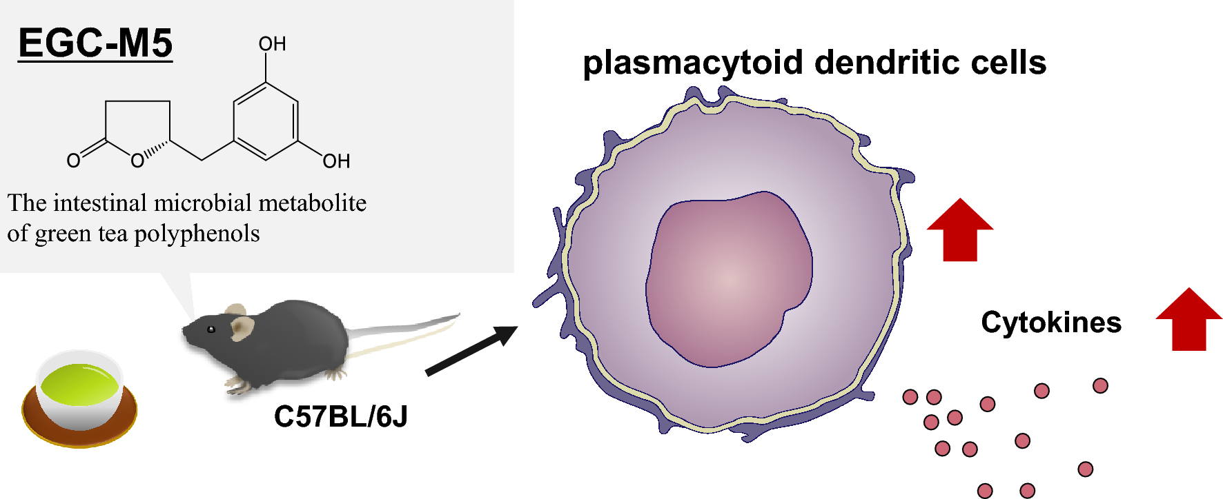
We obtained EGC-M5 through microbial cultivation as previously described [17]. The purity of EGC-M5 was confirmed by high-performance liquid chromatography (HPLC) analysis (at least 95%). Roswell Park Memorial Institute (RPMI) 1640 medium was purchased from Fujifilm Wako Pure Chemical Co., Ltd. (Osaka, Japan). Fetal bovine serum (FBS) was purchased from Sigma-Aldrich (St. Louis, MO, USA). 2-mercaptoethanol and 4% paraformaldehyde (PFA) were purchased from Fujifilm Wako Pure Chemical Co. Ltd. (Osaka, Japan). GM-CSF was obtained from Fujifilm Wako Pure Chemical Co. Ltd. (Osaka, Japan). LPS was purchased from Sigma-Aldrich (St. Louis, MO). Streptomycin and Penicillin G were purchased from Meiji Pharmaceutical Co. (Tokyo, Japan).
In vivo experiment
All animal studies were conducted in compliance with the Law of Act on Welfare and Management of Animals (No. 105), the Standards Relating to the Care and Keeping and Reducing Pain of Laboratory Animals (Notice of the Ministry of the Environment No.88 of 2006), Fundamental Guidelines for Proper Conduct of Animal Experiment and Related Activities in Academic Research Institutions under the jurisdiction (Notice of the ministry of Education, Culture, Sports, Science and Technology No. 71 of 2006), the Kyushu University Animal Experiment Regulations and Administrative Instructions Kyushu University Animal Experiment Regulations. The animal experiments were approved by the Kyushu University Animal Care and Use Committee in Fukuoka, Japan (A23-127–1). Six-week-old C57BL/6J male mice, purchased from Kyudo (Tosu, Saga), were housed in a room with controlled humidity (50–70%) and temperature (25 ± 2 °C) on a 12 h dark–light cycle (light from 8:00 to 20:00). They had access to fresh autoclaved water and MF diet (KBT Oriental, Tokyo, Japan) ad libitum. After 1 week of acclimation, the mice were randomly divided into three groups: Gp1 mice were treated with vehicle (2.5% DMSO in injection water) (n = 8), Gp2 mice were treated with EGC-M5 30 mg/kg b.w. (2.5% DMSO in injection water) orally for 12 days, and Gp3 mice were treated with EGC-M5 100 mg/kg b.w. (2.5% DMSO in injection water) orally for 12 days. On the final day, mice were sacrificed under an isoflurane atmosphere. Organs, including the blood, spleen, and femur, were harvested and analyzed.
Antibodies and flow cytometry
Antibodies for flow cytometry were purchased from BioLegend (San Diego, California, United States). Detailed information is provided in SI Table 1. All antibodies were diluted with 0.1% sodium azide and 2.5% bovine serum albumin (BSA) PBS. Cytometry was performed using the Verse (BD Biosciences). The harvested spleens were placed in a 5 mL dish containing RPMI-1640 medium, and the tissues were crushed using a glass slide to disperse the cells. The tissue fragments were removed by filtering through a mesh, followed by centrifugation for 5 min.
After removing the supernatant, the cells were treated with BD Pharm Lyse™ Lysing Buffer (Becton, Dickinson and Company), which was diluted 10 times with Otsuka distilled water (Otsuka Pharmaceutical). The cells were washed twice with RPMI-1640 medium and the cell count was measured using a hemocytometer. The cell concentration was adjusted to 1 × 10⁷ cells/mL. The cells were fixed in 2% formaldehyde solution for 20 min at 4 °C, centrifuged to remove the supernatant, then incubated at room temperature for 30 min in 5% FBS-TPBS (PBS containing 0.05%Tween-20) to permeabilize the cell membranes. The cells were then stained with 5% FBS-TPBS and analyzed using a flow cytometer, Verse™ (Becton, Dickinson and Company). Plasmacytoid dendritic cells (pDCs) were identified as CD11cint CD3– B220+cells, conventional dendritic cell subset 1 (cDC1) as CD11c+ CD3– CD8+ cells, and conventional dendritic cell subset 2 (cDC2) as CD11c+ CD3– CD11b+ cells.
The femur was placed in a 5 mL dish containing RPMI1640 medium, and the bone marrow was collected using a 2.5 mL syringe and a 26G needle. Tissue fragments were removed by mesh filtration. The supernatant was discarded and the cells were treated with BD Pharm Lyse™ Lysing Buffer. The cells were then washed twice with RPMI1640 medium and counted using a hemocytometer. The cells were fixed in 2% formaldehyde solution for 20 min at 4 °C, left to stand for 30 min at room temperature in 5% FBS-TPBS, and then permeabilized. Samples with obviously abnormal gate conditions were excluded from the analysis.
Ex vivo study
The femur was placed in a 6 well plate containing RPMI1640 medium, and the bone marrow was collected using a 2.5 mL syringe and a 26G needle. Tissue fragments were removed by mesh filtration. Myeloid cells were seeded with or without EGC-M5 in 10% FBS RPMI1640 medium supplemented with Flt-3L (50 ng/mL and 50 μM 2-mercaptoethanol) at a density of 5 × 10⁶ cells/mL. After 5 days of incubation, the medium was removed and the cells were treated with fresh medium containing the same supplements. After 48 h of incubation, the pDC population was assessed using flow cytometry.
qRTPCR
The harvested spleens were homogenized in 700 µL of TRI Reagent (Cosmo Bio, Tokyo, Japan) and added with 200 µL of chloroform (Nacalai tesque, Kyoto, Japan). After centrifugation (12,000×g for 15 min at 4 °C), 300 µL of the aqueous phase was collected and 300 µL of 2-propanol was added and mixed. After further centrifugation at 4 °C at 12,000×g for 10 min, the supernatant was removed and the pellet was washed with 700 µL of 75% ethanol. After another centrifugation (12,000×g for 15 min at 4 °C), the supernatant was removed, and the RNA pellet was dried. The pellet was dissolved in Nuclease-Free Water (NFW) (Ambion, TX, U.S.A.), and the RNA concentration was measured using a NanoDrop 2000 (Thermo Scientific, Kanagawa, Japan). cDNA was synthesized using a Prime Script™ RT Reagent Kit (Takara Bio, Shiga, Japan) in a thermocycler (Astec, Fukuoka, Japan). A volume of 1 µL cDNA was mixed with 2.5 µL of Sso AdvancedTM Universal SYBR® Green Supermix (BIO-RAD, California, U.S.A.), 0.2 µL each of primers F and R, and 1.1 µL of ultra-pure water, and gene expression was analyzed using the CFX96TM Real-Time PCR Analysis System (BIO-RAD, California, U.S.A.). The denaturation temperature was set at 95 °C, the annealing temperature at 60 °C, and the amplification was performed for 50 cycles. The primer sequences are listed below (Table 1).
Statistical analysis
All results are shown as the mean ± standard error (SE). Significant differences were assessed using Student’s t-test and Dunnett’s test using GraphPad 8.0 (GraphPad Software). Statistical significance was set at P < 0.05. Outliers were identified using the ROUT test (Q = 1%).


Comments (0)