
Remember me

Human articular chondrocytes (HCs) were isolated from femoral head cartilage collected during total hip arthroplasty. Informed consent was obtained for all specimens, and the study was approved by the Institutional Review Board (No. H-2107-030-1232). Full-thickness cartilage was minced and digested enzymatically at 37 °C with 0.2% protease (Type XIV; Sigma, St. Louis, MO, USA) for 1 h, followed by 0.2% collagenase (Type IA; Sigma) for 3 h. The resulting suspension was filtered through a 70-μm nylon sieve to remove undigested fragments.
Human osteoblasts (hFOB 1.19, CRL-11372; American Type Culture Collection, Manassas, VA, USA) were cultured in high-glucose Dulbecco’s Modified Eagle’s Medium with nutrient mixture F12 (DMEM/F12; Gibco, Invitrogen, Carlsbad, CA, USA), supplemented with 10% fetal bovine serum (FBS; Gibco) and 1% Penicillin–Streptomycin (Thermo Fisher Scientific, IL, USA). Cells were maintained in a humidified incubator at 37 °C with 5% CO2. When cultures reached 70–80% confluency, cells were detached using 0.25% trypsin–EDTA (Gibco) and passaged. Passages 1 and 2 were used for subsequent experiments.
2.1.2 Preparation of platelet-rich plasma (PRP)Allogenic leukocyte-depleted PRP was prepared by collecting peripheral blood from healthy donors into anticoagulant-treated tubes. Blood was centrifuged at 150g for 10 min to separate red blood cells, buffy coat, and plasma. The plasma layer, rich in platelets and depleted of leukocytes, was aspirated while avoiding the buffy coat. A second centrifugation step was performed at a higher speed if necessary to further concentrate the platelets.
2.2 Scaffold-based co-culture2.2.1 Scaffold fabrication, cell labeling, and seedingPoly-lactic-co-glycolic-acid (PLGA) mesh scaffolds (Ethicon; Johnson & Johnson, USA) measuring 10 × 8 mm, were used. Scaffolds were placed in uncoated 24-well plates, and each scaffold was seeded with 1 × 10⁴ chondrocytes and osteoblasts. Prior to seeding, osteoblasts and chondrocytes were labeled using fluorescent dyes for cell tracking. Osteoblasts were stained with 3,3′-Dioctadecyloxacarbocyanine Perchlorate (DiO, green; Invitrogen), while chondrocytes were labeled with 1,1′-Dioctadecyl-3,3,3′,3′-Tetramethylindocarbocyanine Perchlorate (DiI, red; Invitrogen). After labeling, cells were washed with PBS to remove excess dye before seeding onto the scaffold. To ensure homogeneous cell distribution and prevent cell sedimentation at the bottom of the well, the plates were placed on a bidirectional rotator at 50Hz for 24 h at 37 °C.
2.2.2 PRP application and co-culture conditionsTwo types of scaffolds were prepared: PRP-containing and PRP-free. For PRP-containing scaffolds, PRP was activated by adding 10% calcium chloride before mixing with the cell suspension. The activated PRP was applied to the scaffold before the co-culture process. Both PRP-containing and PRP-free scaffolds were incubated under the same culture conditions described in Sect. 1.1 to maintain cell viability and proliferation, ensuring efficient cell attachment and growth on the scaffold.
2.3 Cell proliferation, phenotype, and morphological analysis2.3.1 Cell adhesion & proliferation analysisCell adhesion and proliferation were assessed using the Cell Counting Kit-8 (CCK-8, Dojindo, Tokyo, Japan), following the manufacturer’s instructions. The cell adhesion assay was performed 24 h post-seeding, where cells were washed with DPBS (GibcoBRL), and 10 μL of CCK-8 solution per 100 μL of media was added. The absorbance was measured at 450 nm using a microplate reader (Spectramax M5, Versamax; Molecular Devices) after 3 h of incubation to form formazan crystals. Cell proliferation was subsequently evaluated at 3- and 7-days post-seeding using the same method.
2.3.2 Phenotype analysis (RT-PCR)To confirm phenotype maintenance during co-culture, RT-PCR was performed at 24 h, 3 days, and 7 days post-seeding. Total RNA was extracted using the RNeasy Mini Kit (Qiagen, Hilden, Germany), and cDNA synthesis was performed using the Transcriptor First Strand cDNA Synthesis Kit (Roche, Basel, Switzerland). Primers targeting collagen II and aggrecan for chondrocytes, and alkaline phosphatase (ALP) and osteocalcin (OC) for osteoblasts were used, with GAPDH as an internal control. PCR amplification was conducted with an initial denaturation at 95 °C for 3 min, followed by 28–32 cycles of denaturation at 95 °C for 15 s, annealing at 55 to 68 °C, and extension at 72 °C for 30 s depending on the target gene. Table 1 provides detailed primer sequences along with the specific annealing temperatures and PCR cycles for each gene.
Table 1 Primer sequences and PCR conditions for RT-PCR analysis2.3.3 Morphological analysis (FE-SEM & fluorescence microscopy)The morphology and spatial distribution of co-cultured chondrocytes and osteoblasts on the PLGA mesh scaffolds were evaluated using field emission scanning electron microscopy (FE-SEM) and fluorescence microscopy.
For FE-SEM analysis, cells on the scaffolds were washed three times with PBS and fixed in 2% glutaraldehyde (Sigma) at 4 °C for 2 h. After fixation, samples were dehydrated in increasing concentrations of ethanol (70 to 100%) and dried in a vacuum. The dried samples were then sputter-coated with a thin gold–palladium layer and imaged using an FE-SEM (JSM-740F, JEOL Ltd., Japan) at 24 h, 3 days, and 7 days post-seeding.
Fluorescence imaging was conducted using a DMi8 inverted fluorescence microscope (Leica Microsystems, Germany) to evaluate the spatial distribution and integration of labeled chondrocytes and osteoblasts on the scaffold. Images were captured at 24 h, 3 days, and 7 days post-seeding to monitor cell localization and potential migration patterns over time.
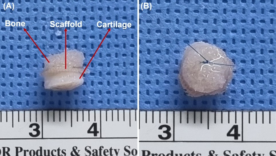
2.4 Gap-mimic construct and in vivo implantation2.4.1 Preparation of the gap-mimic constructWe produced a gap-mimic construct comprising three layers: cartilage, scaffold, and bone (Fig. 1A). Articular cartilage and bone were harvested from the resected femoral head during total hip arthroplasty. Articular cartilage underwent more than three freeze–thaw cycles at -70 °C to eliminate resident cells, ensuring that only the scaffold-seeded cells contribute to the observed effects. Bone tissue was subjected to the same freeze–thaw cycles as cartilage, followed by sterilization via irradiation at 25 kGy to ensure complete devitalization while maintaining the extracellular matrix structure, with all procedures performed at our institution's bone bank. Bone and cartilage tissues were processed into disc-shaped constructs with a diameter of 6 mm.
Fig. 1
Preparation of the gap-mimic construct. A Gap-mimic construct composed of cartilage, scaffold, and bone. B Final assembled construct secured with 6–0 nylon sutures for in vivo implantation
The PLGA scaffold was placed between the bone and cartilage discs and sutured using 6–0 nylon (Fig. 1B). Four experimental groups were prepared: (1) scaffold alone, (2) scaffold with co-cultured cells, (3) scaffold with PRP alone, and (4) scaffold with PRP and co-cultured cells.
2.4.2 In vivo implantationTwenty-four 6-week-old BALB/c-nude mice were used in this study, with approval from the Institutional Animal Care and Use Committee (IACUC approval No. 22-0219-S1A0). Mice were anesthetized with 2–3% isoflurane for induction and 1.5–2.5% for maintenance. Constructs were implanted subcutaneously on both sides of the dorsal skin. Mice were divided into two groups described above based on PRP treatment, each consisting of 12 mice. At 4 and 8 weeks post-implantation, twelve mice were euthanized for construct retrieval and analysis.
2.5 Evaluation of gap healing in vivoTo evaluate gap healing, three assessments were conducted: macroscopic analysis to examine gross structural bonding, quantitative analysis to measure the degree of attachment, and histological analysis to assess qualitative features of cellular invasion at the bone-cartilage interface (Fig. 2).
Fig. 2
Evaluation of gap healing at the bone-cartilage interface. A Macroscopic analysis of bisected constructs assessing gross bonding at the bone-cartilage interface. B Quantitative analysis of attachment, where the total construct diameter Ltot is 6 mm. The degree of attachment was measured as the percentage of the attached length Latt relative to Ltot, using a 10 × magnification microscope. C Histological analysis (H&E staining, × 200) showing bone-cartilage interdigitation. The image represents a case with evident cellular invasion (arrow) at the interface
Gap healing was first evaluated through macroscopic analysis (Fig. 2A). After removing the nylon suture, the retrieved constructs were bisected vertically, and the exposed bone-cartilage interface was examined macroscopically to assess the gross bonding. After macroscopic analysis, samples underwent decalcification to facilitate histological processing. Decalcification was performed using 10% EDTA (pH 7.4) at room temperature, with the solution changed every 48 h for 2–3 weeks until complete softening was achieved. The bisected samples were then fixed in 10% phosphate-buffered formalin, embedded in paraffin, sectioned at 5 μm thickness, and stained with hematoxylin and eosin (H&E) for histological examination.
Quantitative analysis was performed to evaluate the degree of attachment (Fig. 2B). The degree of attachment was measured using a 10 × magnification microscope, with the contact length between the cartilage and bone along the bisected 6 mm diameter of the construct quantified. The percentage of the attached length relative to the total diameter was then calculated, providing a quantitative assessment of the physical integration between the bone and cartilage.
Lastly, histological analysis was performed to assess cellular invasion, focusing on bone-cartilage interdigitation as a key indicator of active healing (Fig. 2C). Cellular invasion was considered present if the interface exhibited a serpentine pattern, with cells from the bone or cartilage layer infiltrating the opposing layer. To complement this, fluorescence microscopy was performed using DiO (green, osteoblasts) and DiI (red, chondrocytes) to evaluate the localization and distribution of co-cultured cells within the construct.
2.6 Statistical analysisThe degree of attachment, a continuous variable, and the presence of cellular invasion, a categorical variable, were analyzed to determine if there were statistically significant differences between constructs with and without cells in the PRP (−) and PRP (+) groups. Due to the small sample size, which precludes the assumption of normal distribution, the Mann–Whitney U test was used to compare the degree of attachment (continuous data), while the Chi-square test or Fisher's exact test was applied to evaluate the presence of cellular invasion (categorical data). Additionally, statistical analyses were conducted to assess differences in attachment and invasion between the 4-week and 8-week time points within the PRP (−) and PRP (+) groups.
Comments (0)