Cell culture
Human ADSCs and human FLS MH7A cells were obtained from the Cell Bank at the Chinese Academy of Sciences (Shanghai, China). Besides, human ADSCs were cultured with Human Adipose-derived Stem Cell Complete Culture Medium (#CM-H205, Procell, Wuhan, Hubei, China) supplemented with 10% fetal bovine serum (FBS) (#SH30087.01, HyClone, Logan, UT, USA), 100U/ml penicillin and 100 µg/mL streptomycin (#SH30010, HyClone) at 37 °C with 5% CO2. In addition, MH7A cells were cultured in DMEM (#SH30022.01B, HyClone) containing 10% FBS (#SH30087.01, HyClone), 100U/ml penicillin and 100 µg/mL streptomycin (#SH30010, HyClone) at 37 °C with 5% CO2 as previously described [10].
Preparation of HQT-containing serum
Twelve Wistar rats (male, 8 weeks) purchased from Model Animal Research Center of Nanjing University (Nanjing, Jiangsu, China) were randomly divided into control group and HQT group on average. Rats of HQT group were given 37.6 g/kg HQT from The Second Affiliated Hospital of Guangzhou University of Chinese Medicine by oral gavage once a day for 7 days, while rats of negative control group were given equal volume of physiological saline by oral gavage. During oral gavage, rats were fed with normal diet. Two hours after the last gastric perfusion, abdominal aortic blood of rats was collected into 15mL centrifuge tubes followed by standing at room temperature (RT) for 1 h. Then tubes were centrifuged at 3600 r/minute for 10 min and the supernatant was collected into new centrifuge tubes to inactivate at 56℃ for 0.5 h. Next, the HQT-containing serum was stored at -80℃.
Cell treatments
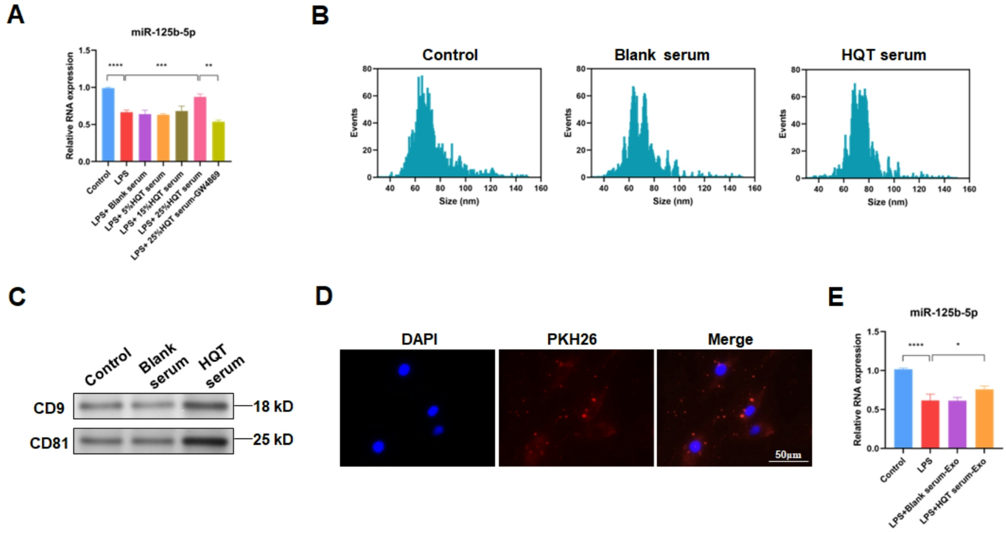
ADSCs were treated with 5%, 15% and 25% HQT-containing serum diluted in culture medium for 48 h, respectively. To block exosome generation and release, ADSCs were treated with exosome inhibitor 10µM GW4869 (#S7609, Selleck, Houston, TX, USA) for 2 h prior to treatment of HQT-containing serum. Moreover, MH7A cells were exposed to 1 µg/mL LPS (#L2880, Sigma, St. Louis, MO, USA) for 24 h to establish RA cell model.
Co-culture of MH7A cells with ADSCs
Co-culture of MH7A cells with ADSCs was performed using a 12-well Transwell filter-plate (#CLS3460, Corning, Corning, NY, USA). Briefly, 2 × 10⁵/mL MH7A cells were seeded at the bottom well containing DMEM while 2 × 10⁵/mL ADSCs were seeded at the upper pore polyester membrane insert (0.4 μm pore size) containing Human Adipose-derived Stem Cell Complete Culture Medium. Then cells were co-cultured for 48 h.
Quantitative reverse transcription-PCR (qRT‒PCR)
Total RNA was extracted from cells or exosomes by TRIzol reagent (#15596026, Invitrogen, Carlsbad, CA, USA). For miRNAs, first-strand cDNA synthesis and qRT-PCR were performed using All-in-One™ miRNA qRT-PCR Detection Kit (#AOMD-Q050, GeneCopoeia, Beijing, China) based on 7500 Fast Real-Time PCR System (Applied Biosystems, Foster City, CA, USA). For mRNAs, first-strand cDNA was synthetized by PrimeScript II 1st Strand cDNA Synthesis Kit (#6210A, Takara, Dalian, Liaoning, China) while qRT-PCR was performed using TB Green Premix Ex Taq™ (Tli RNaseH Plus) (#RR420A, Takara) based on 7500 Fast Real-Time PCR System (Applied Biosystems). Then the amount of target genes was analyzed using 2−ΔΔCt method [23], with U6 for miRNAs or GAPDH for mRNAs as the internal reference as previously described [10]. The primers used in the current study were listed in Table 1.
Table 1 Sequences of primers used for qRT-PCRCell counting kit-8 (CCK-8) assay
In brief, 1 × 10⁴/well ADSCs were collected and seeded into a 96-well plate (#PPP-001-030, Bestopbio, Beijing, China) at 1, 2, 3 day(s) after the treatment of HQT-containing serum, and then 10µL CCK-8 solution (#C0037, Beyotime, Shanghai, China) was added to incubate ADSCs for 4 hours at 37℃. Next, the absorbance at 450 nm was detected utilized a microplate reader (Multiscan MK3, Thermo Fisher Scientific, Cleveland, OH, USA) as previously described [24].
Flow cytometric analysis for cell apoptosis
ADSCs were collected and washed twice with PBS followed by the re suspension into 100µL incubation buffer (10 mM HEPES/NaOH, 140 mmol/L NaCl, 5 mM CaCl2). Then ADSCs were incubated with 10µL Annexin V-FITC (#KGA106, Keygen, Shanghai, China) at RT for 15 min and subsequent moderate propidium iodide (#KGA106, Keygen) at RT for 10 min. Next, ADSCs were analyzed by flow cytometry (BD Calibur, BD Biosciences, San Jose, CA, USA) as previously described [25].
Isolation and analysis of ADSC-derived exosomes
ExoEasy Maxi Kit (#76064, QIAGEN, Hilden, Germany) was used to isolate exosomes from culture medium of ADSCs according to the exoRNeasy Serum/Plasma Handbook. Then the size of exosomes was identified by a nanoparticle tracking analyzer (N30E, NanoFCM, Nottingham, UK). Moreover, the markers of exosomes were detected by Western Blot using CD9 antibody (1:1000, #bs-5660R, Bioss, Beijing, China) and CD81 antibody (1:1000, #bs-2489R, Bioss).
Exosome uptake
To detect exosome uptake into MH7A cells, 10 µg of ADSC-derived exosomes were suspended and stained using 100µM PKH26 (#UR52302, Umibio, Shanghai, China) at 37 °C for 15 min, and then centrifuged at 100,000×g at 4 °C for 70 min to remove unbound probes. After the incubation with PKH26-stained ADSC-derived exosomes, the fluorescence signals of PKH26 in MH7A cells were reported by a fluorescence microscopy.
Enzyme-linked immunosorbent assay (ELISA)
Levels of IL-1β, IL-6 and TNF-α in cellular supernatant of MH7A cells and rat serum were measured by ELISA as previously described [10].
Western blot (WB)
Total proteins were extracted from cells using RIPA Lysis Buffer (#89900, Thermo Fisher Scientific). Then equal amount of protein from different groups were separated by SDS-polyacrylamide gel electrophoresis (SDS-PAGE) followed by the transfer onto PCDF membranes (0.45 μm pore size, # IPFL00010, Millipore, Bedford, MA, USA). After being blocked with 5% non-fat milk for 1 h at RT, membranes were then incubated with primary antibodies at 4 °C overnight. The next day membranes were washed and incubated with appropriate secondary antibodies for 1 h at RT, membranes were extensively washed. Finally, signals of targeted proteins were detected by BeyoECL Moon Kit (#P0018F, Beyotime). The primary antibodies used in this study were listed as follow: CD9 antibody (1:1000, #bs-5660R, Bioss), CD81 antibody (1:1000, #bs-2489R, Bioss), CD63 antibody (1:1000, #bs-1523R, Bioss), NF-κB p65 antibody (1:1000, #bs-5660R, Bioss) and GAPDH antibody (1:10000, #KC-5G5, Aksomics, Shanghai, China).
Knockdown of miR-125b-5p and CD63 in ADSCs
First, short hairpin RNA (shRNA) against pre-miR-125b or CD63 was cloned into pLOV-CMV vector. Then 15 µg pLOV-CMV vector, 5 µg pMDL vector (envelop vector) and 10 µg psPAX2 vector (packaging vector) were mixed and transfected into 293T cells cultured in DMEM using Lipofectamine 2000 (Invitrogen). Viral supernatant was collected, filtered and concentrated at 3th day after transfection. Next, 1mL viral supernatant was used to infect ADSCs for 48 h. Finally, knockdown of miR-125b-5p and CD63 was confirmed by qRT-PCR.
Luciferase reporter assay
Luciferase reporter assay was performed as previously described [10]. Briefly, the CD63 gene promoter was cloned into pmirGLO luciferase reporter vector (Promega, Madison, WI, USA) followed by the transfection into MH7A cells using Lipofectamine 2000 (Invitrogen). Next, relative luciferase activity was detected by Dual-Luciferase Reporter Assay System (#E1910, Promega) at 48 h post transfection, with Renilla luciferase activity as the internal control.
Cycloheximide treatment
When grown to approximately 80% confluence, ADSCs were treated with 50 µg/mL cycloheximide (CHX) (#IC0720, Solarbio, Beijing, China) and HQT-containing serum for 3 h, 6 h and 12 h, respectively. Then ADSCs were washed and collected followed by the detection of CD63 levels using Western blot.
Establish of RA rat model
All animal procedures in this study were performed according to National Institutes of Health guidelines and approved by the Ethics Committee of The Second Affiliated Hospital of Guangzhou University of Chinese Medicine. 25 male 8-week-old Wistar rats purchased from Model Animal Research Center of Nanjing University (China) were randomly divided into control group, RA group, RA + Exosome (Blank) group, RA + Exosome (HQT) group and RA + miR-125b-5p si-Exosome (HQT) group, 5 rats per group. The RA rat model in RA group was established as previously described using collagen-induced arthritis (CIA) model [10]. Besides, rats of RA + Exosome (Blank) group were injected 2 × 10⁹ exosomes derived from ADSCs treated with blank serum into joint, and rats of RA + Exosome (HQT) group were injected 2 × 10⁹ exosomes derived from ADSCs treated with HQT-containing serum into joint, while rats of RA + miR-125b-5p si-Exosome (HQT) group were injected 2 × 10⁹ exosomes derived from miR-125b-5p silenced-ADSCs treated with HQT-containing serum into joint. Exosomes were diluted into physiological saline and injected once per 3 days for 30days. In addition, rats of negative control group were injected with equal volume of physiological saline. After 30 days, arthritis index (AI) was evaluated based on the degree of joint redness and swelling as previously described [10]. Meanwhile, blood was collected and used for subsequent ELISA analysis. Then rats were euthanatized. A flow chart to show all the procedures done within the current study is present in Supplementary Figure S1.
Statistical analyses
All experiments were repeated three times. Statistical analyses for data presented as mean ± standard error of the mean (SEM) was performed by SPSS 21.0 (IBM Corp., Armonk, NY, USA). Comparison of continuous variables between two groups was carried out using the Mann-Whitney U test and Student’s t tests. Moreover, post-hoc Tukey’s test following One way ANOVA was utilized for the statistics among multiple groups. P value < 0.05 was considered statistically significant.


Comments (0)