Materials
Two types of membranes have been tested, pig sublingual mucosa as a biological membrane and Nuclepore™ Whatman® as a synthetic membrane, consisting of polycarbonate with a pore diameter of 0.05 µm (Cytiva, Buckinghamshire, UK). This synthetic membrane has been demonstrated to have similar permeability to that of human mucous membranes, and pig sublingual mucosa [27]. Pig’s tongues were provided under the minutes of the ethical committee and the oversight of this stable. Oral mucosa was dermatomed to a thickness of 500µm (Dermatome GA 630, Aesculap, Germany). Parts of the sublingual oral mucosa were therefore prepared to match the Franz diffusion cells. In additional, to establish the specific thickness, each mucosal portion was then measured with a digital micrometer (MAHR, Göttingen, Germany). Unboiled loin skin from large white Landrace pigs was dermatomed at 500±50 µm (Dermatome GA630, Aesculap, Germany). Animal manipulation was validated by the Institutional Review Board and the Ethics Committee of the Institute, and animal handling was in accordance with the Guide for the Care and Use of Laboratory Animals.
Biocide active of Fungitrol (FUN) was purchase from Troy Chemical (Delft, Netherlands), propiconazole (PRO) from Janssen PMP (Beerse, Belgium) and permethrin (PER) from Tagros (Tamil Nadu, India). The absorption kinetics of three solutions of these biocides at 1% in water:ethanol (Merck, Darmstadt, Germany) (1:1) were evaluated. Physicochemical properties of the three actives are detailed in Table 1. Fungitrol is the compound with the lowest molecular weight and most hydrophilic, while Permethrin is the compound with the highest molecular weight and most hydrophobic, leaving Propiconazole with intermediate characteristics.
Table 1 Physicochemical properties of biocidesPrevious work has demonstrated the effect of various formulations applied on mucosa on permeation against water and various drugs [16, 27]. Thus, in the present work, three formulations with different physicochemical properties were chosen to be tested on the permeation of biocides through the mucosa:
Formulation 1. A hydrophobic formulation with Isopropyl myristate 10% in filant vaseline.
Formulation 2. A hydrophilic formulation with sodium carboxymethylcellulose 4% and glycerin 10% in water.
Formulation 3. A liposomal formulation with Cer3 Cer6 10%. Ceramide 3 (Evonik) 23.7 %wt Ceramide 6 (Evonik) 24.0%, Cholesterol (≥99% Sigma Aldrich) 31.6%, Palmitic acid (≥99% Sigma Aldrich) 23.0%. Total lipid concentration (Ceramide3+Ceramide 6+cholesterol+palmitic acid) 10%.
All liposomes are formed by the thin-film hydration approach. The lipids are dissolved in an organic solvent, using 3 ml of CHCl3:MeOH (2:1) (v/v) (Chloroform (Merck), Methanol LiChrosolv® Reag. (Merck). The solvent is then evaporated in a rotary evaporator at 100 rpm and 50 °C until the formation of a fine lipid coating on the flask walls. The lipid layer is subsequently desiccated and moisturized with a PBS 10% (Sigma Aldrich) urea aqueous solution (Probus, ≥99%) and heated repeatedly until a white, smooth liposomal mixture is formed. The temperature of heating is dependent on the phase transition temperature of the lipids. Liposomes of formulation 3 were previously DLS characterized [28] and it results to present two different size populations (peak1, 502nm (88%) and peak2, 110nm (12%)) with a Z-Potential of −41.4±0.5 (mV) which means to have great stability and good physical properties.
Trans-mucosal Water Loss (TMWL) Water Permeability
Two types of membranes have been used, artificial and biological; Whatman® Nuclepore™ artificial membranes and porcine sublingual mucosa. To study the barrier function of the mucosa, transmucosal water loss was used using a Tewameter TM300.
Trans-mucosal water loss (TMWL) measurements were carried out on the membranes placed in Franz static diffusion cells (3 ml, 1.86 cm2, Lara-Spiral, Counternon, France) as described elsewhere [16]. The Franz cells were acclimatised in a bath thermostated (Julabo, Seelbach, Germany) by stirring the receiving fluid continously. Following 1 h of equilibration, the membrane temperature (32 ± 1 °C) was monitored and a TMWL measurement was performed for each kind of membrane. Then, 70 µL of was deposited on the membranes by triplicate and a subsequent measurement of TMWL was performed 1 h after application. Moreover, the mucous membrane was used as a control without any application.
Permeation Kinetic Diffusion Assay
Before evaluating the effect of the formulations on mucous membranes in the permeation of different biocides, it was considered appropriate to determine the suitability of using a synthetic membrane Whatman® Nuclepore™ for the same purpose. Thus, the different formulations were applied on the synthetic membrane and the permeability of propiconazole through the membrane was evaluated.
In addition, as in the above TMWL study, sublingual mucosa and dermatomed porcine skin was used at a thickness of 500±50µm. Diffusion kinetics studies were conducted with the same manual Franz static diffusion cells (3 ml, 1.86 cm2, Lara-Spiral, Counternon, France). TEWL, for skin and mucous membranes were determined before the start of the test with a Tewameter TM 300 (Courage&Khazaka, Cologne, Germany).
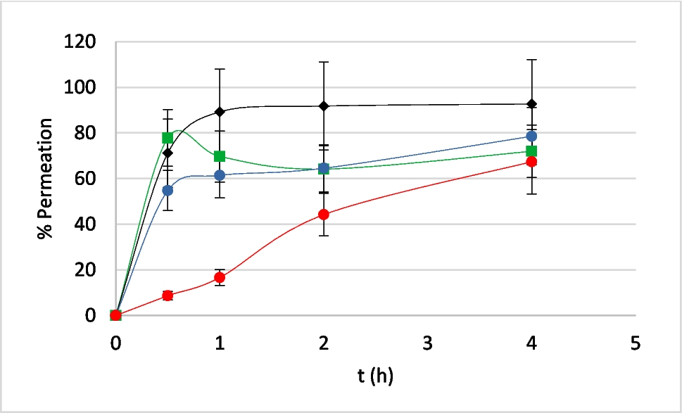
Permeation of three biocides were also determined under unmodified and modified membranes or mucoses. A volume of 70µL of each formulation was placed on the modified membranes or mucous membranes and within 1 h an infinite dosage (300µL) of the testing biocide sample was applied. The testing biocide solution was made with fungitrol (FUN), propiconazole (PRO) and permethrin (PER) dissolved in ethanol at a concentration of 1% for each active ingredient (Merck, Darmstadt, Germany). An aliquot of 200µL of receiving fluid was taken at various time intervals (30 min, 1 h, 2 h, 4 h) and immediately replaced the same volume with fresh fluid. The receiving fluid (RF) used was EtOH:H2O (75:25), assuring sink conditions of the compounds. The recirculating bath system was at 43 °C to obtain a membrane surface temperature of 32 ± 1 °C. The aliquots were appropriately diluted and filtered (0.45 µm, Cameo, Sigma-Aldrich, St Louis, USA) before their analysis with HPLC/DAD.
The release of the active ingredient was evaluated through the cumulative amount released (Qn, μg/cm2), which corresponds to the cumulative amount of active ingredient quantified in the receiving liquid per surface area of the sample [29], with the following equation (1):
$$Qn=\frac_^(Ci\times Vs)}$$
(1)
where: Qn is the cumulative amount of active ingredient released at time n (μg/cm2); Cn is the concentration of active ingredient in the sample (μg/mL); Vc is the volume of the vertical diffusion cell (7 mL);); \(_^Ci\) is the sum of the active concentrations (µg/mL) determined in sampling intervals 1 to n-1; Vs is the volume of the sample and A is the surface area of the sample (1.86 cm2).
Analytical Measurements HPLC/DAD
The collected samples were analysed by reversed-phase HPLC, using Agilent1620 Infinity II LC System HPLC (Waldbronn, Germany) with a quaternary pump (G-7111B), autoinjector (G-7167 A), thermostat multicolumn (G-7116 A) and VWR diode-array detector (G-7115 A). The software used was OpenLab CDS v.2.3.53. For the authentication of the analytical methods, the guidelines established by the International Conference on Harmonisation [30] were followed. The ICH directions were applied to develop the calibration curve, the quantitation limit (LoQ) and the detection limit (LoD). The analytical conditions and the HPLC-DAD methodology for the three biocides are listed in Table 1. (Table 2)
Table 2 Method conditions used in the HPLC/DAD analysis for Fungitrol, propiconazole and permethrinStatistical Analyses
Statistical analyzes were conducted with the non-linear regression software STATGRAPHICS + 5 (Statgraphics Technologies, Inc., Virginia, USA). The Kruskal-Wallis non-parametric test was employed when the data presented an abnormal distribution. The permeation parameters of the three biocides measured for each membrane were then compared with pig skin. Significance was tested for the 0.05 (p) probability level. The results are reported as average ± standard deviation (SD) (Table 2).


Comments (0)