
Remember me
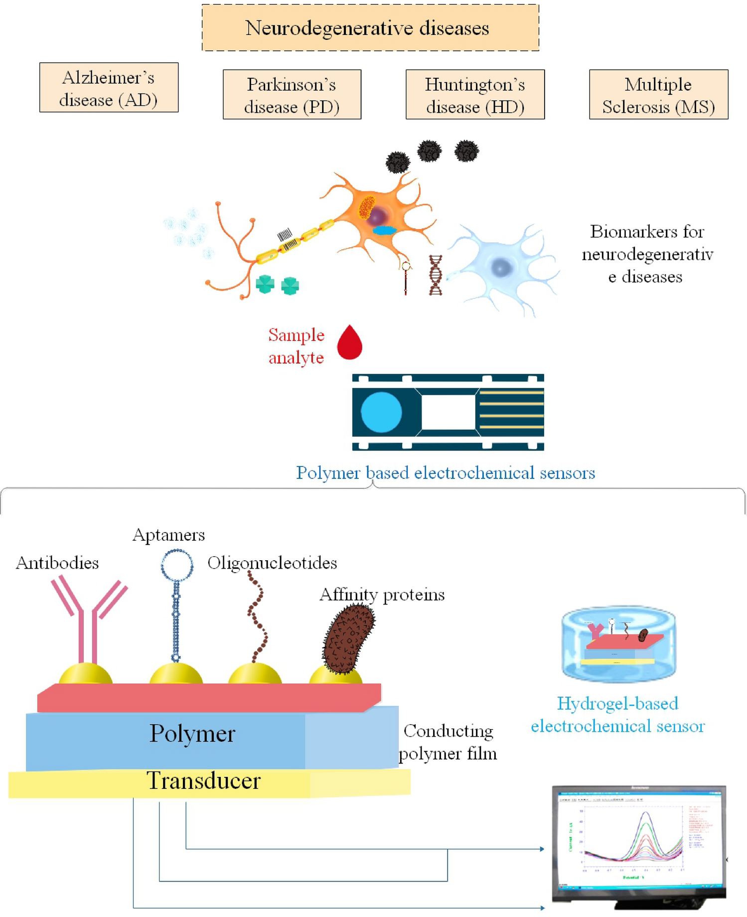

Due to their unique properties, such as compatibility, conductive nature, electron promoter, and inexpensive cost, polymer-based electrochemical sensors contribute to producing electrochemical sensors and biosensors. Electrodes modified with a single material have not yet hit the market because of issues with sensitivity, selectivity, surface poisoning from adsorbed intermediates, and interference from other species. One attractive feature of metal NPs and polymers frameworks modified electrodes as electrochemical sensors or biosensors is that they avoid these difficulties. A biosensor's selectivity in an electrochemical biosensor depends on the recognizing element, the host matrix, and the interaction between the two. Obtaining the required sensitivity and stability is made easy with metal NPs and polymers frameworks, since polymers act as selective adsorbents for biomolecules, and MNPs mediate the redox reactions of biomolecules (Prakash et al. 2013).
Polymeric NPs (PNPs) are a new kind of optically active nanomaterial that has just come to light due to its many desirable properties, including photostability, minimal cytotoxicity, rapid emission rate, high fluorescence brightness, biocompatibility, and a high and adjustable fluorescence quantum yield. Sensing and biosensing, controlled drug delivery, nanomedicines, catalysis, wastewater treatment, and many more biological and medical applications use PNPs, which are synthesized either from conjugated or nonconjugated polymers or by polymerizing appropriate monomers (Bharadwaj et al. 2022). One of the primary neurotransmitters in the CNS, dopamine (DA), is essential for operating dopaminergic (DArgic) neurons. One of the main characteristics of age-related ND, such as PD, is the gradual loss of this particular population of cells. Although this method has medium- to long-term adverse effects, the mainstay of PD symptomatic treatment has been the injection of L-DOPA, an amino acid precursor of DA that passes the BBB, whereas DA does not. DA-nanoencapsulation treatments have been extensively investigated as potential substitutes for DA replacement to overcome this restriction. Overcoming DA's low encapsulation yield and/or poor biodistribution/bioavailability remains challenging today. Researchers presented the synthesis of a family of PNPs inspired by neuromelanin. Researchers' approach was based on the reversible coordination complexation of DA to iron metal nodes polymerized with a bis-imidazole ligand, which encapsulates DA inside NPs. In addition to being straightforward and affordable, the researchers' approach yields DA loading efficiencies of up to 60%. Compared to free DA, DA nanoscale coordination polymers (DA-NCPs) demonstrated improved absorption by BE(2)-M17 DArgic cells, decreased toxicity, and slower breakdown kinetics in vitro. When the particles were directly infused into the rat ventricle in vivo, they quickly spread throughout the healthy rat brain, raising striatal DA levels. More notably, the frequency and length of apomorphine-induced rotations were considerably reduced after 4 days of nasal administrations with DA-NCPs equal to 200 μg of the free drug per day than in the vehicle or DA-treated rats used for comparison. Researchers showed the benefits of using nanostructured DA for DA-replacement treatment (García-Pardo et al. 2021).
The target analyte must be specifically and selectively recognized in most analytical procedures. Biomolecules such as Abs, enzymes, and receptors have met these criteria. Unfortunately, in certain situations, biomolecules cannot be used; for example, proteins will become insoluble in organic solvents and very acidic or basic environments. Not to mention how expensive and time-consuming it is to prepare and isolate these biomolecules, which limits their use. This led to the development of synthetic receptors that function similarly to their biological counterparts. Using MIP is one way to get these synthetic receptors. MIPs can also be used to create analyte-specific recognition sites on a synthetic polymer. The artificial recognition sites are designed to match the target analyte in size, shape, and chemical function, resulting in highly selective and specific identification. MIP is a polymer with a unique binding site that can only bind to specific analytes. Its excellent stability, ease of synthesis, and potential to reduce costs led to its extensive use as a receptor rather than an Ab or enzyme. Repurposing MIPs allows for the creation of more promising MIP-NPs. There are practical uses for MIPs in electrochemical sensors, and they have even been tested on human samples. Integrating MIP-NPs with electrochemical sensors has allowed for the real-time monitoring of several compounds of biological significance, including pharmaceuticals, insecticides, environmental toxins, secondary metabolites, and more (Fauzi and Saputri 2019).
Researchers created an electrochemical sensor based on MIP-NPs for the sensitive and selective detection of diazinon (DZN) pesticides. Suspension polymerization was employed to generate the diazinon imprinted PNPs, which were then used to alter the carbon paste electrode (CPE) composition to manufacture the sensor. The SWV and CV techniques were used for electrochemical experiments. Researchers demonstrated that compared to the CPE-based non-imprinted PNPs (nano-NIP-CP), the nano-MIP-CP had a much greater adsorption capacity for diazinon. With two linear ranges of 2.5 × 10⁻⁹ to 1.0 × 10⁻⁷ mol L−1 and 1.0 × 10⁻⁷ to 2.0 × 10⁻⁶ mol L−1, the suggested sensor demonstrated exceptional sensitivity (95.08 μA L μmol−1) for diazinon, along with a LOD 7.9 × 10⁻¹⁰—10 − L−1. The sensor detected diazinon in apple fruit and well water samples, with recovery values ranging from 92.53% to 100.86% (Motaharian et al. 2016).
Conducting polymers (CPs) are among the most practical materials when designing new sensor platforms. The possibility of using the polypyrrole (PPy) polymer to make ultrasensitive biosensors has prompted research into the material. Biodetection systems are significantly more effective and reliable when they have specific properties, such as high conductivity, fast electron transfer at the electrode/solution interface, charge storage capacity, redox reversibility, relative biocompatibility, and functional groups for directed biomolecule anchoring. An appealing prospect for new biotechnological uses, this organic polymer is structurally flexible, environmentally stable, inexpensive, and easy to synthesize. Researchers have shown that PPy is an excellent material for a substrate or matrix to electrodeposit AuNPs with low molecular aggregation and maximal surface area (Avelino et al. 2021).
A new, cost-effective electrochemical sensor for measuring quorum signaling molecules (AHLs) in Gram-negative bacteria was created by combining magnetic NPs with MIP technology. Through surface polymerization, magnetic molecularly imprinted polymers with the ability to selectively absorb AHLs were effectively produced. Researchers used electrochemical measurements to characterize the particles after depositing them onto the surface of a magnetic CPE. Researchers recorded the oxidative current signature that is typical of AHL using DPV. An innovative method for monitoring quorum signaling molecules in Gram-negative bacteria has been developed using an electrochemical sensor based on Fe3O4@SiO2-MIP. Its real-time detection capacity, high specificity, outstanding repeatability, and strong stability make it a promising tool for clinical diagnosis and food analysis (Jiang et al. 2016).
Highly specialized sensors might be made from polymer-based sensors with the proper adjustments or features. Polymers may be used directly in sensing or by immobilizing specific receptors on them. Polymers' ease of functionalization, biocompatibility, extended lifespans, softness, lightweight, and economical manufacturing are only a few advantages of employing them as sensors. Significant changes have been made to improve performance and transfer lab-derived sensors into device marketing applications. However, there are issues with polymer-based sensors: Finding the appropriate precursors is the first step, which is followed by improving and creating the reaction processes and procedures that enable the acquisition of a detectable signal. (2) Looking for new materials to use as polymer matrices. (3) Creation of ion-selective and pH-based sensors. (4) Conducting trials to automate the acquisition of polymer-based sensors. To solve these challenges, multidisciplinary cooperation among chemists, engineers, and computer scientists will accelerate the adoption of large-scale manufacturing and development (Alam et al. 2022).
Polymer-Based Electrochemical Sensors in Diagnosis of ADA vital biological sensor for ACh detection is produced by immobilizing the enzymes acetylcholinesterase (AChE) and choline oxidase (ChO) on the surface of iron oxide (Fe2O3) NPs and poly(3,4-ethylenedioxythiophene) (PEDOT)-rGO nanocomposite modified fluorine doped tin oxide (FTO). Researchers used SEM, EIS, and CV to assess nanocomposites' qualitative and quantitative properties. This biological sensor showed a wide linear range of 4.0 nM to 800 μM, with a detection limit (based on S/N ratio) of 4.0 nM and a response time of less than 4 s. Over a long storage period, the sensor demonstrated excellent sensitivity, selectivity, and stability. The biosensor has shown a low detection limit, unprecedented for previously reported ACh sensors, and high sensitivity. The creation of Fe2O3 NPs/rGO/PEDOT modified FTO electrodes for assessing ACh levels in blood samples has broadened the use of biosensors since neurotransmitter detection is a significant problem for individuals with AD or memory loss (Chauhan et al. 2017).
Molecular imprinting technology uses MIPs to provide a personalized capacity for molecular recognition of template molecules. The cross-linked polymer matrix may remember the template molecule by attaching it to specific locations. After removing the template molecule, binding sites sized and shaped similarly to the original imprinted template molecule are revealed. When MIPs reabsorb, they very selectively attach to the template molecule. In MIPs, immunosorbents play a pivotal role in the retention mechanism that relies on molecular recognition. Because of their many advantages over Ab-based routes, MIPs are often referred to as synthetic Abs. These include improved handling, stability (against acids, bases, ions, and organic solvents), reduced cost, high-pressure resistance, a broad working temperature range, and ease of preparation. Drug delivery, cell recognition, and protein recognition are just a few areas of medicine where MIPs are an adequate substitute for biological entities. Researchers targeted a biosensor for β-amyloid detection based on molecularly imprinted polypyrrole (MIPPy). Researchers used an artificial receptor called an imprinted polypyrrole. It was created by electropolymerizing pyrrole on carbon electrodes that had been screen-printed and then subjected to β-amyloid. Β-amyloid provides a molecular template inside the polymer. Using a ferro/ferricyanide marker, the biosensor was assessed by CV. Researchers turned the biosensor's performance-affecting parameters, such as the number of electropolymerization cycles and the time it took for β-amyloid to attach, to attain the highest level of biosensor sensitivity. Researchers derived the Freundlich constant as 0.22 ng mL−1 and the exponent as 10.60, respectively, by evaluating the β-amyloid binding affinity with the biosensor surface using the Freundlich isotherm. A 1.2 pg mL−1 detection limit was shown using the biosensor. A biosensor was used to determine the concentration of β-amyloid in artificial CSF (Vais et al. 2021).
P-tau proteins are promising indicators for the diagnosis and prognosis of AD. Researchers developed a novel voltammetric sensor that employs a vanadium MXene polydopamine (VxPDA) redox-active composite and a Tau-441-specific polyaniline MIP (PANI MIP) for the sensitive detection of Tau-441 in plasma and interstitial fluid (ISF). The VxPDA/PANI MIP sensor offers a broad detection range of 5 fg/mL to 5 ng/mL in ISF without redox mediators, with a lower LOD of 2.3 fg/mL. In addition, a portable device that employs this approach can detect Tau-441 in fake serum with high sensitivity and specificity within a therapeutically relevant range. This sensor's potential for minimally invasive, early AD diagnosis in clinical settings is highlighted by its quick detection time (about 32 min) and inexpensive cost (around £20/device). This development attempts to ease the shift away from intrusive CSF-based AD diagnosis methods (Arjun et al. 2024).
Tau protein is a biomarker of AD, and researchers created a new electrochemical biosensor based on an MIP for its impedimetric measurement. Hyperphosphorylated Tau protein (Tangles) is a potential biomarker for Alzheimer's diagnosis, as shown by a recent association between AD symptoms and the presence of Tau proteins in an aggregated form. The MIP was created by electropolymerizing 3-aminophenol (AMP) with CV, while the protein template (p-Tau-441) was present. The MIP was immediately constructed on an SPCE. Proteinase K's proteolytic activity in urea broke down the p-Tau-441 protein attached to the polymeric backbone, and it was subsequently removed to leave empty spaces. EIS assessed the respective imprinted and non-imprinted electrodes' performances. In PBS buffer with a pH of 5.6, the MIP-based sensors' detection limit was 0.02 pM. The created platform produced serum samples with acceptable outcomes and high selectivity. Due to its ease of fabrication, quick reaction time, and low cost, the biosensor that the researchers developed is a promising tool for on-site Tau protein screening and an appealing addition to clinically proven approaches (Ben Hassine et al. 2021).
Tau-441, a protein with 441 residues, is an NFT component essential for diagnosing AD. These tangles begin in the entorhinal cortex and expand as AD worsens. The existence of AD biomarkers in ISF and blood is suggested by the combination of the tau proteins found in plasma by Zetterberg and colleagues and the fact that the brain and skin share an ectodermal origin. The researchers showed the creation of an MIP-based sensor surface for sensitive Tau-441 detection from ISF. Aniline was electropolymerized using CV in the presence of the Tau-441 template to form the biosensor surface. For comparison, a non-MIP (NIP) was created using a pH 7.4 phosphate-buffered saline (PBS) solution. Tau-441 was detected in PBS and ISF as part of sensor performance evaluations for the developed MIP sensor. As the concentration of Tau-441 increased, the MIP's current decreased linearly, suggesting that Tau-441 had successfully bound to the polymeric backbone of the MIP. With a LOD of 587 pg/mL (12.2 fM/L) in ISF, the sensor detected Tau-441 in the range of 50 fg/mL to 500 ng/mL (1.22 fM/L to 12.2 nM/L). Future research will examine microneedle patches and MIP-modified electrodes for detecting Tau protein in blood and ISF (Norman et al. 2024).
An MIP electrochemical sensor for identifying amyloid-β42 (Aβ42) as an AD biomarker was demonstrated by researchers. A straightforward ultrasonic technique created a nitrogen-doped carbon-dot-graphene (NCD-G) nanohybrid. Researchers used PPY as an imprinted polymer for electropolymerization using an Aβ42 template. The NCD-G and MIP were altered on an SPCE without a binding agent. SEM, transmission electron microscopy (TEM), UV–Vis spectroscopy, XPS, CV, and SWV were used to examine the morphology and electrochemical characteristics of MIP/NCD-G modified SPCE. Under ideal circumstances, the developed sensor showed a 1 pg/mL LOD and an excellent linear response from 5 to 70 pg/mL. This sensor performs exceptionally by increasing effective surface area, electrical conductivity, and electrocatalytic activity. This MIP sensor achieves exceptional selectivity and sensitivity by combining a nanohybrid with a molecularly imprinted method. Additionally, it was miniaturized and had remarkable repeatability, which might be used as a basis for future portable medical devices (Pakapongpan et al. 2024).
Screen-printed graphene electrodes (SPGEs) coated with ultra-thin layers of polymerized 1,5-diaminonaphthalene (pDAN) have been used to create an immunosensor with high sensitivity detection of beta-amyloid peptides, which are a trustworthy biomarker for AD. DAN was electropolymerized to apply a continuous polymer coating of regulated thickness to the graphene screen-printed electrodes. Raman and XPS were used to analyze the surface properties of virgin graphene and graphene electrodes treated with polymers. Researchers looked at how the polymer's thickness affected the electron transport rates. By functionalizing the pDAN-modified SPGE with the anti-beta-amyloid Ab used as the peptide bioreceptor, an immunosensor for the specific detection of beta-amyloid peptides Aβ(1–42) was created. The immunosensor demonstrated 1.4 pg mL−1 and 4.25 pg mL−1 detection and quantification limits, respectively, and was used to select Aβ(1–42) with a linear range of 1 pg mL−1 to 1000 pg mL−1. The biosensor's ability to analyze human plasma that has been tampered with was further verified. Using a low-cost SPGE platform, the immunosensor allows for the quick, accurate, precise, repeatable, and highly sensitive detection of Aβ(1–42), opening the door to research-based real-time investigations and diagnostic ex vivo applications (Abbasi et al. 2021).
There has been a lot of focus on chitosan NPs in recent decades due to their potential as polymeric and bio-based NPs. As nanocarriers, they might encapsulate items like active chemicals or medications, transport them to a targeted location, and then release them at a regulated rate (Yanat and Schroën 2021). The wide range of chitosan's properties—which include electrical, photothermal, catalytic, antimicrobial, drug degradation, pollutants removal, biosensing, and so on—makes it the ideal "smart" nanostructure material. Due to its expanding range of uses, chitosan has recently become a highly regarded research material (Raja 2020). Because of its concentrated presence, CSF has traditionally been the primary site of detection for AD biomarkers such as amyloid plaques. Nevertheless, the BBB makes it more difficult to detect these markers in the blood, leading to lower concentrations. Researchers devised a novel strategy to overcome this obstacle and find relevant AD biomarkers in blood plasma, such as amyloid plaques and apolipoprotein E4 (ApoE4). This is achieved by adding a chitosan-coated immobilization matrix of Au nanostars (AuNSs) to an SPCE. Electrical and morphological studies, UV–Vis spectroscopy, CV, and Nyquist plots all pointed to the 0.5% chitosan solution having the best dispersion and conductivity. Clinical studies that followed assessed electrical responses to quantify levels of amyloid-β 42 (Aβ42) and APoE4 in plasma samples taken from human blood. Strong linear correlations and minimum error bars in the DPV responses showed peak currents proportionate to biomarker concentrations. Minimal interference across biomarkers was seen in cross-reactivity experiments using mixed solutions of amyloid-β 40 (Aβ40), Aβ42, and ApoE4, demonstrating the proposed sensor’s excellent specificity. This enhanced gadget shows exceptional promise as a proof-of-concept system, providing a less intrusive, more economical, and less complicated way to identify and monitor the advancement of AD. With such a large surface binding area, researchers' effective technique opens up exciting new possibilities for improving AD diagnoses (Shin et al. 2024).
To build a flexible dual-mode platform for the sensitive profiling of AChE inhibitors—an essential component in the search for AD treatments—researchers created a novel bioinspired hydrogel scaffold that could be used to integrate hybrid nanoflowers (HNFs) into the sensing interface in real time. Researchers' approach incorporates AChE-specific aptamers into the hydrogel after magnetic bead imprinting using Strep-Tactin-coated magnetic beads (STMBs), shaping it, mimicking the tenacious anchoring of real tree roots. Improved binding integrity of HNFs with sensing interfaces results from this configuration's stable and biomimetic environment, which promotes HNF development. Researchers evaluated galantamine's inhibitory efficacy using the platform's dual-mode detection capabilities, which integrate colorimetric and electrochemical sensing. Additional evidence of the hydrogel’s usefulness and affordability comes from its remarkable reusability, which preserves more than 95% of its original activity even after repeated applications, and its long-term stability, which maintains 91% of its original performance. Overall, this hydrogel scaffold takes inspiration from nature and provides a fresh approach to biosensing with high efficiency and reliability for HNF-based biosensors. It has enormous promise for use in several fields of medicine and cutting-edge biosensing technology (Liu et al. 2025).
One of the biomarkers associated with AD is tDNA, and a very sensitive electrochemical biosensor for its measurement is detailed here. Through the use of catalytic hydrolysis by alkaline phosphatase (ALP), electroactive molybdophosphate anions were precipitated on a GCE, while it was still in place. After that, tDNA recycling amplification is performed. The four DNA strands that comprise X-shape DNA (X-DNA) are designated S1, S2, S3, and S4. The work of DNA polymerase allowed them to be further stretched in four directions. Both X-DNA motifs that were produced were subsequently polymerized. At the same time, ALP is encased in a network of hydrogels to create a porous material of the ALP@DNAhg type. Reducing the amount of GO and functionalizing it with AuNPs (Au@rGO) was used to modify the GCE. Strand displacement capture of ALP@DNAhg initiates tDNA recycling assembly for signal amplification. Massive quantities of ALP are rendered immobile as a consequence of this. Once molybdate (MoO42−) and pyrophosphate have been introduced, phosphate may be produced by catalyzing the hydrolysis of pyrophosphate by ALP. When it comes into contact with molybdate, it will create phosphomolybdate anions (PMo12O403−), which are redox active. The detection limit is 3.4 × 10⁻³ pM, and the amperometric signal is concentration-dependent within the 1.0 × 10⁻² to 1.0 × 10⁴ pM range for tDNA (Hua et al. 2019).
To better understand the function of sialic acid (SA) in the degenerative process of AD, it is crucial to conduct sensitive and targeted monitoring of SA in the CNS. Researchers developed an electrochemical biosensor using a new stimuli-responsive copolymer to detect SA in mouse brains with high sensitivity and selectivity. It is worth mentioning that the copolymer enhanced the detection sensitivity to 0.4 pM by converting SA recognition into a conformational transition and wettability switch. This allowed redox labels and targets to be more easily accessed and enriched on the electrode surface. The proposed approach enhanced sensing signals and showed good selectivity toward SA compared to possible interference molecules coexisting in the intricate brain system. This was made possible by the directional hydrogen-bonding interactions in the copolymer and the high affinity between phenylboronic acid (PBA) and SA. The electrochemical biosensor, which exhibited exceptional analytical capabilities, was effectively used to assess the dynamic shift of SA levels in real-time brains of mice administered AD in conjunction with in vivo microdialysis. The first-ever report of the precise concentration of SA in several brain areas of live AD mice is a significant step forward in our knowledge of SA's function in physiological and pathological processes (Ding et al. 2017).
Polymeric-Based Electrochemical Sensors in Diagnosis of MSIt is crucial to diagnose MS, a neurological inflammatory disease that progresses over time. Short non-coding RNAs, also known as miRNAs, have emerged as promising predictive biomarkers for the early diagnosis of a wide range of disorders due to their aberrant expression. Regarding MS, miR-155 is a top biomarker. To detect miR-155, the researchers used a non-label electrochemical nano biosensor that was sensitive, inexpensive, and non-invasive. An aptamer-modified graphite sheet (GS) substrate containing a nanocomposite of SWCNTs and PPY serves as the miR-155 capture probe in the biosensor. Researchers conducted all electrochemical experiments using Fe(CN)63-/4-present as a redox probe. The biosensor can detect concentrations as low as 10 aM and has a dynamic range from 10 aM to 1 µM. Additionally, the biosensor that was created was effectively tested on human serum. The suggested biosensor shows promise as a tool for early MS detection due to its simplicity of manufacture, almost cost-effectiveness, high selectivity, repeatability, and reproducibility (Shariati et al. 2022).
A new class of biomarkers called TDP-43-associated proteinopathies, frontotemporal lobar degeneration (FTLD), and amyotrophic lateral sclerosis (ALS) has just come to light. Unfortunately, there are several drawbacks to the current detection systems, including their complexity, high cost, and reliance on human operators. Researchers introduced a new electrochemical biosensor for TDP-43 detection incorporated into a lab-on-a-chip (LoC) platform. To immobilize TDP-43 Abs specifically, the sensor uses transducer functionalization using electrosynthesized PPY derivatives containing carboxylic groups. One indirect method for detecting and quantifying TDP-43 is differential pulsed voltammetry (DPV). The chip is sensitive to concentrations of TDP-43 protein ranging from 0.01 ng/mL to 25 ng/mL, has a linear detection range, and responds quickly. Its LOD is 10 pg/mL. Additionally, TDP-43 can be used as a POC diagnostic tool due to its effective detection in complex matrices such as serum from both healthy persons and ALS patients. There is hope for improving the diagnosis and treatment of ND using an electrochemical biosensor built onto a chip because of its high sensitivity, fast response, and robust performance (Turco et al. 2024).
Researchers are beginning to explore the biological uses of hydrogels, which are also finding more and more use in fabricating electrochemical sensors. An electrochemical sensor generates a sensing signal by interacting with a sensing element and the analyte of interest. These sophisticated sensors convert raw data into usable electrical signals that scale with the concentration of the analyte of interest, allowing for both quantitative and qualitative analyses. Hydrogels may serve as platforms for biomolecules to maintain their biological activity because of the one-of-a-kind microwater environment they produce. Concurrently, the three-dimensional structure of hydrogels allows them to possess an exceptionally high specific surface area. Hydrogels are, therefore, mainly used as a platform for biomolecule immobilization in electrochemical sensor research. The remarkable sensitivity with which these biomolecular hydrogels can detect analytes makes them invaluable in the MS diagnostic process (Fu et al. 2019; Abune et al. 2021; Liu et al. 2022).
Normal and abnormal physiological and pathological processes rely on matrix metalloproteinase-9 (MMP-9). MS is a chronic autoimmune disorder that affects the CNS. This enzyme serves as a peripheral biomarker for neuroinflammation in MS. The current method for tracking MS subclinical disease activity involves costly MRI examinations. Detecting MMP-9 using a disposable, low-cost sensor device appropriate for home monitoring of inflammation might be a practical alternative to expensive MRI scans. By doing so, researchers can anticipate when anti-inflammatory treatments will not work, allowing for earlier therapeutic intervention to reduce neuronal damage and avoid impairment. The details of creating a disposable sensor that can detect MMP-9 quickly and efficiently are detailed here. Electrodes were coated with oxidized dextran and then cross-linked with peptides that included particular cleavage sites for MMP-9 to create biosensors. Impedance measurements were used to track the films' breakdown when exposed to the enzyme. Within 5 min of adding MMP-9, the sensor would typically show a noticeable change in impedance, indicating a fast reaction. MMP-2 is a protease that may impede MMP-9 detection; however, the sensors exhibited a completely insignificant response. Calibration curves were constructed using the most sensitive and selective peptide sequence, Leu–Gly–Arg–Met–Gly–Leu–Pro–Gly–Lys. The detection range of MMP-9 was 50 to 400 ng/ml, which is clinically relevant. On electrode surfaces of varying roughness, two distinct hydrogel degradation processes were discernible; both processes seemed appropriate for tracking MMP-9 activity. The sensor materials may be readily modified to react to different proteases by choosing peptide cross-linkers with appropriate cleavage sites (Biela et al. 2015).
Polymer-Based Electrochemical Sensors in Diagnosis of PDAmong neurological diseases, PD is characterized by profound impairments in both motor and non-motor functions. One protein identified as a very sensitive PD biomarker is PARK7/DJ-1. For this reason, a MIP-based sensor based on the DJ-1 protein was used. The researchers detailed creating and improving an SPCE surface, including MIPPy, for identifying the DJ-1 protein. To make the MIPPy DJ-1 imprinted sensor, molecular recognition-capable nano matrices were synthesized by electropolymerizing pyrrole (Py) monomer on an SPCE electrode using CV. In addition, the MIPPy sensor was compared with the AuNP-attached impedimetric immunosensor. Using CV, EIS, and SEM, MIPPy/immunosensors were characterized. The MIPPy-based sensor showed analytical performance similar to the DJ-1 immunosensor, with a LOD 1 nM and a linear range of 1 nM to 500 nM. Using these MIPPy/immunosensors, the DJ-1 analysis was conducted satisfactorily in control and over-expressed N2A cells. Additionally, there was a strong association between the MIPPy-based sensor, the AuNP/PPy immunosensor, and the Western blot (Kumar et al. 2022).
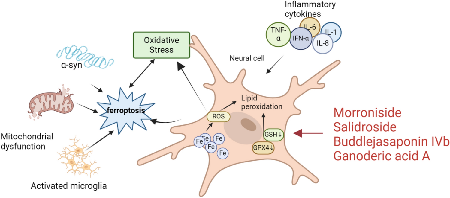
A molecularly imprinted electrochemical sensor for sensitive detection of α-Syn was built using this material in conjunction with graphene nanosheets (GS) as electrode modification materials. Electropolymerization of pyrrole was used to synthesize the MIP sensor, which was then characterized using SEM, CV, and DPV. CMP–GS nanocomposites substantially raised the MIP sensor's sensitivity by enhancing the GCE's electroactive surface, signal intensity, and electron transport rate. Under ideal circumstances, the sensor's linear range extended from 1 × 10⁻⁴ to 8 ng mL − 1, and there was a significant improvement in detection limits compared to previous approaches, reaching as low as 3.5 × 10⁻⁵ ng mL−1 at S/N = 3. A possible approach for detecting α-Syn in the blood of PD patients might be the sensor, which exhibited great sensitivity, minimal interference, and good stability. In addition, the researchers developed a novel approach to using CMPs in MIP electrochemical sensors (Ma et al. 2020).
Researchers created and studied electrochemical sensing and biosensing, a novel conductive ink incorporating carbon black into a poly(vinyl alcohol) matrix. To improve electrical conductivity and reaction kinetics, Palladium NPs were added to the devices after they were evaluated using morphological and electrochemical methods. The metal deposition characteristics were examined using chemometrics, and the sensor was used to identify PD biomarkers, including α-Syn and adrenaline. The neurotransmitter showed a linear behavior of 0.75 to 100 μmol L−1, and the device showed a LOD of 0.051 μmol L−1. Then, synthetic CSF samples were used to evaluate the three-electrode setup. The apparatus was then altered with specific Abs to measure α-synuclein with EIS. For α-Syn concentrations in phosphate buffer, a linear range of 1.5 to 15 μg mL−1 was found, with a computed LOD of 0.13 μg mL−1. When the suggested immunosensor was used on blood serum samples, the linear range of α-Syn was between 6.0 and 100.5 μg mL−1, with a LOD of 1.3 µg mL−1. Its potential applicability to complicated matrices is shown by the fact that both linear curves attend the range for the actual diagnosis (Orzari et al. 2024).
Clinical care for PD may be enhanced by a portable test that quickly determines levodopa levels. To improve the electrochemical detection of levodopa, researchers added polymers to screen-printed electrodes (SPEs). CV applied a thin coating of polyaniline on the surface of the electrode. Electrode conductivity was unaffected by the extensive surface covering seen by SEM. Researchers measured levodopa at physiologically appropriate concentrations by discriminating between a common interferent (ascorbic acid) and a structurally related molecule (L-tyrosine) using DPV measurements with the electrodes modified with polyaniline. The polymer layer did not allow for the separation of dopamine and levodopa. Nevertheless, these two molecules are identical except for an amino acid moiety in dopamine and a free amine group in levodopa. According to density functional theory calculations, Aniline created a hydrogen bond between the monomer's amino group and the meta-hydroxyl group found in dopamine and levodopa, with comparable binding energies. Therefore, SPEs functionalized with polymers are a valuable tool for measuring PD-relevant chemicals; nevertheless, to get selective detection, more optimization is required (Noguchi et al. 2022).
Among the first-generation selective adenosine A2A receptor antagonists, istradefylline (IST) stands out. It is now available to PD patients who are having "off" periods as an auxiliary therapy, according to a recent FDA approval. Scientists detailed the process of creating a new electrochemical sensor and used it to test plasma samples for IST using an anodic stripping square wave voltammetric (ASSWV) technique. The sensor, which is composed of zirconium (Zr) NPs on top of a pencil graphite electrode (PGE) surface, was created by layering an acid orange 10 (AO10) polymer platform on top of the ZrNPs. Equipped with SEM, EIS, X-ray powder diffractometry, and CV, the sensor was characterized. The sensor was then used to create an ASSWV approach based on oxidation for IST estimation. By following the ICH standards for analytical method validation, researchers improved the ASSWV technique and confirmed its analytical performance. This method's linear range was 25 × 10−9—170 × 10−9 M. Quantitation was 21 × 10−9 M, and the LOD was 6.9 × 10⁻⁹ M. It was proven that the ASSWV approach was accurate and precise. With recovery values of 96.0–97.9%, the approach achieved selectivity. The quantification of IST in human plasma samples was effectively accomplished using the suggested ASSWV technique. To sum up, the ASSWV technique that has been suggested is the first electrochemically viable strategy for IST estimation in plasma samples from humans. The approach contributes significantly to IST pharmacokinetic research, therapeutic monitoring, and safety profile refinement (Ali et al. 2025).
Researchers have proposed a surface-imprinted polymer (SIP) EIS biosensor to detect the aggregation of α-Syn. This biomarker manifests in saliva and blood during the first stages of PD when the BBB diminishes. Polycaprolactone (PCL) is stamped onto interdigitated EIS electrodes using a low-temperature melt stamping process to create the surface-imprinted polymer stamp. The end product is a small-footprint biosensor that is ideal for non-invasive monitoring of the disease biomarker and is cost-effective. The sensors were tested with α-Syn dilutions in deionized water and matrix solutions with constant ionic content and decreasing α-Syn concentrations to eliminate the concentration-related background effects. The gadget's answer verified that it specifically targeted the monomeric α-Syn protein. The sensor has a linear detection range of 5 µg/L and a LOD of 5 pg/L. In the future, researchers' approach to measuring α-Syn monomers for PD patients has excellent promise since it encompasses the normal range of α-Syn in saliva. As a possible tool for ongoing monitoring of the disease biomarker, the sensor's capacity for repeat sensing was shown by regenerating the SIP surface and reusing it (Massey et al. 2024).
Scientists examined the sensitivity of electrochemical screen-printed sensors made using graphene-based conductive PEDOT: PSS (G-PEDOT: PSS) and Polyaniline (G-PANI) inks applied to a working electrode for the detection of various dopamine concentrations. The suggested inks' microstructures were examined using an SEM characterization approach. Researchers evaluated the developed electrodes' dopamine detection effectiveness by investigating CV electrochemical approaches using a ferri/ferrocyanide redox pair. Dopamine detection using screen-printed electrodes has been improved with G-PANI ink regarding LOD and stability. Additionally, researchers investigated electrochemical analysis for dopamine-specific detection independent of ascorbic acid (Ghosh, et al. 2022) (Table 2).
Table 2 The potential of the Polymer-based electrochemical sensor in detecting several NDs
Comments (0)