
Remember me
We describe two families comprising three individuals affected with a severe neurodevelopmental disorder. Parents in both families are healthy and unrelated; the lack of relatedness was confirmed with Somalier [55] (relatedness score between parents of Family A: -0.014, and between parents of Family B: 0.007, cutoff < 0.05). Neither family reported a history of neuromuscular disorders, although the maternal grandmother of Family A had recurrent pregnancy loss of unknown aetiology. Both families are Nepalese: Family A is reportedly of South Asian/Indo-Aryan descent while Family B originates from Eastern Nepal with no known Indian heritage.
Case report—family AProband AII-1 (male) was the first pregnancy for mother AI-2, complicated by insulin-treated gestational diabetes. A first trimester screen showed low aneuploidy risk and the morphology ultrasound was normal. A growth ultrasound at 31/40 weeks revealed foetal growth restriction (estimated foetal weight < 5th percentile) and small cerebellum (transcerebellar diameter < 5th percentile). A TORCH screen for antenatal infections and a foetal echocardiogram were normal. Reduced foetal movements were noted for several weeks prior to delivery.
Following spontaneous labour onset, AII-1 was delivered via caesarean section at 33 + 2/40 weeks due to foetal distress and breech presentation. Birth weight, head circumference and length were 1238 g (< 3rd percentile), 28.2 cm (7th percentile), and 41.5 cm (25th percentile), respectively. Abnormal hip and knee position was noted, with suspected congenital hip dislocation. APGAR scores were low (3, 5 and 6 at 1, 5 and 10 min, respectively), requiring ventilatory support and neonatal intensive care. Chest X-ray showed lung hypoplasia, and a single dose of surfactant and caffeine was administered.
Bedside cardiac ultrasound showed a structurally normal heart with normal outflow tracts and a bidirectional patent ductus arteriosus. Suspected late-onset sepsis (coagulase-negative Staphylococcus) was treated with antibiotics. AII-1 received phototherapy for physiological jaundice (bilirubin 168 μmol/L), albumin for hypoalbuminaemia (11 g/L) and a single packed red cell transfusion for anaemia with persistent borderline thrombocytopenia (platelet count 79–120 × 109/L).
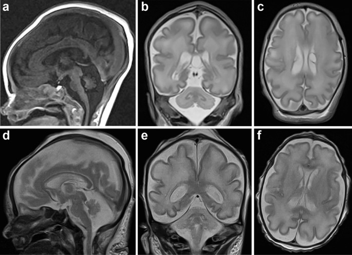
Initial postnatal metabolic tests, including very long chain fatty acids, transferrin isoforms, lysosomal enzymes, glycine and ornithine, were normal, as were electromyography and ophthalmological assessment. Creatine kinase (CK) was elevated (798 U/L, single measurement, normal < 180 U/L). Skeletal survey showed gracile long bones. BRAINZ monitoring and electroencephalogram (EEG) at corrected age of 35/40 weeks’ gestation showed diffuse cerebral dysfunction and EEG seizures, which were treated with phenobarbitone. Repeat EEG 1 week later confirmed findings. Cranial ultrasound suggested lissencephaly. Brain magnetic resonance imaging (MRI) showed a hypoplastic cerebellum (transverse diameter < 2 SD below normal), cerebellar vermis, and pons; bilateral choroid plexus cysts were noted (Fig. 1a–c). Spine MRI found no cord abnormalities but suggested an epidural hematoma without spinal cord compression.
Fig. 1 The alternative text for this image may have been generated using AI.
MRI of proband AII-1 and BII-1. a Midline sagittal FLAIR, b coronal T2 and c axial T2-weighted brain MRI of AII-1 at 7 days of age (corrected gestational age of 35/40 weeks), demonstrated a mildly small pons and pan-cerebellar hypoplasia. A cavum septum pellucidum and vergae, along with bilateral choroid plexus cysts, are visible in (c) (left: 9 × 6 mm; right: 6 × 3 mm; anteroposterior × transverse; mild gliotic lining of cysts observed during autopsy). d Sagittal, e coronal and f axial T2-weighted brain MRI of BII-1 at 12 days of age (corrected gestational age of 33 + 5/40 weeks) showing cerebellar vermian height, width and transcerebellar diameter are all more than 3 standard deviations below the mean for a 33-week-old corrected age neonate
Neurologically, AII-1 had limited respiratory effort, minimal spontaneous antigravity movements, brisk reflexes with clonus, thin overlapping fingers, and arthrogryposis with finger, elbow, hip, knee and ankle contractures. Knees were fixed in hyperextension and ankles were in dorsiflexion. Given persistent ventilator dependence and poor prognosis, intensive care was redirected to palliation and AII-1 died at 33 days postnatal (corrected gestational age 37 + 5/40 weeks).
After the birth of a healthy male sibling (AII-2), the third pregnancy (AII-3, female) showed growth restriction and clenched hands at 20 + 5/40 weeks. Continuing growth restriction, cerebellar hypoplasia and multiple upper and lower joint contractures were confirmed at 25 + 4/40 weeks, and amniocentesis was performed. Given phenotypic resemblance to AII-1 and poor prognosis, the pregnancy was terminated at 26 + 6/40 weeks’ gestation.
Autopsy of AII-1 and AII-3 showed global, symmetric growth restriction. AII-1 weighed 1975 g at 37 + 5/40 weeks’ gestation, (expected for 33–34 weeks) and AII-3 weighed 602 g at 26 + 6/40 weeks’ gestation (expected 894 ± 135 g). Internal organs were small but normally positioned and formed (Table S3).
Both siblings had distinctive facial features including high-arched palates and retro/micrognathia (AII-1 is shown in Fig. 2a; AII-3 had a downturned mouth and protuberant globes, not shown). Arthrogryposis with fixed upper and lower limb joint deformities was present in both siblings (Fig. 2b, c showing AII-1). Congenital hip dislocation was seen in AII-1 and hip adduction in AII-3.
Fig. 2 The alternative text for this image may have been generated using AI.
Pathology and histopathology of Family A and B. a Facial features of AII-1 at a corrected age 37 + 5/40 weeks’ gestation included micrognathia, a narrow mouth, and a high, narrow arched palate. b, c Fixed deformities of upper and lower limbs were evident; elbows mildly flexed, wrists hyperextended and fixed, fingers flexed mainly at the metacarpophalangeal and proximal interphalangeal joints, fisted hands with adducted thumbs, and boutonnière deformity of the 3rd and 4th fingers. Lower limbs show hypoflexed hips with congenital dislocation, fixed flexion deformities of the feet and ankles, and bilateral club and rocker-bottom feet. H&E-stained skeletal muscle of d AII-1 (psoas) and e AII-3 showed marked variability in myofibre diameter with both atrophic (yellow arrow) and hypertrophic fibres [2.5–22.0 μm in AII-1; 7.5–30.9 μm in AII-3; normal ~ 7 μm at 20/40 weeks’ gestation; [1], and internalised nuclei (AII-1: not quantified; AII-3: 10% of fibres). Scale bar for E and F is in F. Liver Masson Trichrome staining of f AII-1 and g BII-1 (40 × magnification). Liver histology of AII-1 showed irregular portal fibrosis, mild intrahepatocytic and bile canalicular cholestasis. Liver histology of BII-1 showed acute canalicular cholestasis with bile duct plugging, mild portal inflammation with ductular reaction and portal/periportal fibrosis with focal portal-portal bridging. h Lateral and i inferior view of the brain of AII-1 showed symmetrical cerebral hemispheres with sulcal and gyral development appropriate to gestational age. Notably, there is reduced size of the brainstem (2.5 g; 1.3% of total brain weight, normal: 2.4%) and cerebellum (4 g; 2.1% of brain weight, normal: 4.1%). H&E of the cerebellar cortex of j AII-1 and k BII-1 (200 × magnification) showing preserved lamination with normal external granular and molecular layers but a reduced number of Purkinje cells. l The dentate nucleus of AII-1 had a simplified, nodular architecture, reduced neuronal staining and increased surrounding glial cells. m The inferior olives showed poor demarcation from adjacent tissue, reduced neuronal staining and somewhat increased glial cell number on GFAP stain
Muscle atrophy was noted, more prominently affecting distal limbs in AII-3. Most muscles appeared macroscopically normal, but in AII-1, the quadriceps was fatty and ill-defined. Histology showed areas of myofibre diameter variability and an increased number of internal nuclei in both siblings, and angulated fibres in AII-3 (Fig. 2d, e).
Liver pathology in both siblings included a macroscopically fibrous appearance with portal fibrosis, ductular reaction (liver-injury-induced reactive bile duct proliferation), extramedullary haematopoiesis, and a mixed chronic inflammatory infiltrate (Fig. 2f). AII-1 showed mild intrahepatocytic and bile canalicular cholestasis, with bile ducts occasionally plugged (Fig. 2f). Early cholestasis and bile plugs were also seen in AII-3.
Brain weights were reduced: 190 g in AII-1 and 91.9 g in AII-3, compared to expected weights of 343.6 ± 37.9 g and 124.9 ± 18.2 g, respectively. Although the brainstem and cerebellum were macroscopically relatively normal, they were reduced in size (Fig. 2h, i). The cerebellar cortex appeared normal, but Purkinje cells were reduced or lacking (Fig. 2j showing AII-1). The dentate nucleus and inferior olivary nucleus (olives) were abnormally simplified in both siblings (Fig. 2l, m), with olives showing minimal convolution and a simplified “U” shape (Fig. 2m). AII-3 showed multiple discontinuous peripheral nodules and AII-1 showed fewer neurons and an increased number of glial cells surrounding the dentate on GFAP staining (Fig. 2m). Brain pathology raised the possibility of pontocerebellar hypoplasia (PCH), particularly olivopontocerebellar hypoplasia (OPCH).
Case report—family BThe affected child (BII-1) was the first pregnancy for his mother (BI-2) from a naturally conceived dichorionic, diamniotic twin pregnancy, with foetal demise of the co-twin at ~ 10 weeks. A 22-week ultrasound was unremarkable; however, foetal growth restriction (< 5th percentile) was evident by 31/40 weeks. Ongoing severe growth restriction, abnormal Doppler ultrasound and reduced foetal movement prompted an emergency Caesarean section delivery at 32/40 weeks’ gestation. Birth weight was 1.2 kg (5th percentile), head circumference was at the 3rd percentile, and length < 5th percentile. BII-1 was born in poor condition with hypotonia. Minimal spontaneous movements and respiratory effort (APGAR scores: 1 and 2) necessitated intubation and ventilation.
BII-1 had posteriorly rotated ears, synophrys, a broad nasal bridge, a low anterior hairline, and bilateral single palmar creases. Hands were clenched with camptodactyly and overlapping fingers. He had fixed talipes with overlapping toes. Both hips were dislocated. A small phallus and small testes were noted. Renal ultrasound showed mild bilateral pelvicalyceal dilatation. Echocardiogram showed right heart dilatation, mild tricuspid regurgitation, pulmonary hypertension, and a patent ductus arteriosus. Cranial ultrasound revealed a markedly small/hypoplastic cerebellum (<< 5th percentile) and right-sided grade I intraventricular haemorrhage. Brain MRI, though limited by artefact, showed PCH (Fig. 1d–f).
Ventilation requirements remained high over 6 weeks, progressing from conventional ventilation to high-frequency ventilation: all attempts at weaning were unsuccessful. He had marked tracheobronchomalacia. Care was redirected to palliation and BII-1 died on day 45 (corrected age: 38/40 weeks’ gestation).
Autopsy of BII-1 confirmed growth restriction (weight: < 5th percentile, normal length) and a markedly small brain (220 g; normal range 368.91 ± 39 g). Organ weights (heart, liver, thymus, adrenal glands) were < 5th percentile. Abnormal ear morphology, fixed foot contractures with external rotation and rocker-bottom appearance, loosely clenched hands, micropenis and bilateral femoral dislocation were confirmed. Liver histology showed acute canalicular cholestasis with bile duct plugging. There was mild portal inflammation with ductular reaction. Collagen stain demonstrated portal and periportal fibrosis with focal portal–portal bridging (Fig. 2g). Some of the changes were thought to arise secondary to sepsis; however, the degree of fibrosis in particular was unusual and may relate to the patient’s underlying genetic condition. Histological analysis of the cerebellar cortex revealed patchy reduction/absence of Purkinje cells (Fig. 2k).
Genome and RNA-seq identified a deep-intronic variant in CDK5RAP3Family A: Chromosome microarray for AII-1 and AII-3 was unremarkable. No causative variants were identified with genetic diagnostic testing: Myotonic Dystrophy Type-1 testing (Victorian Clinical Genetics Services, VIC, Australia), SMN1-deletion testing (South Eastern Area Laboratory Services, NSW, Australia), Neuromuscular Panel (NSES v2; including arthrogryposis, congenital muscular dystrophy and myopathy genes; PathWest Laboratory, WA, Australia) and Epileptic Encephalopathy Panel (v2.2, 80 genes; Children’s Hospital at Westmead, NSW, Australia). A PCH Panel (Table S4, Children’s Hospital at Westmead, NSW, Australia) identified heterozygous variants of uncertain significance in SEPSECS (NM_016955.3:c1027-56_1027-53del) and TSEN54 (NM_207346.2:c.767G > A), which were not pursued due to the absence of biallelic variants and likely autosomal recessive inheritance.
A family history indicative of a monogenic disorder prompted trio genome sequencing (AII-1, AI-1, AI-2) and RNA-seq (AII-1). No segregating, plausible variant(s) were identified in known Mendelian genes. The only plausible candidate was a homozygous deep-intronic variant, chr17(GRCh38):g.47974691G > A; CDK5RAP3 NM_176096.3:c.334 + 243G > A (ClinVar Reference VCV004070917.1), predicted by SpliceAI to activate a cryptic donor splice-site.
RNA-seq (AII-1) confirmed CDK5RAP3 c.334 + 243G > A caused PE/intronic sequence inclusion, encoding a premature termination codon (PTC, NP_788276.1;p.V112Gfs*8), predicted to trigger NMD (Fig. 3a). Minimal canonical splicing was detected, indicating the c.334 + 243G > A allele is hypomorphic (Fig. 3a). Low levels of residual CDK5RAP3 splicing likely permitted survival of AII-1 until birth, in contrast to the embryonic lethality of mouse Cdk5rap3 knockout [58, 69]. Pathogenicity of CDK5RAP3 c.334 + 243G > A was further supported by the absence of individuals with homozygous loss-of-function variants in CDK5RAP3 in gnomAD v4.1.0, by neurodevelopmental disorders linked to other UFMylation complex components, and by familial segregation (Fig. 3b, c).
Fig. 3 The alternative text for this image may have been generated using AI.
Segregating, biallelic deep-intronic CDK5RAP3 variants cause aberrant splicing. a An RNA-seq of AII-1 (bottom/red, quadriceps muscle, age: 33 days) and a paediatric disease-control (top/blue: male, quadriceps muscle, age: 8 years) revealed aberrant inclusion of a deep-intronic PE, containing a PTC (red hexagon), between exons 4 and 5 in CDK5RAP3 transcripts. Sashimi plot loops represent split reads; only 4 reads support canonical exon 4–5 splicing, while 157 reads support splicing from exon 5 to the PE acceptor site. The variant strengthens the PE donor (SpliceAI Donor gain D-score = 0.42) promoting mis-splicing. b Family A pedigree prepared in accordance with Ref. [5] and c Sanger sequencing traces showing segregation of the CDK5RAP3 NM_176096.3:c.334 + 243G > A variant in homozygous affected siblings (AII-1, AII-3, filled symbols) and heterozygous, unaffected family members (AI-1, AI-2, AII-2, AII-4, half-filled symbols). Age-at-death is indicated for affected individuals. d Analysis of the shared haplotype harbouring the NM_176096.3:c.334 + 243G > A variant in both families. Colours represent the alternative nucleotide for 118 high-quality variants called in the region of homozygosity detected by exome in AII-1 (GRChg38 chr17:45,983,659–49,424,990) for each individual with exome data. Reference nucleotides are shown in white. Only variants with a genotype quality score > 35 for all individuals were included for analysis
Family B: SNP microarray showed no copy number change of significance. The Trio exome was non-diagnostic. Research reanalysis of the trio exome data identified the homozygous CDK5RAP3 c.334 + 243G > A variant in BII-1. Both parents were heterozygous for this variant.
Evidence for a shared founder allele in South Asian populationsBecause the same variant was present in both families, we investigated the surrounding genetic region for a shared ancestral origin. Across 118 single nucleotide variants (SNVs) in both pedigrees (AII-1, AI-1, AI-2; BII-1, BI-1, BI-2), we identified a shared homozygous region of ~ 3.6–3.7 Mb (Fig. 3d), consistent with inheritance from a common ancestral haplotype and founder event many generations ago. Although both families are Nepalese, their differing ancestry, with only Family A reporting Indian heritage, argues against recent relatedness. Population data further support the likelihood of a South Asian founder allele: in gnomAD v4.1.0, eight of ten heterozygous individuals have South Asian ancestry.
CDK5RAP3 c.334 + 243G > A profoundly reduces canonical transcript and protein levelsRT-PCR spanning CDK5RAP3 exons 4–11 was performed on mRNA from skeletal muscle (AII-1, AII-3), umbilical cord (AII-3), and amniocytes (AII-3). Note, no tissue was available for family B. Compared to controls, AII-1/ AII-3 specimens showed: i) undetectable canonical exon 4–11 splicing (full-length transcripts NM_176096.3 and NM_001278197.2; Fig. 4a, blue arrow); ii) inclusion of a 108-bp PE from intron 5 (red arrow in Fig. 4a and red transcripts in Fig. 4c); iii) aberrant splicing from exon 5 donor to the PE cryptic acceptor (chr17(GRCh38):47,974,688), retaining most of intron 5, with or without additional retention of introns 7 and/or 8 (Fig. 4a, c grey transcripts). CHX-treatment of AII-3 amniocytes increased PE-inclusion transcripts (Fig. 4a), consistent with NMD targeting PE-inclusion transcripts encoding a PTC.
Fig. 4 The alternative text for this image may have been generated using AI.
CDK5RAP3 transcript and protein expression studies. a RT-PCR spanning CDK5RAP3 exons 4–11. Two amplicons are detected in controls: canonically spliced (823 bp product, blue arrow; NM_176096.3 and NM_001278197.2 transcripts) and larger transcripts retaining intron 5 or introns 5 and 7 (“intron retention”, grey bracket; NM_001278217.2, NM_001278216.2 and NM_001278198.2). In AII-1 and AII-3, several CDK5RAP3 transcripts retaining intronic sequences were present: (1) canonical transcript containing a 108 bp PE between exons 5 and 6 (“Variant-PE”, red arrow); (2) transcript containing 108 bp PE plus an additional 470 bp intronic sequence 3’ downstream and sometimes retaining intron 7 (grey bracket). b qPCR showing canonical splicing is reduced to 4.4% in AII-1 muscle and to 1.6% and 2.3% in AII-3 amniocytes treated with DMSO or CHX, respectively, compared to control. (***p = 0.0002, ****p < 0.0001 one-way ANOVA with Šídák’s multiple comparisons test). Treatment: CHX, DMSO (D) or untreated (–), NTC = negative template control, M = size marker, U.C. = umbilical cord. c Schematic of NCBI annotated CDK5RAP3 transcripts (top) and aberrant transcript detected in AII-1 and AII-2 (bottom) with introns shown as lines and exons as boxes. The open reading frame (ORF)/coding sequence is shown in colour or dark grey, while 5’ and 3’ untranslated regions (UTR) are shown in light grey. Red hexagon = termination codon. d Western Blot using antibodies targeting a CDK5RAP3 N-terminal (PA5-89,007) or C-terminal (HPA022141) epitope. Full-length CDK5RAP3 (red arrow; predicted 57 kDa, observed 65 kDa) is detected with both antibodies. Additional bands (white arrow), consistent with alternative isoforms lacking the N-terminus, are detected only with the C-terminal antibody. Antibody validation confirmed PA5-89,007 has a low affinity for these proteins (Fig. S3). Ages: Proband: AII-1: 37 + 5/40 weeks’ gestation, AII-3: 26 + 6/40 weeks’ gestation; controls: C1: 4 m male, C2: 5y female, C3: 18/40 weeks’ unknown gender, C4: age/gender unknown, C5: 37/40, C6: 28/40, C7: 19/40, C8: 51 d, C9: 27/40 weeks’ gestation
CDK5RAP3 shows complex alternative splicing, with transcripts retaining introns 5, 7, and/or 8 also present in controls (Fig. 4a–c grey transcripts). We examined the abundance of CDK5RAP3 transcripts in GTEx Project V9 long-read transcriptomics data [[25]; Fig. S1; skeletal muscle, lung, liver, brain]. Notably, NM_001278217.2 expression was higher than the MANE Select (canonical) transcript NM_176096.3. Alternative transcripts NM_001278216.2 and NM_001278217.2 were particularly highly expressed in cerebellar hemispheres.
qRT-PCR confirmed that canonical exon 4–5–6 splicing was reduced in AII-1 skeletal muscle and AII-3 amniocytes to < 5% of control levels (Fig. 4b). Both siblings showed an overall reduction of all CDK5RAP3 transcripts, suggesting that the apparent enrichment of intron 5-retaining transcripts in AII-1/AII-3 specimens observed by RT-PCR reflects increased relative amplification due to canonical isoform loss (Fig. S2).
Western blot using N- and C-terminal CDK5RAP3 antibodies (Fig. S3 for epitopes) confirmed that full-length CDK5RAP3 protein was reduced to < ¼ control levels in AII-3 amniocytes and below detectable levels in proband skeletal muscle and umbilical cord (Fig. 4d). A ~ 5–10 kD smaller protein product (white arrow, Fig. 4d) was detected by the C-terminal CDK5RAP3 antibody in proband skeletal muscle and amniocytes, potentially representing an alternate isoform translated from intron 5-retaining transcripts with an initiation methionine in exon 6 (Fig. 4b, grey transcripts). Western blot across developmental stages (13/40 weeks’ gestation to 31 years) suggests a ~ 5–10 kD smaller isoform may be physiologically present in skeletal muscle < 28/40 weeks’ gestation (Figs. 4d and S4).
Putative retained CDK5RAP3 isoforms show poor UFL1-bindingCDK5RAP3 forms part of the UFM1 ribosome E3 ligase (UREL) complex, directly interacting with UFL1 to facilitate UFMylation of the 60S ribosomal subunit protein RPL26 [64]. This modification is essential for 60S subunit release from the SEC61 translocon (Fig. 5e) during ribosome stalling or normal termination of translation [44].
Fig. 5 The alternative text for this image may have been generated using AI.
UFMylation and UPR assessment in CDK5RAP3 deficiency. a Co-IP to determine whether wt full-length CDK5RAP3, or shorter isoforms derived from ORF3 (NP_001265146.1) and ORF1, can complex with UFL1. FLAG-tagged CDK5RAP3 wt, ORF3 and ORF1 are detected with FLAG and CDK5RAP3 antibodies (arrows). Probing for UFL1 reveals only full-length wt CDK5RAP3 can co-precipitate UFL1, indicating interaction. In contrast, UFL1 co-precipitation was significantly reduced with ORF3 and ORF1 (9.8 ± 3.7% and 1.7 ± 2.8%, respectively; ***p < 0.0001 one-way ANOVA; quantification across 3 experiments in (b). Bar graphs show mean ± standard deviation. c Schematic illustrating proteins predicted to arise from wt and variant CDK5RAP3 gene sequences. d Western blot of UFMylation pathway components (UFM1, UFBP1, RPL26) in AII-3 amniocytes showed increased UFMylation of UFBP1 and decreased di-UFPylation of RPL26, indicating dysregulation of the pathway. e Schematic illustrating complex formation of UFMylation proteins (UFBP1, UFM1, CDK5RAP3, UFL1) with the 60S ribosomal complex (including RPL10A and RPL26) at the ER membrane. Illustration was generated in Biorender and is based on crystal structures from RCSB PDB, 8QFC [44] and 6R7Q [59]. f RT-PCR analysis of XBP1 non-canonical splicing using control (C4, C) and AII-3 skeletal muscle (SkM) or amniocytes treated with DMSO (–) or 2.5 μg/ml Tunicamycin ( +) for 5 h to induce the UPR. Primer information is in Table S2. Top: primers amplifying both spliced (XBP1s) and unspliced (XBP1u) products. Middle: forward primer bridging the excised region, amplifying only XBP1s. Bottom: forward primer within the excised region, amplifying only XBP1u. +ve positive control fibroblasts with UPR activated, -ve: negative control fibroblasts with no UPR activation, NTC no template control
Although CDK5RAP3 c.334 + 243G > A nearly abolished canonical protein (NP_788276.1) production, the 5–10 kD smaller observed protein product may represent the annotated alternative isoforms NP_001265146.1, unaffected by c.334 + 243G > A (Fig. 4c, d). NP_001265146.1 shares the C-terminal 395 amino acids with NP_788276.1 (Fig. S3), but has a short, distinct N-terminus (24 versus 111 amino acids).
Co-immunoprecipitation experiments compared the ability of wt, NP_001265146.1 (open reading frame-3; ORF3) and truncated mutant (ORF1) CDK5RAP3 isoforms to bind UFL1. Wt CDK5RAP3 bound UFL1 as expected [39], while NP_001265146.1 showed a markedly reduced UFL1 co-precipitation and ORF1 failed to pull down UFL1 (Fig. 5a–c).
AII-3 amniocytes have a UFMylation defectTo determine whether CDK5RAP3 deficiency caused a UFMylation defect, we investigated known UFMylation substrates RPL26 and UFBP1 in AII-3 amniocytes. RPL26 is UFMylated on Lys132 and Lys134 [64, 65]. While mono-UFMylated RPL26 levels were unchanged, di-UFMylated RPL26 was reduced. UFMylated UFBP1 was enhanced in AII-3 amniocytes compared to control (Fig. 5d).
Constitutive unfolded protein response (UPR) was not activated in skeletal muscle and amniocytes from affected individualsDespite previous reports of ER stress in CDK5RAP3-deficient U2OS cells and hepatocytes [35, 69], we found no evidence of abnormal UPR activation in skeletal muscle or AII-3 amniocytes. XBP1 splicing, a surrogate marker of ER unfolded protein induced stress and UPR induction [71], was not elevated at baseline in proband tissues (Fig. 5f). Upon tunicamycin treatment to elicit ER stress, XBP1 splicing patterns were as expected and comparable between control C4 and AII-3 amniocytes (Fig. 5f), indicating preserved UPR responsiveness.
Global proteomic profile alterations due to CDK5RAP3 deficiencyTo assess the impact of the c.334 + 243G > A variant on the global proteome, we performed quantitative mass spectrometry comparing AII-3 and primary amniocytes derived from healthy control C4 (‘Proteomics Dataset 1’, Table S5, 4 replicas each). We identified 6826 unchanged, 188 significantly upregulated and 288 significantly downregulated proteins (Fig. S5a) with CDK5RAP3 among the significantly reduced proteins (log2FC: -1.9; 26.8% of control, q < 0.00001) consistent with Western blot results (Fig. 4d).
Given CDK5RAP3 is a UFMylation substrate adaptor, we examined UFMylation pathway components (Fig. S5b, Table S6). KIF11 (aka EG5), a recently identified UFMylation substrate [40], was significantly upregulated (log2FC: 1.4, q < 0.001). Other substrates EIF6 [60], SLC7A11 [68], P4HB [81] and UFL1 showed modest changes below the significance threshold (> 0.50; log2FC of 0.34, 0.36, -0.38 and -0.28, respectively; q < 0.0001). Confirmatory Western blot did not demonstrate a reduction in UFL1 levels in AII-1 and AII-3 (Fig. S6).
Pathway enrichment analysis using Metascape [80] revealed the GO cellular components (CC) “ECM” to be the most significantly downregulated summary term, represented by specific pathway terms such as “ECM organisation”, “Non-integrin membrane-ECM interactions”, and “Assembly of collagen fibrils and other multimeric structures” (Fig. S5c; Table S7).
Interestingly, ECM, via different subsets of ECM proteins, also appeared among upregulated pathways, represented by GO CC “External encapsulating structures”, GO Molecular Functions (MF) “S100 protein binding”. Several cell cycle and mitotic progression pathways and cellular components pathways were also significantly upregulated (e.g. GO biological processes (BP) “Mitotic cell cycle”, reactome gene sets “Mitotic prometaphase” and “condensation of prometaphase chromosomes” together with “Golgi-to-ER retrograde transport”, which share overlapping protein components [e.g. KIF11, KIF4A/B, and KIF21A]; Fig. S5d). Additional themes included cytoskeletal organisation and cell attachment, heat stress, and growth factor signalling. Overall, proteome findings highlight global ECM and cell attachment dysregulation in AII-3 amniocytes. Themes align with prior studies linking CDK5RAP3 to cell cycle regulation, cell growth and cancer-related processes such as metastasis and growth factor signalling [41, 42, 49, 63, 67, 78] as well as the role of UFMylation in microtubule network and centrosome organisation [35].
ASO restoration of CDK5RAP3 protein confirms modulation of ECM organisation, cell adhesion and migrationTo mitigate caveats from studying one affected cell line, we developed a complementation assay to identify pathways specifically disrupted by CDK5RAP3 deficiency and restored upon re-expression. A targeted antisense oligonucleotide (ASO-T) was designed to block the cryptic donor site activated by c.334 + 243G > A. ASO-T or a scrambled control ASO (ASO-S) was introduced via nucleofection into control C4 and AII-3 amniocytes, and harvested after 72 h. ASO-T-mediated restoration of canonically spliced CDK5RAP3 transcripts and protein to control levels was confirmed by RT-PCR, qRT-PCR, Western blot, and mass spectrometry (Fig. 6).
Fig. 6 The alternative text for this image may have been generated using AI.
An ASO targeting the variant PE donor restored full-length CDK5RAP3 transcript and protein expression. Transcript/protein impact of CDK5RAP3 PE donor targeting ASO (ASO-T) and scrambled control ASO (ASO-S) nucleofected into AII-3 and C4 primary amniocytes was assessed with a RT-PCR and Western blot (C-terminal antibody HPA022141). ASO-T restored canonical CDK5RAP3 splicing, and rescued full-length CDK5RAP3 protein expression in AII-3 amniocytes. ASO-S had no effect. b Quantitative RT-PCR (***p < 0.0001, pairwise t-tests between samples) and c mass spectrometry (***q < 0.0001, *****q < 0.00001) confirmed restoration of CDK5RAP3 transcript levels and protein levels, reaching 118.0% and 91.4% of control levels, respectively
Proteomic/phosphoproteomic principal component analysis (PCA) showed overlapping ASO-T and ASO-S clusters for control cells, indicating minimal off-target effects (Fig. S7; Proteomics Dataset 2’ and ‘Phosphoproteomics Dataset’ in Table S8–S9). In contrast, proband ASO-T and ASO-S clusters were distinct, with ASO-T-treatment shifting profiles towards the control along PC1.
We identified an “ASO-T Rescue Set” of 57 proteins and 505 phosphopeptides whose expression was partially or fully restored towards control levels after 72 h of ASO-T treatment in proband cells (Figs. 7a and 8a; Table S8–S9). Pathway analysis using the ‘ASO-T Rescue Set’ resulted in the detection of only 32 up- and no downregulated reactome/KEGG/GO pathways (Table S10, Fig. 7b). Identified pathways related to ECM, cell adhesion (cell–matrix, cell–substrate, ECM–receptor interactions), cytoskeletal networks, neurodevelopmental signalling (e.g. PDGF signalling, axon guidance, and neuronal migration) as well as scavenger receptor-mediated uptake, NCAM1 interactions, and protein digestion and absorption.
Fig. 7
Comments (0)