This study evaluated the mechanical behavior through FEA analysis of a premolar submitted to revascularization and apexification processes. The revascularization group showed higher tensile stress distribution inside the tooth. In addition, this technique presented the lowest compressive stress distribution value in the alveolar bone and periodontal ligament. Therefore, the tested hypothesis was accepted.
Tensile and compressive stress are different forces that can be analyzed by FEA. Tensile stress is the force that acts on a structure, causing stretching or extension in it. Tensile stress is generally developed in the horizontal direction, while compressive stress causes compression or shortening and is developed in the vertical direction [29]. Tensile stress is represented by positive values, and compressive stress is represented by negative values. Both forces are defined by the ratio between the applied force and the cross-sectional transversal area [30].
The experimental models in this study showed an immediate effect of revascularization and apexification endodontic procedure treatments on immature teeth. Previous studies have already shown that the fracture resistance of roots is directly associated with the remaining tooth structure and dentin amount [31, 32]. Healthy teeth have a sufficient dentin wall to provide adequate resistance to these elements [33]. However, immature teeth submitted to revascularization or apexification present thin root walls due to the non-deposition of secondary dentin. As a result, they become susceptible to fractures [34]. These studies corroborate our results since the IT showed higher tensile stress values when compared to the R and AX groups (Table 2).
The R, AX, and AC groups showed tensile stress distribution in the distal region due to the force application in the distal sulcus occlusal surface. This stress distribution can also be observed on the cervical surface of the AX and R groups (Fig. 3). These groups presented a lower amount of dentin, which is not favorable to its mechanical strength according to the results found by Valera et al. (2004) [34].
The minimum principal stress observed in dentin (–349.84 MPa) is higher than the typical compressive strength reported in the literature [35, 36]. This outcome may be attributed to local numerical stress concentrations at the boundary conditions and the idealized geometric simplifications inherent in the finite element model. In this context, these values represent localized peak stresses rather than realistic clinical magnitudes.
The finite element analysis demonstrated that the revascularization (R) technique resulted in slightly higher tensile stresses within coronal dentin (203.75 MPa) compared to apexification (203.14 MPa) (Table 2). In this way, the revascularization procedure increases stress within the dentin, which may elevate the risk of fracture in structurally compromised teeth [37]. Conversely, revascularization showed markedly lower compressive stresses (for alveolar bone: –28.53 MPa) compared with AX (for alveolar bone: –33.27 MPa), indicating that R may protect supporting tissues and reduce the risk of periradicular damage or resorption, which also can be favorable for long-term tooth support [38]. Clinically, this means that while revascularization may biologically favor continued root development and periradicular tissue preservation, it also increases stress concentration in coronal dentin, potentially elevating the risk of fracture in structurally compromised teeth. Apexification, conversely, may reduce dentin stress but offers less protection to supporting tissues. Within this perspective, revascularization can be preferred for preserving periapical integrity. However, careful case selection and reinforcement strategies (e.g., restorative approaches to support thin dentin walls) should be considered to mitigate the higher dentin stresses.
Some limiting factors in the revascularization technique can be highlighted, such as its technical execution to stimulate bleeding, clot stabilization, and application of a cervical stop [39,40,41,42]. However, the revascularization technique has shown high success rates in clinical trials, showing the apexification process as a viable treatment option. The outcome success rate of apexification with MTA and revascularization is shown in the literature to have values of 80.77% and 76.47%, respectively [43].
A study by Ali et al. (2019) [41] showed that bovine incisors treated by the apexification technique with MTA did not affect the mechanical strength when compared to a healthy tooth [44]. On the other hand, Mello et al. (2020) [45] evaluated the mechanical performance of teeth after apexification and revascularization, and they detected no statistically significant difference between these groups [45]. Such discrepancies may stem from differences in experimental design, including tooth type (bovine incisors versus human premolars), analysis methods (finite element simulation versus mechanical testing), and loading configuration. These methodological distinctions may explain why the current findings diverge from previous in vitro studies, highlighting the need for further investigations to validate the mechanical behavior of immature teeth under different treatment protocols.
Silujjai & Linsuwanont [43] reported that the increase in root width development after MTA apexification reached rates of 3.30%, while the revascularization technique showed 13.75%. Moreover, rates of 8.55% were found for apexification of root length development, while 9.51% has been reported for revascularization [46]. These rates were updated by Caleza-Jimenez et al. [4], who reported an increase of 3.36% in dentin thickness and 0.29% in root length for the apexification technique, while revascularization increased dentin thickness by 34.57% and 12.75% for root length [46]. However, the revascularization technique in our study showed higher tensile stress distribution when compared to apexification. On the other hand, apexification showed the furthest compressive stress distribution from critical stress (Table 2).
Although revascularization promotes dentin apposition and increased root thickness, the newly formed tissue may present lower mineralization and reduced elastic modulus compared to mature dentin [47]. Consequently, the mechanical continuity between the old and the newly deposited dentin may be compromised, concentrating stresses along the junction area. Furthermore, the irregular dentin deposition pattern reported in revascularized roots may alter stress distribution, explaining the higher dentin stress observed despite the apparent geometric reinforcement.
Silujjai & Linsuwanont [40] suggest that the content within the root canal may be associated with the root mechanical strength, which corresponds to the different mechanical behavior of the groups in this study [43]. The 5 mm of MTA adopted to the revascularization and apexification models followed the same amount used by Darak et al. (2011) [19]. The literature shows that the use of gutta-percha to fill the root canal may not provide additional mechanical performance in roots after MTA apexification. However, revascularization may improve the development of biological tissues around the periapical root area, thereby favoring the mechanical strength of the roots [48, 49].
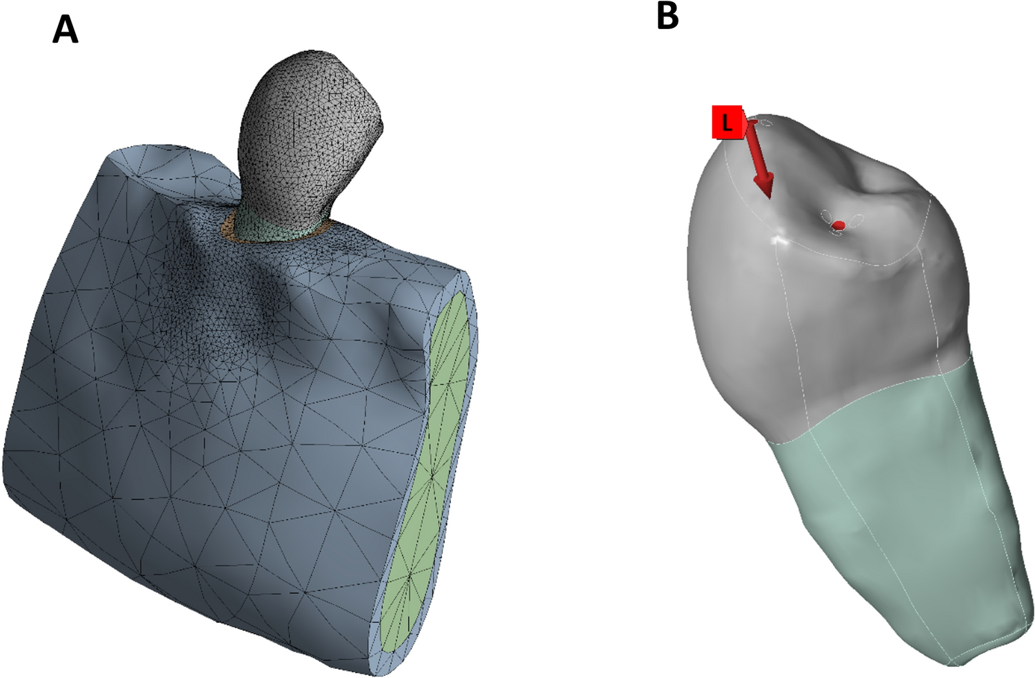
The failure criterion adopted for the bone is based on the Minimum Principal Stress. In Fig. 4, the color map highlights the compression regions with reddish and orange tones. Since it is common to associate warmer colors with the most critical results, we chose to invert the color scale to maintain this convention. In this way, warm colors continue to represent the most critical areas. Negative values indicate compressive stress, while positive values correspond to tensile stress. According to Papavasiliou et al. [50], the physiological limit,s for compressive stress of cortical bone are 140 to 170 MPa and 72 to 76 MPa for tensile stress [50]. Our results for the alveolar bone stress distribution (Fig. 4) showed compressive stress from –28.53 to –33.27 MPa and tensile stress distribution values of 2.73 to 3.27 MPa which are between the physiological limits of cortical bone.
It is important to explore how the periodontal ligament (PDL) behaves since this structure is submitted to dynamic loads (tensile and compressive stress) during mastication and orthodontic treatments, for example [51]. Additionally, there is limited data on the behavior of the periodontal ligament (PDL) in revascularization and apexification techniques simulated through FEA, which motivated this study to gain a deeper understanding of its behavior [52]. Tensile stress causes a rupture of collagen fibers, which leads to damage in the periodontal ligament [53]. In this sense, Fig. 5 and Table 2 show that the revascularization technique produces less tensile stress distribution compared to the apexification technique, resulting in less damage to the PDL.
Furthermore, the revascularization technique showed higher mechanical strength. These findings suggest that compared to the healthy group, both revascularization and apexification can be performed without inducing bone resorption, as their stress values remain below the critical thresholds reported for dentin [54] and alveolar bone [55] in the literature.
Within the limitations of this study, the dimensions of the access cavity, along with the volume of dentin and enamel removed during endodontic access preparation, play a critical role in influencing the fracture resistance of the tooth, and it can vary [56]. Moreover, although the adoption of a 10% threshold for mesh convergence has been employed in previous dental FEA studies [57, 58], it is considered less stringent than the conventionally recommended 1–5% variation in peak stress values between successive mesh refinements. The use of a broader tolerance may introduce a higher degree of numerical uncertainty, particularly in regions of elevated stress concentration, such as coronal dentin. In addition, calcifications can occur after revascularization treatment, but they were not simulated in this study due to their high variability [14]. Another limitation of FEA is that it assumes material properties to be homogeneous, linear elastic, and isotropic. While these modeling assumptions are common in FEA studies, they may still affect the clinical translatability of our findings.
The simplification of material properties as isotropic, linearly elastic, and homogeneous can lead to under- or over-estimation of absolute stress values, particularly in tissues such as the PDL and blood clot. Such an assumption disregards their inherently viscoelastic and time-dependent behavior, which could influence local stress and strain distributions. Nevertheless, because the present model simulated a single static loading condition, the use of elastic parameters provides a reasonable first approximation of their short-term mechanical response. In addition, the static loading approach does not reproduce fatigue or dynamic effects that may contribute to long-term failures in vivo. Consequently, although our results reliably capture comparative trends between apexification and revascularization, caution is warranted when interpreting absolute stress magnitudes in a clinical context. Practically, this suggests that while revascularization appears favorable for reducing periradicular (PDL and alveolar bone) stresses, apexification may better preserve dentin integrity. Nonetheless, true clinical outcomes will ultimately depend on biological adaptation, loading variability, and restorative strategies.
Despite these limitations, FEA remains a practical tool, offering reduced computational time and more efficient analysis for simulating and studying the behavior of complex systems that are difficult to replicate in vitro.
The lack of scientific findings comparing the mechanical strength after the revascularization and apexification procedures in immature premolars encouraged our research group to conduct this study. This paper intends to help clinicians better understand the mechanical behavior after performing these techniques. Nevertheless, more studies are needed to complement our findings.

Comments (0)