To the best of our knowledge, studying the histologic findings in the buccal mucosa due to the use of SFZN individually and in combination with ESOm has not been described previously. Thus, this study provides a novel histologic description of the effect of these drugs on buccal mucosa, providing a valuable nidus for future research.
In our study, male rats were chosen over female rats to avoid any possible modulatory effect of estrogen [22]. The buccal mucosa could be used to monitor early genotoxic events, and 90% of all cancers appear to be epithelial in origin [23].
The pyknotic nuclei and vacuolation detected in the SFZN Gp of our study are in line with the findings by Heikal et al. (2023) [24], which were detected in the renal tissue in response to SFZN administration and were attributed to increased reactive oxygen species (ROS), which could have been exaggerated in the SFZN group of our study due to the downregulated GSH levels, as proved by our investigations. Thepleomorphic nuclei detected in our study in SFZN Gp and ESOm Gp are similar to what was reported in epithelial cells of smokers [25].
The possible similarity between SFZN and smoking is that both have an impact on tissues through the occurrence of hypoxia, as smoking is related to high levels of serum carboxyhemoglobin [26], and SFZN is related to high levels of serum methemoglobin [27].
The nuclear changes were detected along the whole epithelial thickness of the SFZN Gp and were confined to the superficial layers in the ESOm Gp, which highlights the possible protective effect of ESOm on buccal tissue when administered in combination with SFZN.
The irregularities detected in the BM of SFZN Gp could be due to the altered architecture of the epithelium, visible in our histological results, as mentioned by Meinl and Brunner (1980) [28], and could be due to variable changes detected in our study, which require further investigations at the electron microscopic level to elucidate the nature of these irregularities.
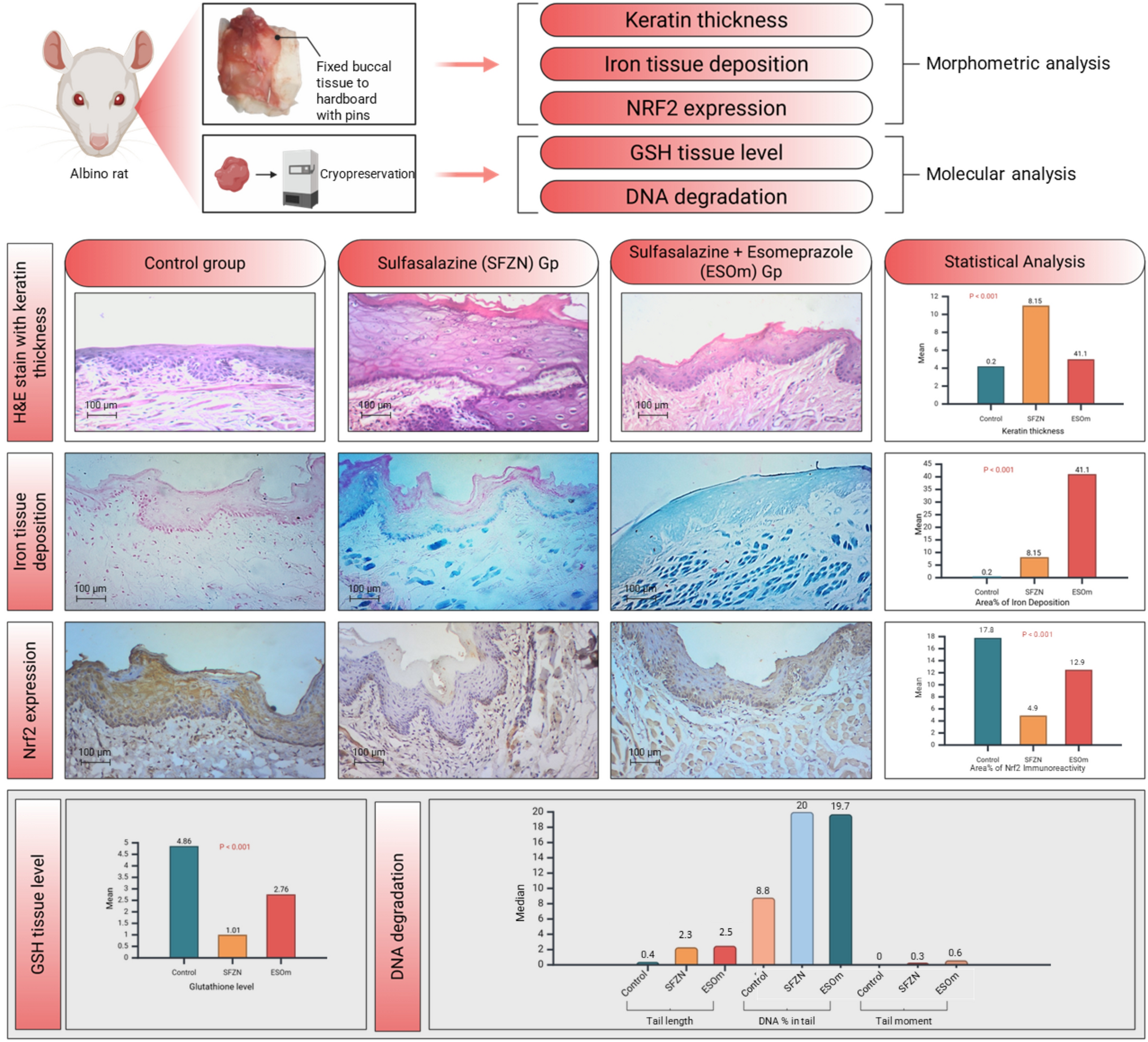
Some of the histologic findings detected in SFZN Gp, including premature keratinization and increased keratin thickness, resemble the signs of architectural and cytological features of oral epithelial dysplasia described previously [29]. Our novel results offer valuable resources and insight for future research in the field, as these findings were restored in the ESOm Gp, which showed epithelial, LP, and BM architectures that were comparable to those of the control Gp.
Cell enlargement with marked rounding is commonly observed in most ferroptotic cells and iron deposition in the tissues is a hallmark of ferroptosis [4]. We found characteristic rounding of the basal cells and significant iron deposition, detected in the SFZN Gp and ESOm Gp according to the results of our study using Mallory Prussian blue special stain.
According to our observations, there was increased spacing between the collagen fibers in the SFZN Gp, which may reflect degradation of collagen fibers that were not observed in the control Gp. Collagen degradation and vacuolation occur post-mortem due to a lack of oxygen, leading to protein denaturation and collagen degradation [30]. Tissue hypoxia, as a subsequent result of hypoxemia resulting from methemoglobinemia [27], could be a reasonable explanation for the LP spacing detected in our results.
Scoring of the histological findings was not within the scope of our study as we relied basically on other parameters concerned with changes at the molecular level (GSH tissue level, iron deposition in the buccal mucosa, immunohistochemical reaction to Nrf2 levels and DNA degradation analysis). Further studies are required to correlate the results of our study with different histological scoring systems.
Nrf2 has been described as the superstar of ferroptosis [31]. Our results reflected downregulation of Nrf2 in the SFZN Gp, even significantly less than that detected in the ESOm Gp. This reflects that ESOm administration rescued the level of Nrf2 to some extent, reflecting that SFZN disables or has a negative regulation on the Nrf2 machinery, a hallmark of ferroptosis. This could explain the histological findings in the SFZN, reflecting impaired epithelialization represented by the excessive keratin thickness and increased number of stratum granulosum layers. Deficient Nrf2 mice have been found to show excessive keratinization [32].
However, these results are contradictory to the results published previously [33], which revealed that using SFZN with high glucose induced endothelial dysfunction reduced the damage in the vascular endothelium by activating various mechanisms, including the Nrf2 antioxidant pathway.
This finding may highlight that the structural difference between the epithelium and the endothelium might be the cause behind this contradictory finding, suggesting that a comparative study of the two tissues under the same circumstances is mandatory to elucidate the effect of SFZN.
The Nrf2 expression detected in ESOm Gp, which was statistically downregulated compared to the control group, could have been maintained through KEAP1 deactivation [32]. Further investigations are required to reveal the exact mechanism.
Depletion of GSH levels is a hallmark of ferroptosis [4] and carries disequilibrium of the cellular antioxidant defense mechanism [2]. The results of our study reflected a significant depletion of buccal tissue GSH in SFZN Gp. This aligns with what was reported previously [1], regarding renal GSH depletion due to the use of SFZN.
On the other hand, the depletion of GSH in the ESOm Gp compared to the control Gp was still significantly higher than that of the SFZN Gp. This reflects to some extent that ESOm, in combination with SFZN, rescued the GSH level, and further investigations are required.
A contradictory result that requires further investigations to reveal the exact mechanism is the significantly higher tissue iron deposition in the ESOm Gp in relation to SFZN Gp and to the control Gp.
The comet assay has been widely used in human biomonitoring to investigate the geno protective and genotoxic effects of various agents and was considered an easy method, especially of the buccal mucosa, as it has a proven sensitivity to detect low levels of DNA damage and is superior to other methods [20, 34].
The genetic makeup of eukaryotic organisms is encoded in double-stranded DNA. Single-stranded DNA, which is more vulnerable to damaging attacks by endogenous or exogenous chemicals, is important for the replication, recombination, and transcription processes; therefore, special single-stranded DNA-binding proteins participate in the protection of it [35]. Single-stranded DNA damage negatively affects the normal replication machinery [36]. We found single-stranded DNA damage in SFZN Gp and ESOm Gp, which reflects the hazardous effect of these drugs at the nuclear level.
Single-stranded DNA damage can activate certain damage response mechanisms, triggering cell cycle checkpoints and preventing potentially damaging DNA propagation [36]. Therefore, further research and follow-up studies are needed to reveal the association between single-stranded DNA damage detected in our study and possible repair mechanisms to deeply investigate the exact extent and effect of SFZN and ESOm on DNA damage and to better interpret the results of our study and its clinical relevance.
Moreover, a future study using neutral comet assay conditions and multiple software packages rather than a single software would be recommended to yield a more accurate interpretation of the results.
The aforementioned data highlight the importance of further future investigation of the NRF2\KEAP1 signaling pathway and its exact relation to using SFZN and ESOm at different concentrations and durations.

Comments (0)