
Remember me

Tendon injuries, including tears and ruptures, are a common form of tendinopathy and are among the most prevalent orthopedic injuries [1]. Annually, millions of people worldwide suffer from tendon injuries, which are common among both athletic and non-athletic populations [2]. The etiology of tendon injuries involves factors such as sports, trauma, overuse, inflammation, age, and genetics [3]. These injuries present significant clinical challenges in patients due to the tendon’s inherent limited regenerative capacities, which are generally associated with the tendon’s hypocellular and hypovascular nature [4], native healing processes, and post-injury response to rehabilitation [5].
The Achilles tendon (AT) is one of the most commonly injured tendons [2, 3, 6]. Anatomically, the AT is composed of the tendons of the lateral gastrocnemius, medial gastrocnemius, and soleus muscles, and is among the thickest and strongest tendons in the body; these features enable the AT to resist large tensile forces. However, the anatomical location and AT ruptures have increased over the past four decades and continue to rise [7]. As the AT connects the soleus and gastrocnemius muscles to the calcaneus bone and is vital to mobility, injury to the AT can result in severe debilitation in affected patients [7]. Injuries can lead to pain, swelling, loss of function, and immobility, which may result in decreased daily activity and restrictions on sports activities [8]. The severity of Achilles tendinopathy can be measured based on the severity of pain, alterations in daily living activities, and limitations or elimination of sporting activities [9]. Furthermore, severe AT injuries can be classified for athletes according to the resulting loss of function, > 30-day time loss, and presence of surgical injury [10].
Current treatments for tendon injuries include conservative methods such as pain management, physiotherapy, cryotherapy, and rehabilitation exercise strategies. If these strategies fail or are not indicated because of the cause and/or severity of the tendon injury, there are surgical treatment options that include direct suturing, implantation of tendon grafts (autografts, allografts, xenografts), implantation of synthetic materials, and the use of biologic therapies (cellular, acellular) [5]. However, current methods and therapies often result in clinical failure, leading to reinjuries, scar tissue formation, and difficulties in restoring functional and biomechanical properties [11]. For example, in a study involving 762 patients, up to 3.7% of surgically managed AT injuries failed to fully recover, and up to 9.8% of conservatively managed ATs failed to return to normal function [12]. Therefore, developing a functional, practical, and lasting treatment for tendons is necessary.
Tissue engineering is emerging as a promising field for AT research. It involves stem cells, biologics, and scaffolds made from natural, synthetic, or composite materials. The goal is to develop a construct that restores tendon functionality by mimicking both the mechanical and biological characteristics of the tendon. This approach aims to recreate the AT microenvironment and enhance healing by utilizing stem cells and biologics. This review investigates tissue engineering constructs for AT injuries, including their components—cells, biologics, and material scaffolds—as well as their biomimetic features and translation to the clinic.
1.1 Achilles tendon anatomy, composition, and biomechanicsThe AT is a connective tissue that is located in the posterior aspect of the lower leg and connects the gastrocnemius and soleus muscles to the point of the calcaneus bone. The tendon components that comprise the AT originate from the posterior aspect of the leg, arising from the lateral gastrocnemius, medial gastrocnemius, and soleus muscles, which insert onto the point of the calcaneus bone. The anatomical locations of AT injuries may be described based on their location: intramuscular region, non-insertional region (middle tendon region), and pre-insertion site (about 2 cm above the calcaneus) [13].
Each tendon in the AT is composed of highly organized fiber bundles encased in a thin layer of connective tissue called the epitenon. These bundles include primary, secondary, and tertiary fibers, all composed of collagen fibers formed from fibrils with chains of tropocollagens. [14] The fibrils form tendon fascicles, which are bundled together by the endotenon layer, and this entire structure is covered by the epitenon layer [15]. These layers allow the vasculature to traverse and supply nutrients. In addition, unlike most flexor tendons of the hand and foot, the AT is not housed within a synovial sheath at any point along its course [16]. The AT primarily consists of Type I collagen (60–85%); it includes lesser quantities of collagen types III, IV, V, and VI; and contains elastin fibers and proteoglycan content [17]. Tenoblasts and tenocytes, constituting 90–95% of cells within the tendon, secrete the extracellular matrix (ECM), which contains non-collagenous glycoproteins such as fibronectin, laminin, thrombospondin, and tenascin-C [18].
The AT is the strongest, largest, and thickest tendon in the human body, having a length of about 150 mm, a thickness of 5 to 7 mm, and a width of approximately 20 mm [19]. Biomechanical assessment of the AT demonstrates tissue properties, including tensile strength, Young’s modulus, and elasticity. The AT transmits the strongest forces across the ankle to extend the foot [20]. The characteristics of the biomechanical forces exerted in the typical AT in people includes: average ultimate tensile stress of 100 MPa; the Young’s modulus range 1 to 2 GPa; range for strain at failure is 4–10%; stiffness between 17 to 760 N/mm; and elasticity around 6% of input energy during the load-bearing phase of walking [13, 21]. These measurement results can vary based on the analysis methods used, such as ultrasound force and location [13], as well as individual factors like age, gender, height, weight, physical condition, and activity level.
1.2 Achilles tendon defect, healing and repair: mechanism and current methodsHealing remains a major clinical challenge for AT injuries, mainly due to the hypovascular and hypocellular nature of the tendon, which, as noted above, limits its intrinsic capacity for healing and repair. Indeed, during the healing process, AT injuries are often complicated by maladaptive outcomes collectively referred to as tendinopathies. These include the formation of non-tendinous tissues—such as mineralized deposits, cartilage-like tissue (chondrogenesis), and fatty infiltrates (adipogenesis)–as well as the development of fibrotic tissue, adhesions, and scarring. Collectively, these pathological remodeling events disrupt the normal composition and architecture of the extracellular matrix (ECM), compromising the tissue’s ability to restore its native mechanical and functional properties. The outcome often involves incomplete or poor healing, characterized by diminished tensile strength, altered elasticity, and an increased risk of reinjury or chronic pain.
Successful healing of the AT requires more than just structural closure of the injury site; it demands the re-establishment of tendon-specific cell populations (e.g., tenocytes and tendon stem/progenitor cells), the restoration of aligned collagen fibril architecture, and the recovery of biomechanical performance to pre-injury levels. Achieving this balance between biological repair and mechanical resilience is essential to ensure long-term functional outcomes and to reduce the risk of recurrence. Due to these complexities, AT healing is increasingly recognized as not simply a reparative process but a dynamic interplay between cellular, molecular, and mechanical factors, underscoring the need for advanced therapeutic strategies that can guide tissue regeneration toward a true tendon phenotype.
The ECM of the AT, like any other tissue, is a complex network of biological molecules, including predominantly collagen I and other components like proteoglycans and glycoproteins. The ECM provides the tissue signature and support and transmits forces from the muscle to the tendon, contributing towards its normal function. It has been established that the force transmission of the muscle–tendon complex is dependent on the structural integrity between the muscle fibers and the ECM. It is important to note that the arrangement, orientation, density, and length of the collagen and muscle fibers are crucial to the function of the tendon. Hence, tendons are one of those tissues wherein the ECM is dynamic and adapts to the functional needs. This dynamic nature, to some extent, involves turnover in collagen proteins to meet the changes experienced in mechanical loading or inactivity and disuse of the tendon [17, 22].
AT healing occurs in three phases: inflammation, proliferation, and remodeling [23]. These healing phases involve various cells from multiple sources, including inflammatory cells, resident fibroblasts, and tendon or marrow-derived mesenchymal stem cells [24]. When the AT ruptures, an immune response is triggered involving the recruitment of macrophages, neutrophils, and monocytes to the injured tissue [23]. The inflammatory cells release pro-inflammatory cytokines, initiate the repair process, and release growth factors that promote the recruitment of cells, including tendon fibroblasts and mesenchymal stem cells [24]. Additionally, tenocytes adjacent to the site of injury undergo apoptosis, and surrounding progenitor cells and tenoblasts assist the healing process by migrating, proliferating, and differentiating [25]. Following injury, ECM production increases markedly and has a significant role in remodeling the AT. During the repair phase, collagen type III is the most dominant during remodeling [26]. Although specific cellular events and ECM composition may vary depending on the physiology of a given tendon injury, whether acute or chronic – with acute injuries typically characterized by a strong inflammatory response and rapid ECM deposition, and chronic injuries often showing extended inflammation, fibrotic ECM accumulation, and impaired remodeling – the main factors guiding regeneration remain the resident cells, secreted cytokines, growth factors, and the ECM [24, 27].
The natural healing of the AT is relatively slow and often does not fully recover to achieve complete functionality and strength compared to normal tendons [28]. To address this problem, various strategies have been applied: conservative treatment, surgical treatment, and regenerative approaches (cell and growth factor therapy). Conservative methods which are typically non-surgical approaches that reduce pain and inflammation, protect the tendon from further injury, and support the endogenous tendon repair process. These include cast immobilization and functional bracing with early rehabilitation [29]; cryotherapy [30]; extracorporeal shockwave therapy (ESWT) [31]; therapeutic ultrasound [32]; electrotherapy [33], and eccentric exercises [34]. These approaches are usually indicated for mild injuries, and operative methods may be used if conservative methods fail, e.g., after 3 to 6 months, or for more severe cases [35]. In contrast, surgical methods are used when non-surgical, conservative methods are ineffective or cannot be used. Broadly, the approaches include removal of the injured segment of the tendon, primary repair (suture reconstruction) of the AT, or implanting grafts such as autografts (tissue from the patient), allografts (tissue from a human cadaver), and xenografts (tissue from an animal carcass). However, grafts have limitations, such as increased risk of fibrosis, loss of tissue functionality, and failure to restore normal tendon properties. Additionally, autografts have limitations due to donor site morbidity [36], as well as increased likelihood of pain at the harvest site, loss of function in donor site tissues after harvest, and limited graft dimensions that may not meet the requirements of the injury. Limitations and concerns of alternative tendon grafts include graft failure, rejection, infection, and disease transmission [37]. As a result, the functional outcomes linked to tendon grafts are generally poor [38, 39]. Although there are disadvantages to the current surgical methods for AT treatments, operative treatments have fewer post-operative infections and fewer re-ruptures compared to non-operative treatments [40].
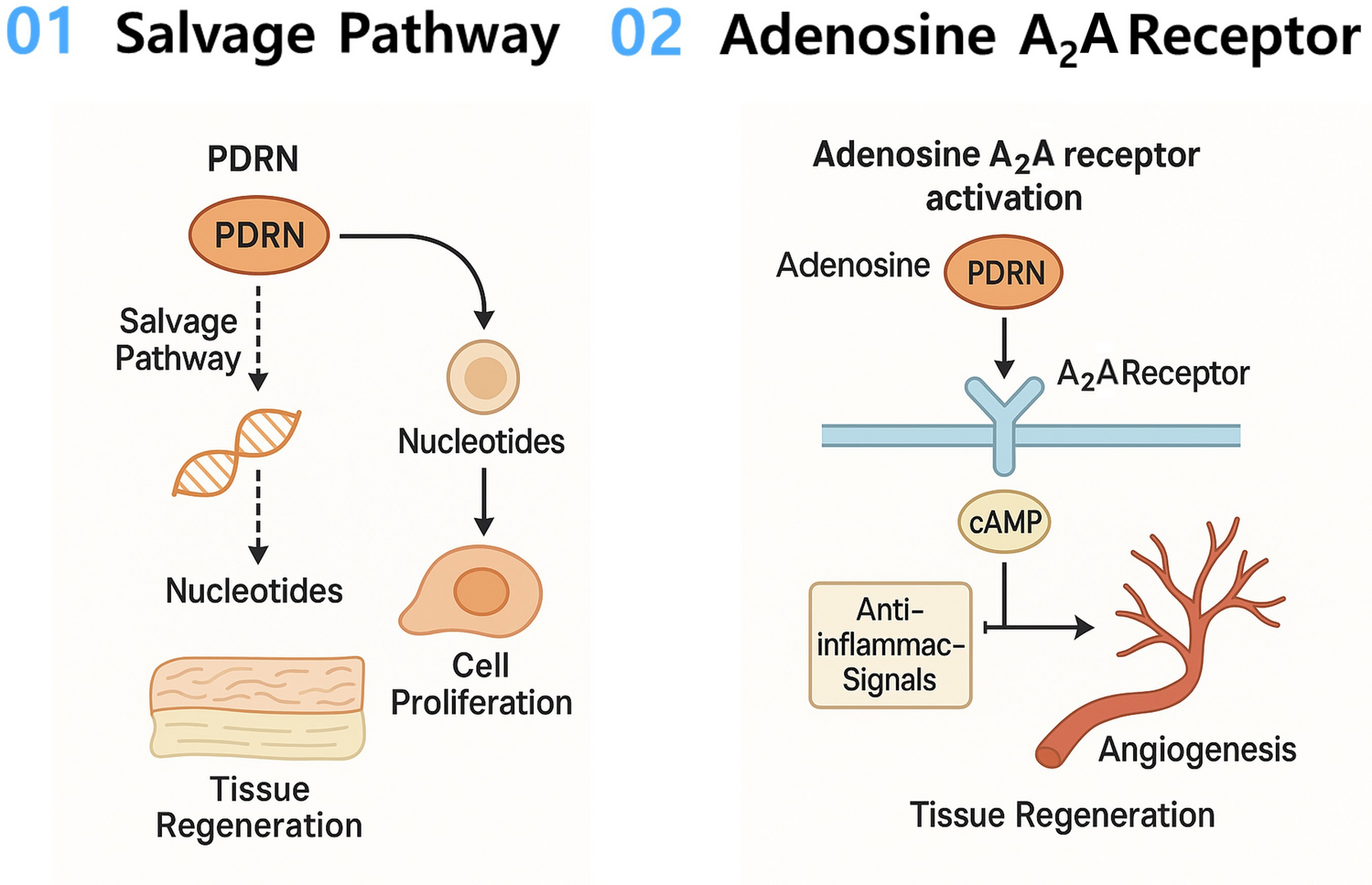
Growth factors and other biologics have emerged as valuable treatments for some Achilles injuries through cell-free approaches that involve injecting substances such as platelet-rich plasma (PRP) and platelet-rich fibrin (PRF), all derived from the patient’s blood and containing various beneficial growth factors [41]. PRP has become a commonly used treatment in clinical practice, including the treatment of AT injuries. Autologous PRF matrices showed promising results after surgical procedures with faster functional recovery in 6 athletes [42]. Similarly, a prospective randomized controlled clinical trial showed similar results after surgical procedures using the end-to-end Krackow suture method, indicating that functional performance was better in the PRP group [43]. While these studies suggest that PRP and PRF treatments improve postoperative functional recovery, their sample sizes are relatively small. To overcome this limitation, larger-scale trials are needed to validate these findings. In a randomized controlled trial using PRP without surgical AT treatment, multiple PRP injections did not show significant tendon healing or improve the outcomes (patient-reported, functional, or clinical) [44]. This study indicates that using PRP alone or alongside surgical treatment can impact the outcome. Growth factors such as TGF-β [45], BMP [46], IGF [47], FGF [48], PDGF [49], and VEGF [50] have been individually studied and applied in tendon regeneration. They play crucial roles in repair and regeneration by recruiting and differentiating cells, synthesizing ECM, and promoting angiogenesis [51]. Among their essential roles and advantages, they have short, effective half-lives and are volatile [51].
Current research in this area is increasingly exploring the potential of cell-based therapies for treating AT injuries, using cells sourced either from the patient’s own body (autologous) or from other donors, including allogenic or xenogeneic sources. The cell types used in these therapies can be stem cells, such as mesenchymal stem cells, or differentiated cells, such as tenocytes [52]. Tenocytes are abundant cells in the tendon microenvironment, playing a crucial role in tendon healing. They create the microenvironment by secreting specific growth factors and rebuilding the ECM during healing. Furthermore, stem cells are extensively studied for their potential to repair and regenerate various tissues. This is due to their multipotent or pluripotent features, which allow them to differentiate into various cell types. Different types of stem cells have been studied for AT regeneration, including tendon-derived (TDSCs) [53], adipose-derived (ADSCs) [54], bone marrow-derived (BMSCs) [55, 56], and induced pluripotent stem cells (iPSCs) [57]. Some stem cells, including mesenchymal stem cells, possess properties beyond their multipotency, including immunomodulatory and self-renewal capabilities. Although MSCs offer benefits, stem cells must be properly characterized and tested before clinical use. Particularly, the allogenic and xenogeneic stem cells, which are not derived from the patient’s tissues. Transplanting these cells may result in adverse effects, such as rejection through donor-host disease transfer or graft failure. Concerns regarding the use of pluripotent stem cells can include the risk of forming teratomas, necessitating further research to identify an optimal method for ensuring their viability and safe use in cell therapies [58].
1.3 Tissue engineering of the Achilles tendonTissue engineering is an emerging field that aims to improve the regeneration of the AT using various combinations of cell therapy, biological therapy, and biomaterial scaffolds for guided tissue regeneration. During AT healing and regeneration, the quality of tissue repair is influenced by biomechanical forces, tissue structure, endogenous cells, and ECM composition [59]. The goal of Achilles tissue engineering is to optimize tendon healing and repair. This involves using biomaterial scaffolds to provide functionality, as well as incorporating cells and growth factors to create a microenvironment that supports tendon healing.
Tissue engineering is a multidisciplinary field combining expertise from material science, biomedical engineering, and cell biology to develop constructs for tissue repair and regeneration [2]. Understanding tissue characteristics and biomechanics is essential for designing a scaffold that accurately reflects tissue properties, mimics tissue architecture, and restores functionality [60]. One approach to tissue engineering for guided tissue regeneration involves the creation of scaffolds that facilitate the regeneration. These scaffolds can also serve as delivery vehicles for cells, growth factors, or other bioactive particles. To achieve this, various manufacturing methods can be employed, including 3D printing, electrospinning, electrospray, surface deposition, and molding [61, 62]. The selection of materials, solvents, and scaffold fabrication methods influences their properties throughout the entire process. Optimizing polymers, material preparation methods, and manufacturing techniques is essential for advancing guided tendon regeneration. Once these are determined, scaffolds can be developed and tested in vitro to characterize their mechanical properties before they are introduced into animal testing for feasibility and validation studies. This process helps characterize the material and provides an opportunity for further modifications. Following material development, cells and biologics can be introduced to the scaffold in vitro to create an enhanced construct for tissue engineering. Cells and biologics have a crucial role in establishing a tissue-specific microenvironment that serves to create an idealized healing environment for tissue repair and regeneration. The selection of materials, cell types, and beneficial biologics is based on the specific 3D matrix with ECM required for tissue regeneration. Following in vitro cytocompatibility testing, in vivo biocompatibility evaluations are essential to assess whether the scaffold causes cell stress or apoptosis, potentially leading to adverse reactions in the body. Additionally, functionality tests must confirm that the implanted construct performs effectively in vivo.
1.3.1 Biomimetic constructsTendon tissue engineering has used different polymers to develop scaffolds. These polymers are classified into three categories: natural, synthetic, and acellular. Natural polymers are derived from natural sources and utilized in tissue engineering constructs. For example, alginate is a natural polymer derived from the cell walls of brown algae and is used in various regenerative constructs. Additionally, synthetic polymers are created artificially and are commonly used in tissue engineering because they can be tuned and modified to achieve specific desired properties. Furthermore, acellular matrices containing ECM proteins are derived from tissues that are decellularized and processed using special techniques. These matrices can be used as scaffolds to mimic the target microenvironment and are designed to mimic the AT structure. These various polymers can be used alone or combined to create a composite biomimetic scaffold that mimics the properties of AT. Composite scaffolds are then combined with biologics or different cell types to develop biomimetic constructs that enhance tissue repair and regeneration.
1.3.2 Natural materialsNatural polymers are commonly used in tendon tissue engineering due to their biocompatibility and bioactivity. Materials such as collagen, hyaluronic acid, fibrin, chitosan, and cellulose are natural polymers and can be applied individually or as composites [63], though achieving the required mechanical strength, particularly for the AT, remains challenging. Nonetheless, numerous studies demonstrate that scaffold composition and architecture can significantly improve both biological and mechanical outcomes. For example, silk fiber scaffolds, filled with silk fibroin/collagen [64] or aligned collagen fibers, produced by advanced extrusion techniques [65], closely mimic the native tendon’s microstructure and show enhanced in vivo cell response, differentiation, and functional recovery. Due to enhanced biomimicry from aligned fibers, the scaffolds combined with BMSCs exhibited higher tensile mechanical properties, including maximum tensile stress and Young’s modulus, compared to random fibers, 5.02 ± 0.18 vs 1.92 ± 0.17 MPa and 34.52 ± 8.19 vs 10.51 ± 1.42 MPa. These findings demonstrate that not only are the materials used in constructs and their tunability important, but also the way the materials are manufactured, and their architecture must be considered, as this significantly influences mechanical properties, cell response, and functionality. Another study compared chitosan-based asymmetric scaffolds seeded with tendon stem/progenitor cells (TSPCs) with the empty defect and showed reduced peritendinous adhesion, while promoting cell proliferation and tenogenic gene expression, leading to improved mechanical strength (significantly higher Young’s modulus compared to the empty defect) and collagen fiber alignment in animal models [66]. These results demonstrated that the chitosan scaffold, particularly when seeded with TSPCs, enhanced tendon regeneration, showing promise as a therapeutic strategy for AT repair. Another silk fibroin scaffold, created using a combined micellization and fibrillation process to mimic tendon structure with highly oriented nanofibrils, achieved strength (6 ± 1 MPa) and stiffness (12.0 ± 1.3 MPa) that closely match those of the AT [67].
Novel approaches are being explored for Achilles tissue engineering, including ostrich eggshell membrane (ESM) combined with PRP. This combination promoted collagen synthesis, collagen organization in newly formed tissues, and gait recovery in vivo [68] (Table 1). However, the group with only the ESM scaffold, without PRP, showed a higher inflammatory response, greater calcification, and the lowest mechanical properties. Additionally, ESM combined with PRP demonstrated increased ultimate (a.k.a. breaking or failure) strength (48.91 ± 15.56 MPa), but decreased Young’s modulus (20.90 ± 7.53 MPa) compared to the normal tendon, while the measurements between ESM and ESM + PRP did not show significant differences. Furthermore, a novel bioactive hydrogel was produced from salamander skin secretions, and it exhibited both antibacterial and antioxidant properties; the hydrogel reduced peritendon adhesion but did not eliminate it [69]. Moreover, the regenerated tendons in rats showed a modulus closer to normal tissue, but their ultimate tensile strength remained weaker (7.1 ± 1.9 MPa) than that of the normal tendons (14.0 ± 5.8 MPa). There has been significant progress in developing hydrogels that trigger gelation at body temperature when injected, known as thermosensitive hydrogels. For example, in a study, researchers demonstrated that a thermosensitive chitosan/collagen hydrogel combined with MSCs supported regeneration in the AT injury model by promoting anti-inflammatory and pro-repair signaling pathways [70]. In addition to their improvements, the hydrogel + MSCs group showed enhanced tensile strength in a rat model, close to that of the uninjured tendon, and higher than that of the hydrogel-only group.
Table 1 Different materials, including natural, synthetic, or composite, along with various designs and fabrication methods, like printing techniques and the mechanical properties (Young’s modulus, E and Ultimate/Tensile strength, UTS) are employed to replicate the aligned structure and create biomimetic constructs for Achilles tendon regeneration (Permissions have been obtained for the use of images from published articles)Taken together, these advances underscore that the success of tendon regeneration depends not only on the choice of natural polymer but also on scaffold design, fiber alignment, and incorporation of biological cues. While many constructs demonstrate improved mechanical properties and cellular responses in preclinical models, further work is needed to refine these biomaterials for consistent translation to clinical practice.
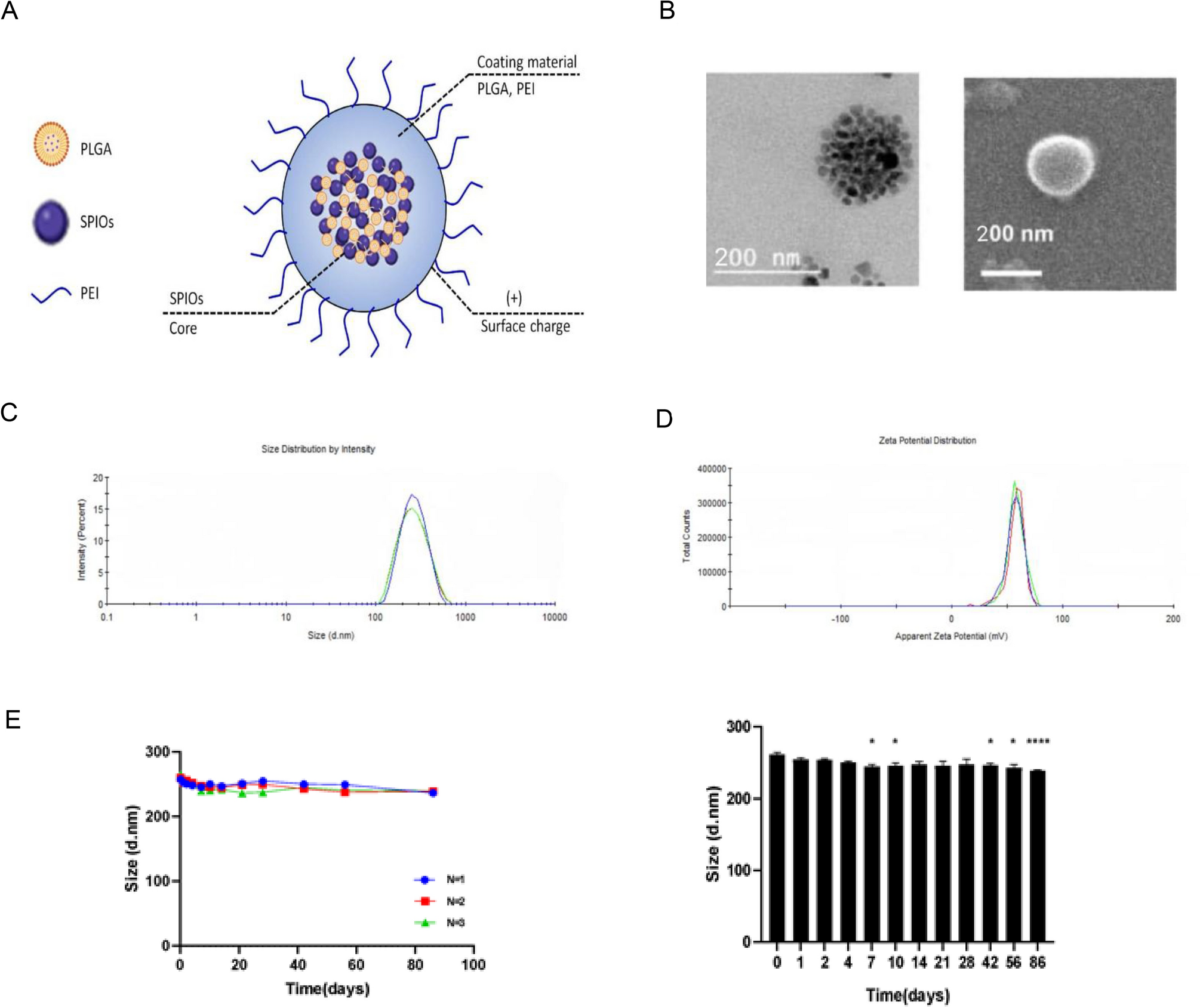
1.3.3 Synthetic materialsSynthetic polymers are widely used in scaffold design for regenerative medicine due to their tunable properties and ability to mimic tissue biomechanics. Materials such as poly(lactic-co-glycolic acid) (PLGA), polylactic acid (PLA), polycaprolactone (PCL), and polyglycolide (PGA) are tested for tendon engineering with attention to biodegradability, toxicity, cytocompatibility, biocompatibility, and biomechanics. Scaffold development follows a staged process from preparation to in vitro and in vivo testing.
Recent advances highlight the value of piezoelectric polymers for their anti-inflammatory properties and aligned scaffold structures for mimicking AT that influence cell response. For example, piezoelectric PCL combined with tetragonal barium titanate nanoparticles (BTO) promoted anti-inflammatory effects, collagen deposition, and functional recovery in AT models [71]. In contrast, disorganized collagen structures, immature collagen, and fibrotic scar tissue were observed in PCL groups. Also, mechanical properties such as tensile strength, Young’s modulus, and maximum load were higher in PLC/BTO (24.66 ± 1.02, 49.59 ± 2.90 MPa, respectively) than in PCL alone (17.81 ± 0.85, 40.93 ± 3.75 MPa, respectively). These findings demonstrate the potential of piezoelectric nanofilms containing BTO to promote tendon regeneration. Another piezoelectric polymer, polyester-based elastomers, enhanced biomechanical strength with an elastic modulus as low as 0.3 MPa and recoverable strain up to 300%, enabling it to withstand Achilles’ motion [72]. They suggested that the scaffold reduced the risk of re-rupture by improving biomechanical properties and demonstrating biocompatibility, with no inflammatory heat response around the implantation site. While another study with hybrid PLA/PGA scaffolds modified with hyaluronic acid also showed improved mechanical
Comments (0)