
Remember me
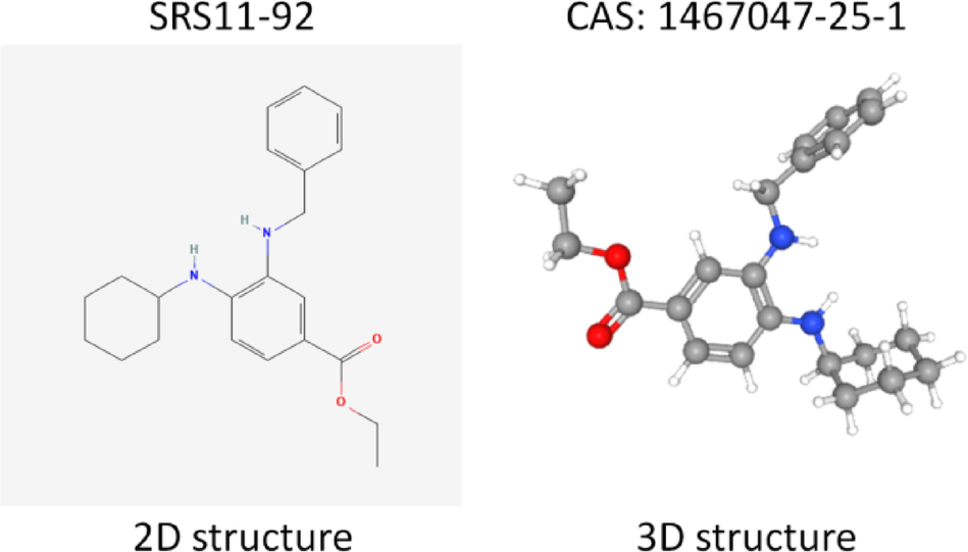
MEM/F12 was purchased from Procell (Wuhan, China). Fetal bovine serum (FBS) and penicillin/streptomycin were obtained from Boster Biological Technology (Wuhan, China). The treatment reagent Aβ25-35 was obtained from MedChem Express (New Jersey, USA). SRS11-92 and ML385 were obtained from Selleck (Houston, USA). The chemical structure of SRS11-92 is shown in Fig. 1. The senescence-associated β-galactosidase (SA-β-gal) staining kit and ROS assay kit were obtained from Beyotime (Shanghai, China). DMSO was purchased from Sigma-Aldrich (St. Louis, MO, USA). The MTT assay kit was obtained from Servicebio (Wuhan, China). ELISA kits for MDA and SOD were purchased from Solarbio (Beijing, China). ELISA kits for GSH, TNF-α, IL-1β, and IL-6 were purchased from Elabscience (Wuhan, China). NE-PER Nuclear and Cytoplasmic Extraction Kit was obtained from Thermo Fisher Scientific (Waltham, MA, U.S.A.). Antibodies for p53, p16, HO-1, GAPDH and β-actin were purchased from Proteintech (Chicago, IL, USA). The antibody for p21 was obtained from Abcam (Cambridge, UK). Antibodies for Nrf2, Histone H3 and NF-κB p65 were purchased from CST (Massachusetts, USA.).
Fig. 1
Chemical structure of SRS11-92
Cell culture and treatmentSTR-authenticated SH-SY5Y cells were purchased from Procell (Wuhan, China) and cultured in MEM/F12 containing 15% FBS and 1% penicillin–streptomycin under 5% CO2 at 37 °C. As described previously (Huang et al. 2024), Aβ25-35 was diluted to 1 mM with sterilised deionised water and then incubated for 1 week at 37 °C to induce aggregation. For experiments, it was diluted in culture medium to the indicated concentrations; vehicle (deionised water) served as control. Cells were incubated with SRS11-92 and/or ML385 at the indicated concentrations.
Cell viability analysisSH-SY5Y cells were seeded into 96-well plates at a density of 5 × 10³ cells/well and incubated for 24 h. Cell viability was assessed using the MTT assay according to the manufacturer’s instructions. To evaluate Aβ25-35 cytotoxicity, cells were treated with 0, 2.5, 5, 10, 20, and 40 μM for 48 h. To determine SRS11-92 toxicity, cells were treated with 0, 0.25, 0.5, 1, 2, 4 μM for 48 h. To assess the protective effect of SRS11-92 against Aβ25-35-induced cytotoxicity, cells were pretreated with SRS11-92 (0, 0.25, 0.5, 1, 2, 4 μM) for 4 h, followed by exposure to 20 μM Aβ25-35 for an additional 48 h. After treatment, the medium was removed, and cells were incubated with 10 μL of MTT working solution at 37 °C for 4 h. Formazan crystals were dissolved in DMSO, and absorbance was measured at 570 nm using a microplate reader. Cell viability was expressed as a percentage relative to the vehicle control.
Morphological observationsSH-SY5Y cells in the logarithmic growth phase were seeded at 1 × 10⁵ cells/mL in 6-well plates and allowed to adhere for 24 h. Cells were then treated with SRS11-92 (0, 0.25, 0.5, 1, 2, or 4 μM) for 4 h, followed by exposure to 20 μM Aβ25-35 or vehicle for an additional 48 h. Cell morphology was assessed, and images were acquired on a phase-contrast microscope (Olympus, Tokyo, Japan).
ROS generation analysisROS levels were measured by dichlorodihydrofluorescein diacetate (DCFH-DA) using a ROS Detection kit (Beyotime, Shanghai, China) as previously described (Xie et al. 2023). After treatments, cells were incubated with 10 μM DCFH-DA solution at 37 °C for 30 min, rinsed with PBS to remove non-specific staining, and imaged under a fluorescence microscope (Olympus, Tokyo, Japan).
Intracellular superoxide dismutase (SOD), reduced glutathione (GSH), and Malondialdehyde (MDA) determinationMDA, SOD, and GSH levels were determined using the respective assay kits according to manufacturers’ protocols. A bicinchoninic acid (BCA) protein assay kit (Beyotime, Shanghai, China) was used to measure the protein content. Absorbance was measured with a microplate reader as above.
Inflammatory cytokines assayCulture supernatants were collected after 48 h and clarified by centrifugation. TNF-α, IL-1β, and IL-6 were measured by ELISA according to the manufacturer’s instructions.
Total and nuclear protein extractionSamples were washed with ice-cold PBS. For whole-cell lysates, cells were extracted in RIPA buffer with protease and phosphatase inhibitors. For subcellular fractionation, nuclear and cytoplasmic extracts were prepared using the NE-PER nuclear and cytoplasmic extraction kit (Thermo Fisher Scientific, Waltham, MA, USA) according to the manufacturer’s instructions. Lysates were cleared by centrifugation (12,000 g, 10 min, 4 °C). Protein concentration was determined by the BCA assay prior to adding sample buffer. Equal amounts of protein were mixed with loading buffer, heated at 95 °C for 5 min, and used immediately or snap-frozen at − 80 °C.
Western blottingA total of 20 μg protein was separated by 8–12% SDS-PAGE, transferred to PVDF membranes, and blocked with 5% skimmed milk for 1 h at 37 °C. Membranes were incubated with primary antibodies overnight at 4 °C. After washing with TBST, membranes were incubated with HRP-conjugated secondary antibodies for 1 h. Signals were detected using a Bio-Rad ChemiDoc XRS + system with a high-sensitivity ECL kit, and quantitative analysis was carried out using ImageJ software.
Animal treatmentHomozygous 3xTg-AD mice (APPswe /PS1m146v/TauP301L) were obtained from Jackson Laboratories, with age-matched wild-type controls (hybrid progeny of C57BL/6 J × 129S1/SvImJ) (Hamilton et al. 2022). Procedures complied with institutional and national guidelines and were approved by the relevant ethics committees. Mice were group-housed at 23 ± 2 °C, 60–70% humidity, 12-h light/dark cycle, with ad libitum food and water. In this study, mice were 12 months old at baseline. Animals were randomly assigned to WT + Vehicle, WT + SRS11-92, AD + Vehicle, and AD + SRS11-92. SRS11-92 was dissolved in 5% DMSO and 95% corn oil and administered intraperitoneally at 2 mg/kg. Vehicle groups received equal volumes of solvent. After behavioural testing, mice under deep anaesthesia were killed by transcardial perfusion with pre-cooled 0.9% physiological saline. Brains were removed and stored at − 80 °C. Serial coronal frozen sections (20 μM) were cut using a cryostat (CM1950, Leica Biosystems, Wetzlar, Germany) and stored at − 80 °C.
Novel object recognition (NOR) testingThe NOR test was conducted according to established protocols with minor modifications (Cantarella et al. 2015). Before testing, mice were acclimatised to the behavioural room. Habituation: mice explored an empty 40 cm × 40 cm × 40 cm arena for 10 min. Training (24 h later): two identical objects were placed and mice explored for 10 min. Testing (1 h later): one familiar object was replaced with a novel object; exploration continued for 5 min. Outcomes (total distance, average speed, number of object-exploration bouts, and time spent exploring novel and familiar objects) were recorded with ANY-MAZE (Stoelting Co., USA). Preference index = A/(A + B) (A: time with the novel object; B: time with the familiar object). Discrimination index = (A − B)/(A + B).
Nissl stainingCryosections were equilibrated to room temperature and incubated in pre-warmed 0.1% toluidine blue O solution (acetate buffer, pH 4.0) at 60 °C for 10 min. Sections were differentiated in 95% ethanol containing 0.1% acetic acid for 15 s, rinsed, dehydrated through graded ethanol, and cleared in xylene. Sections were mounted with neutral balsam. Bright-field images were captured under identical settings.
SA-β-gal stainingSA-β-gal staining was performed on cells and/or coronal sections using the manufacturer’s kit (pH 6.0). Samples with fresh SA-β-gal staining solution were incubated overnight at 37 °C in a non-CO₂ incubator. Images were acquired on a light microscope. For cells, the percentage of SA-β-gal-positive cells was calculated as (blue-stained/total) × 100%.
Statistical analysisAll experiments were conducted at least in triplicate. Results are presented as mean ± SD and analysed using GraphPad Prism 10. Comparisons among three or more groups were performed using one-way ANOVA followed by Tukey’s post hoc test. p < 0.05 was considered statistically significant.
Comments (0)