
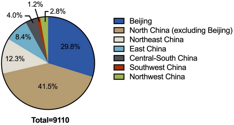
Remember me


The gene TFRC controls transferrin receptor-1 (TfR1) expression. Previous investigations identified pancreatic tissue antibody biomarkers using quantitative serum proteomics. TfR1 protein on cancer cell surfaces increases with TFRC expression, which is strongly related to poor prognosis [7, 12].
The TCGA database has just four normal pancreatic tissue cases. Thus, the expression differential of TfR1 was examined by comparing genetic data from 177 pancreatic cancer patients in the TCGA database with normal pancreatic data from 169 cases (165 GTEx cases and 4 TCGA cases). Figures 1A and B show the analysis results. The expression of TFRC mRNA in pancreatic cancer tissues was substantially higher than in normal tissues (P < 0.05). ROC curve research showed that TFRC's AUC was 0.936, with a 95% confidence interval of 0.905 to 0.962. TFRC expressions accurately distinguish pancreatic cancer disease from normal groups.
Fig.1
A Differences in the expression of TFRC gene between pancreatic cancer group and normal group (TCGA combined with GTEx analysis); B ROC assesses the diagnostic efficacy of TFRC gene expression in tumor versus normal groups (TCGA combined with GTEx analysis)
In pancreatic cancer, TFRC mRNA expression was raised, and its ROC curve showed its accuracy in predicting prevalence. Immunohistochemistry, Western blot, and immunofluorescence assays were used to confirm the enhanced protein expression of TfR1 in clinical samples and in vitro cultivated cell lines.
Expression of TfR1 in pancreatic cancer tissues and cellsFigure 2 shows TfR1 immunohistochemical staining in pancreatic cancer and adjacent tissues, showing abundant TfR1 protein in the cell membrane of cancerous tissue, which has a brownish-yellow or tan granular morphology and some cytoplasmic expression. Staining of neighboring tissues showed minimal TfR1 protein expression.
Fig. 2
Transferrin receptor-1 (TfR1) (IHC × 40, IHC × 200). A. the picture shows the negative expression of antibodies to paracancer, and picture B. shows the high expression of antibodies to paracancer; C. High expression of antibodies in well-differentiated cancer tissues, and high expression of antibodies in poorly differentiated cancer tissues in Fig D
After finding that tissue detachment had occurred in 7 cancer and 9 paracancerous tissue samples during the baking and dewaxing procedures of the IHC experiment, this study comprised 83 pancreatic cancer sites and 81 paracancerous cases. Immunohistochemical staining findings were categorized by high and low expression levels to reduce false positives and bias. The high expression rate of TfR1 in pancreatic cancer tissues was 30.1% (25/83), compared to 11.1% (9/81) in paracancer tissues, being statistically significant (P = 0.003). See Table 1 for information.
Table 1 Expression of transferrin receptor-1 in pancreatic cancer (%)Five pancreatic cancer cell lines (PANC-1, SW1990, KP4, Mia paca2, Bxpc3) and one normal pancreatic ductal epithelial cell (HPDE6-C7) were compared to see if TfR1 is substantially expressed in each. Western blot and immunofluorescence assay revealed a substantial difference (P < 0.05) in TfR1 expression between normal pancreatic ductal epithelial cell lines and five pancreatic cancer cell lines (Figs. 3 and 4).
Fig. 3
Western blot detection and gray value analysis of TfR1 expression levels in normal pancreatic ductal epithelial cells compared with five pancreatic cancer cells (Note: *P < 0.05; **P < 0.01; ***P < 0.001, ##: vs. PANC-1)
Fig. 4
Immunofluorescence detection and gray value analysis of TfR1 expression levels of five pancreatic cancer cells compared with normal pancreatic ductal epithelial cells (Note: *P < 0.05; ** P < 0.01; P < 0.001)
Immunohistochemical staining, Western blot, and immunofluorescence assays showed that pancreatic cancer clinical samples and cell lines had much higher TfR1 protein expression than normal pancreatic tissues and cells. This finding supports TCGA database predictions and emphasizes the importance of studying TfR1 and pancreatic cancer clinicopathology.
Correlation analysis of TfR1 expression with clinicopathological characteristics in pancreatic cancer patientsClinical features of 83 pancreatic cancer patients were categorized and analyzed according to the 2022 criteria for diagnosis and therapy [16]. The results showed a strong association between TfR1 expression and M, TNM, and tumor maximum diameter. With statistical significance (P < 0.05), TfR1 expression increased with later M stages, advanced TNM stages, and tumors larger than 4 cm in diameter. All statistically significant results (p < 0.05) are highlighted in bold within the Table 2.
Table 2 Correlation analysis between TfR1 expression and clinicopathological data of pancreatic cancer patientsRelationship between TfR1 expression and Prognosis in Pancreatic cancer tissuesFigure 5 shows survival differences between high and low TfR1 expression groups using the Kaplan–Meier curve and Log-rank test. In the TCGA sample, the TFRC mRNA high expression group had a significantly poorer overall survival rate than the low expression group (P = 0.005). Overall survival (OS) and progression-free survival (PFS) trends of 83 patients in the clinical sample matched the TCGA database, indicating statistical significance (P < 0.001). This shows that high TfR1 expression may indicate a poor prognosis.
Fig. 5
K-M survival curves in pancreatic cancer for TfR1: A. OS survival curve of TCGA-samples, B. OS survival curves of clinical samples, and C. PFS survival curves of clinical samples
TfR1 expression enhanced disease progression and death. By the final follow-up date, 27 patients were alive and 56 had died, with elevated TfR1 protein levels associated with a higher death rate (P = 0.009). The median OS for 83 pancreatic cancer patients was 22.00 months. The median OS for the TfR1 low expression group was 37.50 months, while the high expression group was 9.00 months. The high-expression group had considerably shorter survival times (P < 0.001). Table 2 shows that patients with higher TfR1 expression had a significantly lower progression-free survival (PFS) of only 6 months, like overall survival (OS) (P < 0.001).
Cox regression analysis of prognostic factors for overall survival in patients with pancreatic cancerOf the 83 pancreatic cancer patients studied, 56 died and 27 survived at the last follow-up. Cox regression was used to analyze the connection between TfR1 expression levels in pancreatic cancer tissues and clinicopathological characteristics, with overall survival as the result. Univariate Cox regression analysis found that nine clinical features, including pathological classification, follow-up grade, adjuvant chemotherapy, TNM stage, tumor diameter, duodenal/gastric invasion, CA125, and TfR1 expression levels, significantly correlated with patient survival (P < 0.05). See Table 3 for information, where significant results are highlighted in bold. The nine clinical parameters strongly linked with overall survival (OS) were used as independent variables in multivariate Cox regression analysis. The study found that pathological categorization, follow-up grade, adjuvant chemotherapy, and TfR1 expression level are independent risk factors for OS prognosis (P < 0.05). Patients with high TfR1 expression exhibited a 2.913-fold higher risk of death than those with low TfR1expression, as shown in bold in Table 4.
Table 3 Univariate Cox regression analysis of prognostic factors for overall survival in patients with pancreatic cancerTable 4 Multivariate Cox regression analysis of prognostic factors for overall survival in patients with pancreatic cancerCox regression analysis of prognostic factors for progression-free survival in patients with pancreatic cancerProgression-free survival (PFS) is the time from randomization to tumor progression or death from any cause. Of 83 pancreatic cancer patients, 21 had no disease progression, 19 with low TfR1 protein expression, and 2 with high expression. A total 62 patients experienced disease progression or death. Of these, 39 with low TfR1 expression and 23 with high expression. TfR1 expression was linked to a higher risk of disease progression, with statistical significance (P < 0.05) as shown in bold in Table 2.
Progression-free survival (PFS) was used to assess the association between TfR1 expression levels in pancreatic cancer tissue and clinicopathological characteristics. We performed univariate and multivariate Cox regression analysis. Univariate Cox regression analysis revealed a significant association between seven clinical characteristics and progression-free survival (P < 0.05): pathological categorization, follow-up grade, TNM stage, maximum tumor diameter, gastric/duodenal invasion, and TfR1 expression. Multivariate Cox regression analysis of the seven clinical parameters related to PFS showed that pathological classification, pathological grade during follow-up, maximum tumor diameter, and TfR1 expression level are independent risk factors for PFS prognosis (All significant results are highlited in bold within the Tables 5 and 6).
Table 5 Univariate Cox regression analysis of prognostic factors for progression-free survival in patients with pancreatic cancerTable 6 Multivariate Cox regression analysis for prognostic factors for progression-free survival in patients with pancreatic cancer
Comments (0)