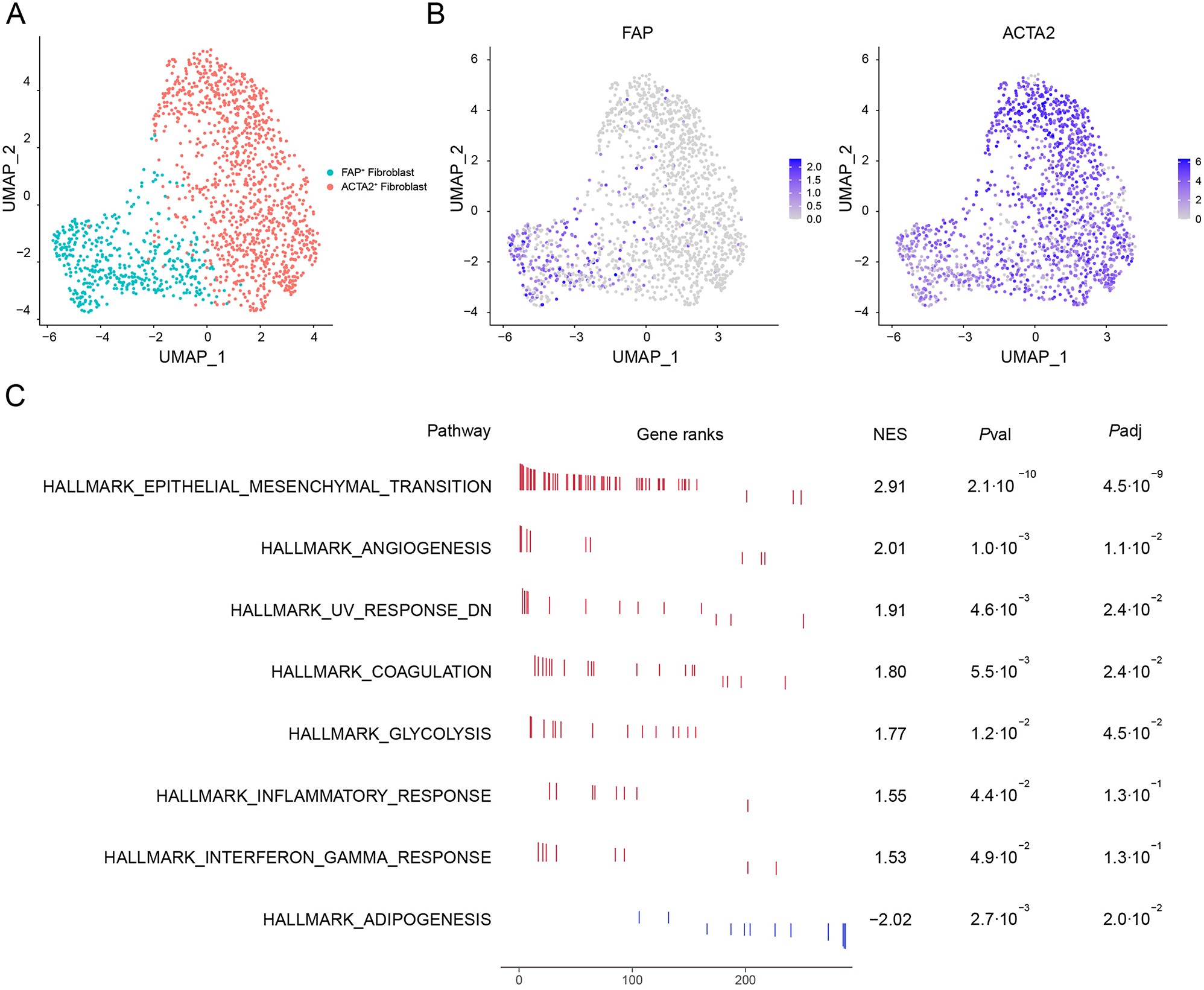
The Sedlis criteria form the basis for determining postoperative adjuvant therapy administration in early- to mid-stage cervical squamous cell carcinoma. However, these criteria remain controversial, as some scholars argue that adjuvant radiotherapy does not significantly improve prognosis in patients with intermediate-risk factors [3]. This retrospective analysis evaluates the prognostic impact of key pathological risk factors following cervical cancer surgery.
Lymph node metastasis represents an independent prognostic factor at all stages of tumour progression. This clinical observation can be explained by several mechanisms: when tumour cells disseminate through the lymphatic system, local treatment approaches (including surgery and postoperative radiotherapy) frequently prove inadequate for complete eradication. Residual tumour lesions may persist beyond the surgical lymphadenectomy field or outside radiotherapy target volumes, ultimately leading to treatment failure. Current research indicates that expression patterns of multiple biomarker genes show strong correlation with lymph node metastasis, suggesting that lymphatic spread in cervical cancer involves mechanisms beyond simple tumour progression. These biomarkers facilitate lymph node metastasis through distinct molecular pathways [4,5,6,7,8]. Consequently, the development and progression of lymph node metastasis follow independent biological mechanisms, further confirming its status as an independent prognostic factor.
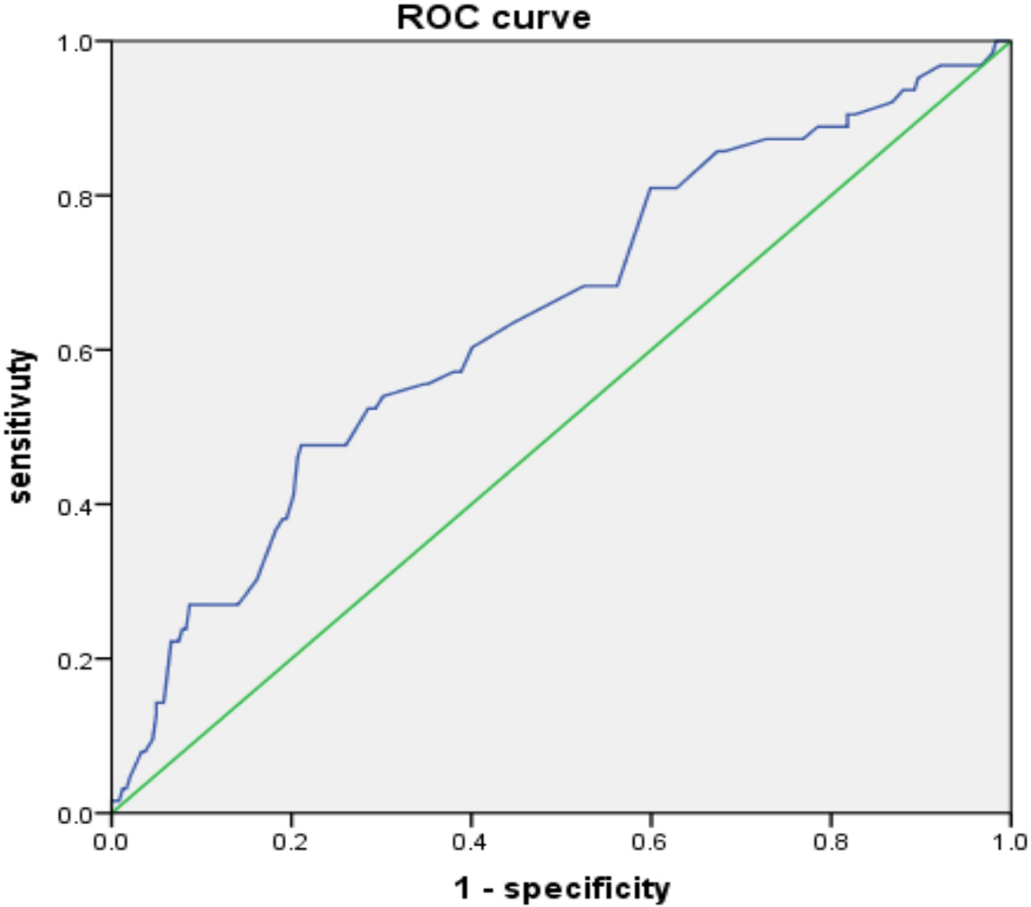
This retrospective analysis identified tumour three-dimensional volume as an independent prognostic factor for 5-year survival in cervical cancer patients with full-thickness stromal infiltration (reaching the serosa). Existing literature supports this finding: one study utilising pre-treatment PET-CT measurements of tumour volume during concurrent chemoradiotherapy similarly established tumour volume as a key prognostic determinant [9]. Although that study involved advanced-stage patients undergoing chemoradiotherapy rather than surgery, its findings corroborate that three-dimensional tumour volume exerts greater prognostic influence in deeply infiltrative, late-stage disease. Additional studies have confirmed maximum tumour diameter, volume and vaginal involvement as significant predictors of recurrence rates [10], consistent with our findings. In cases of full cervical wall infiltration, the serosal barrier redirects tumour spread towards cervical, vaginal and uterine body tissues rather than uterine invasion. Extensive stromal and muscular layer infiltration elevates lymph node metastasis potential, adversely affecting survival outcomes. Obtaining clear surgical margins remains critical for recurrence prevention, as larger tumour volumes complicate resection, increase residual tumour likelihood and amplify recurrence risk [11]. For patients with 100% cervical wall infiltration, three-dimensional tumour volume demonstrates superior prognostic value over maximum diameter alone, substantially increasing five-year mortality risk.The data distribution of higher-volume tumor subgroups shows non-normality, likely caused by the presence of some patients with disproportionately large tumor sizes.
Previous studies have demonstrated that vascular invasion is an independent prognostic factor affecting overall survival, particularly in patients without lymph node metastasis [12, 13]. However, these studies primarily refer to vascular invasion distant from the primary lesion, rather than vascular involvement directly adjacent to it. The aforementioned research identifies vascular invasion as an independent risk factor for prognosis, specifically in cases of satellite vascular invasion, defined by the presence of tumour cell nests within vessels at a discernible distance from the primary lesion. Conventional vascular invasion refers to vascular infiltration confined to the primary tumour lesion, which has a relatively limited impact on prognosis. This retrospective analysis revealed that the prognostic significance of vascular invasion differed between the two groups, as the pathologists in our hospital were unable to specify the number and types of vascular involvement. Extensive vascular invasion and microscopic single-vessel invasion exert distinct effects on patient prognosis. The former is more likely to progress to lymph node metastasis or distant metastasis, significantly influencing patient outcomes. Furthermore, when tumour cells invade blood vessels at a distance from the primary lesion, this suggests strong metastatic and invasive potential, directly impairing patient survival. Conversely, vascular invasion restricted to the peritumoral region indicates a more limited capacity for long-distance invasion or metastasis, resulting in a weaker impact on survival. This retrospective analysis did not permit a detailed assessment of the extent or distance of vascular invasion from the primary lesion. Consequently, vascular invasion could not be established as an independent prognostic factor in either patient group. Nevertheless, we observed that vascular invasion remains a key determinant of prognosis in the first group. Similarly, in the second group, vascular invasion was associated with reduced survival rates. Although this study demonstrates the impact of vascular invasion on prognosis in the results, the lack of stratified statistical analysis limits the precise quantification of its impact on survival, as the authors acknowledge.
The presence or absence of vaginal invasion serves as a criterion for distinguishing between stage I and stage II patients. Some scholars have suggested that comparing AJCC T2 staging with T1 staging represents an independent prognostic factor [14]. AJCC T2 staging corresponds to FIGO stage II. Tumours originating from the cervix that extend into the vagina indicate greater stromal and muscular layer infiltration, increasing the likelihood of vascular invasion and lymph node metastasis, which adversely affects patient prognosis. However, when tumour infiltration is superficial, even with vaginal extension resulting in upstaging from stage I to stage II, the extent of stromal and muscular infiltration remains limited. In such cases, the risk of peripheral tissue infiltration is low and the likelihood of complete resection through radical surgery is higher. Consequently, the impact on overall patient survival remains relatively minor.
Furthermore, we observed that tumours exhibiting endophytic growth patterns with ulcerative or erosive morphology and perineural invasion were associated with poorer prognosis. Although these patients demonstrated lower 5-year survival rates, the differences did not reach statistical significance. In both groups, the majority of cases exhibited endogenous, nodular growth patterns without nerve invasion. When subgroup analyses were performed, the limited sample sizes resulted in insufficient statistical power to demonstrate significant differences.
Pathological grading is also a significant factor influencing patient survival, although it is not an independent prognostic indicator. Other researchers have similarly included pathological grading in their survival models, consistent with the findings of this retrospective analysis. While it contributes to prognosis, it does not function as an independent predictive factor [15]. Higher pathological grades generally indicate greater tumour invasiveness, characterised by a tendency for deeper tissue infiltration and vascular or lymphatic invasion, which further adversely affects patient survival. Therefore, although pathological grading may provide supplementary prognostic information, its ability to independently predict survival remains limited.
Shortcomings of this article:
1.
This study represents a retrospective analysis and does not include data on certain novel biological markers that may influence prognosis. Since most cases lacked measurements for these indicators, the accuracy of the survival model may be compromised.
2.
The proposed model requires validation with additional datasets; however, a small sample size may introduce significant bias, whereas large-scale validation may not be feasible in the short term. In future research, the authors aim to validate the model using new data and, if necessary, collaborate with other treatment centres to achieve this.
3.
Certain variables, such as nerve invasion, were observed in relatively few cases across both groups, increasing the risk of statistical deviation. While these findings may provide reference value, their reliability remains limited.
4.
The sample size remains modest, particularly in the second cohort, where mortality events were infrequent.
5.
For patients with vascular involvement, neither the number nor the status of affected vessels was specified, nor were stratified analyses conducted. Consequently, the precision of vascular survival assessment is diminished.
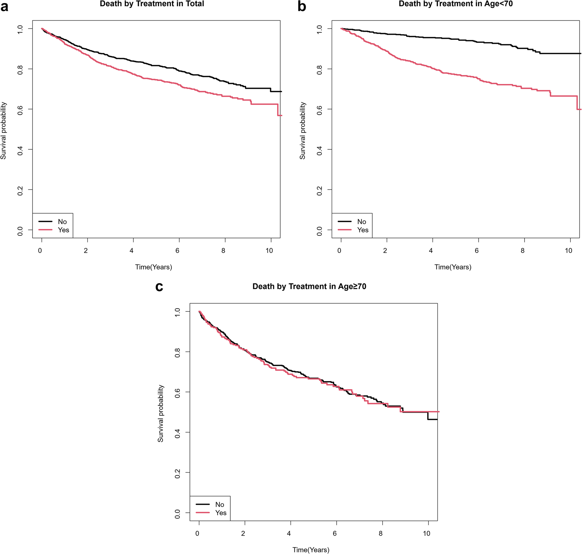
Lymph node metastasis persists as an independent prognostic factor irrespective of tumour infiltration depth. In cases demonstrating deeper infiltration (such as serosal involvement), tumour volume, vaginal involvement, vascular invasion and pathological grading demonstrate enhanced prognostic significance. For these patients, postoperative concurrent chemoradiotherapy should be contemplated even when conventional high-risk factors are absent, with the objective of reducing recurrence rates and improving survival outcomes. Additional studies employing stratified analyses of adjuvant therapies are required to corroborate these findings.


Comments (0)