
Remember me
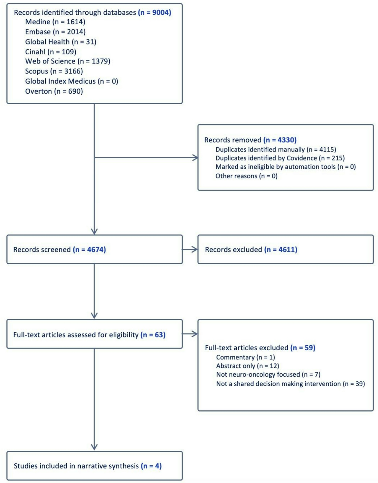
The systematic search across all databases resulted in 9004 records. After the removal of duplicates in EndNote, a total of 4889 records were imported into Covidence. Further duplicates were identified and removed in Covidence, which resulted in 4674 original articles. The screening and selection processes are shown in the PRISMA flowchart (Fig. 1).
Fig. 1
PRISMA (Preferred Reporting Items for Systematic Reviews and Meta-Analyses) Flowchart
Design and characteristics of included studiesFour studies with a total of 172 patients, with diagnoses including anaplastic oligodendroglioma, anaplastic astrocytoma, high-grade glioma (HGG), low-grade glioma (LGG), unspecified glioma, glioblastoma, and brain metastases, met the inclusion criteria (Table 1).
Table 1 Characteristics of included studiesIn the RCT study conducted by El-Jawahri et al. [11] the impact of a goals in care video on decision-making in comparison to a standard verbal narrative was assessed. Patients with malignant glioma were randomized into two groups: a control group of 27 patients who were only given a verbal narrative and an intervention group of 23 patients who were provided with the video supplement [11]. Outcome measures included a cardiopulmonary resuscitation (CPR) preference assessment, knowledge assessment, the uncertain subset within the Decisional Conflict Scale, and a post-intervention preference for care assessment based on four categories: life-prolonging care, comfort care, basic care, and uncertain [11].
The single arm study by van de Belt and colleagues [12] focused on understanding the value of 3D-printed brain tumor models. A total of 11 adult patients with glioma who completed magnetic resonance imaging (MRI) and diffusion tensor imaging (DTI) were all given a full-scale 3D model of their specific brain tumor [12]. To determine the facilitators, barriers, and effects of the 3D models, semi-structured interviews were conducted with patients [12].
The qualitative study by van Diest et al. [13] explored the use of an online tool on the experiences of patient-proxy dyads. Only adult patients with a confirmed diagnosis of glioblastoma and their proxies were included in the study [13]. The online tool was constructed with the aim of informing patients of the progression of the disease. A total of 15 patient-proxy dyads were provided the online tool without instructions during “think-aloud sessions” [13]. Outcome data were collected by semi-structured interviews from a non-blinded research team [13].
The prospective study by Leu et al. [14] assessed the impact of SDM processes on satisfaction of both patients and medical staff. The patient cohort was divided into two groups of patients with intracranial glioma and metastases who were undergoing treatment considerations [14]. The first group consisted of 22 patients before the introduction of SDM, and the second group consisted of 74 patients after the implementation of SDM [14]. The implementation of SDM included training for medical staff and the use of decision grids tailored for three types of intracranial tumors. Satisfaction for patients was measured using the CollaboRATE questionnaire [15,16,17,18] and the Advancing Quality Alliance (AQuA) questionnaire [19] for medical staff.
Methodology quality assessmentAll included studies demonstrated a clear research question, and the collected data from each study enabled the research questions to be answered. The methodology quality assessment for all four included studies is shown in Table 2.
Table 2 Methodology quality assessment of included studiesIn the study conducted by El-Jawahri et al. [11] a computer-based randomization scheme assigned participants into groups. Baseline characteristics in each group were not comparable, as the intervention group had a higher overall mean age, percentage of male participants, and percentage of participants with an “excellent/very good/good” health status compared to the control group. The study’s assigned intervention was adhered to and completed by all participants. As the outcome assessors were not blinded to assigned interventions, there is a high risk of bias within the study.
Van de Belt et al.’s [12] exploratory approach with semi-structured interviews and the data collection method of audio-recording the interviews and transcribing verbatim audio transcripts, and reaching thematic saturation, was appropriate to understand barriers and facilitators of using a 3D brain tumor model. The study’s findings of participants’ experience with the model were adequately derived and interpreted from the data using existing frameworks to classify barriers, facilitators, and effects. There was coherence between the qualitative sources of participants’ interviews and the research team’s data collection, analysis, and interpretation.
The qualitative approach of the study by van Diest et al. [13] using think-aloud sessions and semi-structured interviews, which were audio-recorded and transcribed verbatim in two phases to reach data saturation, was appropriate to determine patients’ and their proxies’ experiences with using the online tool. Data findings based on patient-proxy dyads’ experiences with the online tool were adequately derived and interpreted using thematic analysis. The patient-proxy dyads’ interviews and the research team’s data collection, analysis, and interpretation were all coherent.
In the assessment of Leu et al.’s study [14] it is unclear if participants were a comprehensive representation of the glioma patient population, as the only demographic data provided are the participants’ age (range and mean age) and glioma type. The CollaboRATE and AQuA questionnaires were appropriate outcome measurements for patient and staff satisfaction. While all patient participants completed the CollaboRATE questionnaire, one staff participant did not complete the AQuA after the introduction of the SDM intervention. The customized decision grids were not validated prior to use in the study and were not accounted for as a confounder in the study design and analysis. The SDM intervention was implemented as intended in the study for all participants.
Narrative synthesis of interventionsSDM Training for Medical ProfessionalsIn the study by Leu et al. [14] one lead neurosurgeon was provided an SDM training by National Health Service (NHS) England, which consisted of traditional classroom teaching, discussions, role playing, and patients’ presentations over a period of two days. After completion of the course, the medical staff were cascade-trained with a foundation in SDM and the techniques to use as standard care by the lead neurosurgeon. The exact details of the SDM training course were not provided. After incorporating SDM as a standard care practice, the medical staff’s satisfaction significantly improved from 61.68 to 90.95% on the AQuA questionnaire [14].
Decision gridsDecision grids for three types of intracranial tumors (HGG, LGG, metastases) developed by Leu et al. [14] were based on the National Institute for Health and Care Excellence (NICE) guidelines [20] and an Option Grid template [21]. Each decision grid was composed of specific treatment options based on the type of tumor. Treatment options provided in grids included Best Medical Care (BMC), resection and biopsy for HGG patients; active surveillance, resection, and biopsy for LGG patients; BMC, stereotactic radiosurgery or radiotherapy, and resection for patients with metastases. Benefits and risks (adjusted based on individual patients’ profiles) were provided for each treatment option in the grids. However, the use of decision grids did not result in a significant improvement in patients’ CollaboRATE questionnaire scores [14]. As the decision grids were used in combination with SDM training of medical staff in the study, it is unclear how the grids can directly impact the process of facilitating the SDM for brain tumor patients.
3D brain tumor modelAs mentioned in van de Belt et al.‘s study [ 12 ] 3D models of specific patients’ brain tumors were constructed using an Ultimaker 3D printer with imported functional MRI and DTI images from patients’ imaging scans. Segmented parts were printed in particular colors to identify certain regions of the brain model, including the tumor, corticospinal tract, areas for speech, language, and motor processing. The associate costs for each 3D model were less than $6 USD, but the cost of the 3D printer used was approximately $1741 USD [ 12 ]. In semi-structured interviews, patients reported that the 3D models provided positive effects of improved communication, coping, acceptance, knowledge, and understanding of risks and benefits. In addition, emotional confrontation was reported as a negative effect [ 12 ].
Glioblastoma progression online toolAn online tool that covers the progression of glioblastoma was designed for adult patients and their proxies within the study by van Diest et al. [13]. The online tool provided information on possible symptoms (initial, during recurrent growth, end-of-life phase), anti-tumor treatment, and palliative treatment. In addition, a line plot of tumor size over time was provided with interrupted lines indicating unpredictability in determining the amount of time for tumor recurrence and end-of-life stage. The Phase 1 group mentioned that the content of the online tool provided positive aspects of useful information with a clear overview [13]. Negative aspects that the Phase 1 group mentioned included lengthy, unclear medical jargon and missing information [13]. After revisions to the online tool based on the Phase 1 group’s feedback were implemented, the Phase 2 group reported that information on treatment options for tumor relapse provided hope, and they valued the topic of psychological aspects [13].
Goals of care videoA video supplement was created by El-Jawahri et al. [11] to depict three levels of care for malignant glioma patients in Boston, USA. The six-minute video provided images and scenes for each type of care using a cinema vérité style. In the control group, the participants’ average age was 56 with a range from 47 to 77 years old. The video intervention group had a slightly younger participants’ age average of 51 with a range of 32–72 years old. The implementation of the video resulted in a higher preference for comfort care (91.3%, n = 21) compared to the control group (22.2%, n = 6).11 A higher knowledge assessment score (5.3) was seen with the use of the video intervention compared to the control group (4.6) [11]. Patients expressed a significantly lower willingness to undergo CPR in the video intervention group (8.7%, n = 2) in comparison to the control group (40.7%, n = 11).11 The majority of participants (82.6%) reported being “very comfortable” with the video and would “definitely recommend” it to other cancer patients (82.6%).11
Comments (0)