
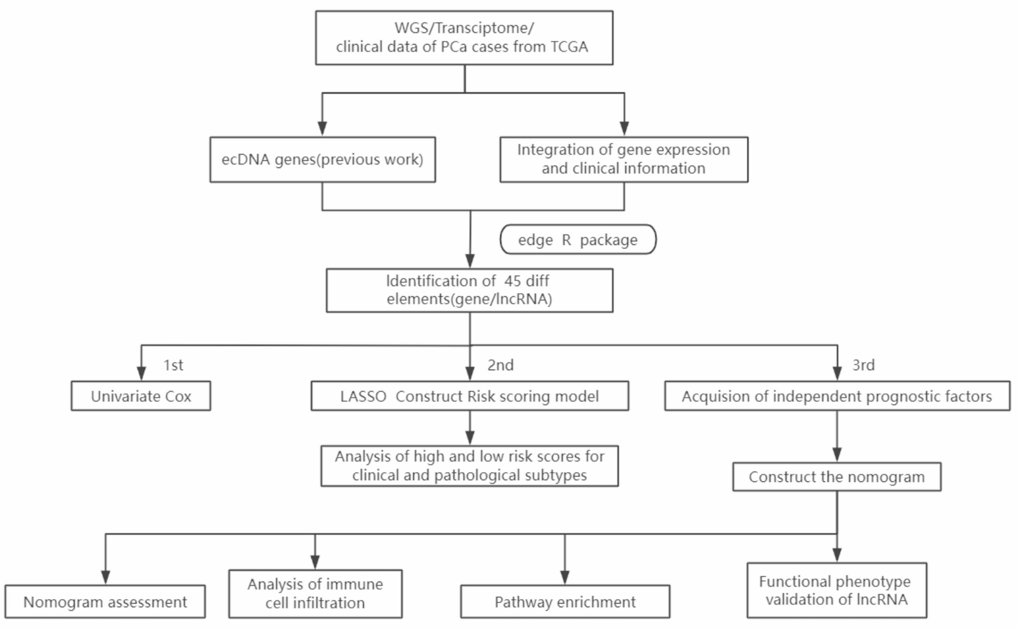
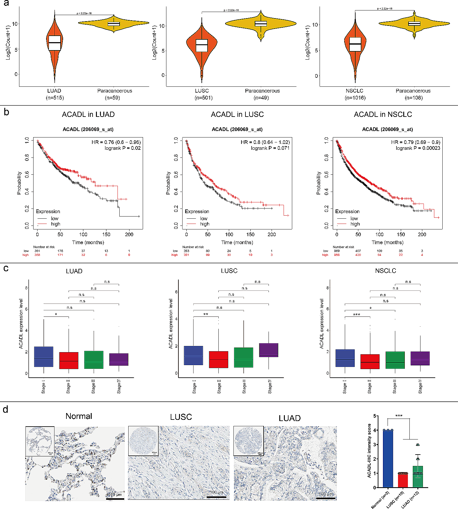
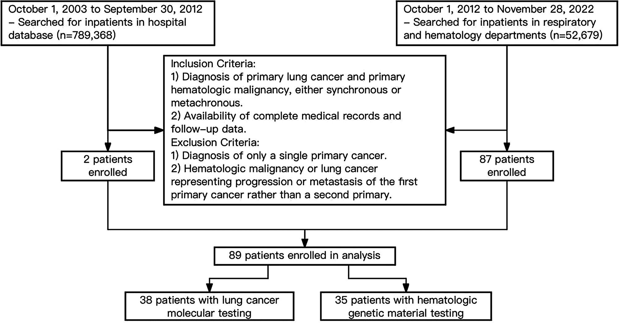
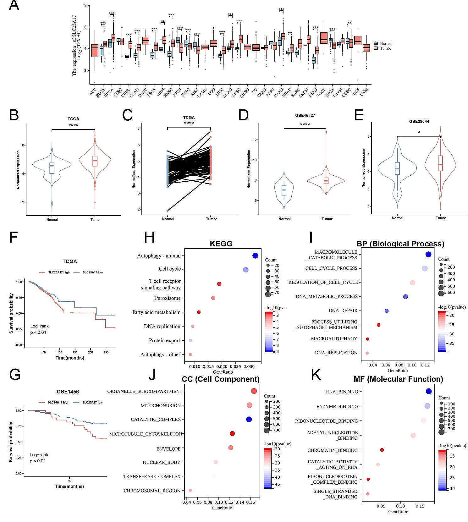
Remember me
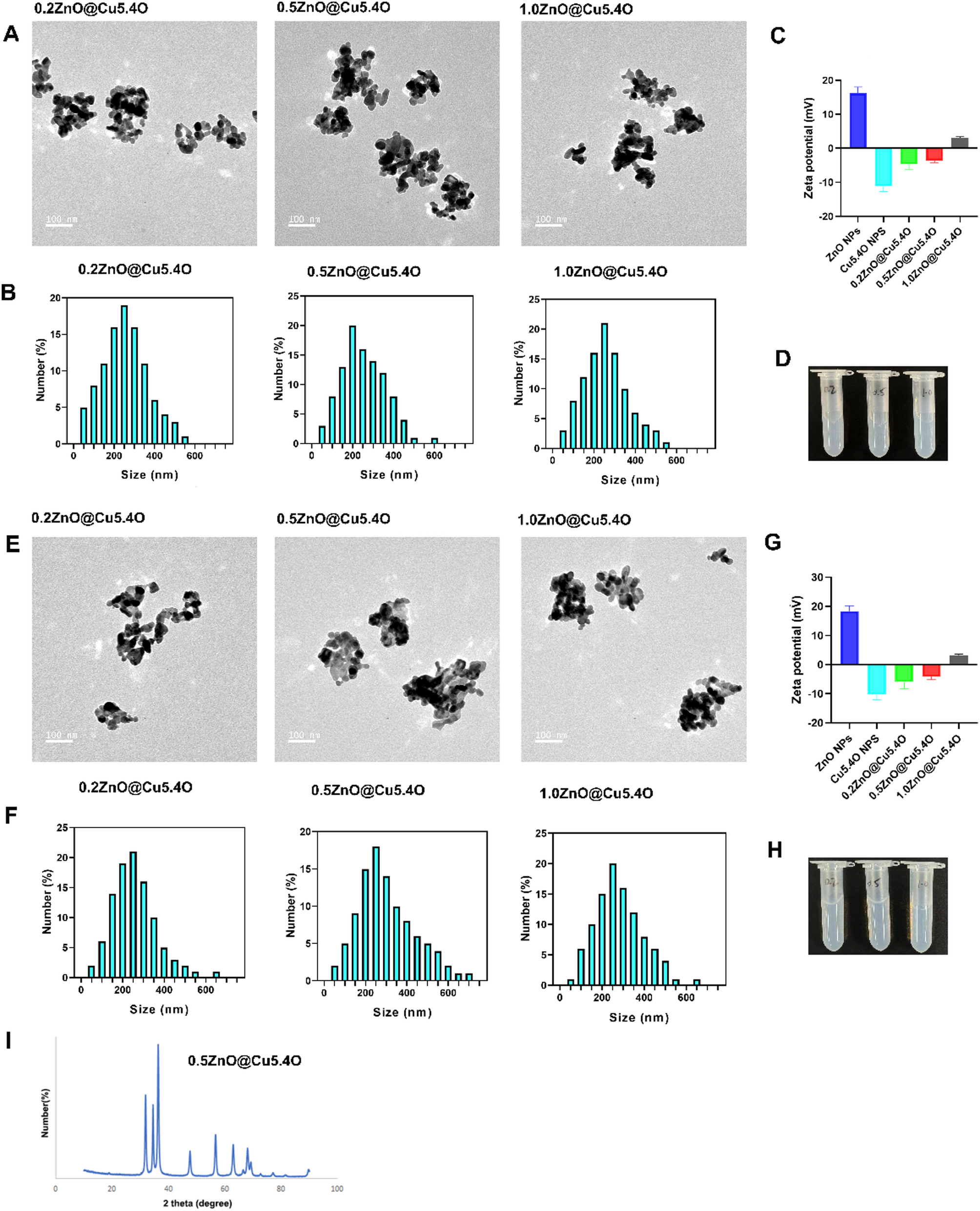

TEM and particle size analysis demonstrated that the synthesized ZnO@Cu5.4O nanoparticles with different ratios were uniformly dispersed in aqueous solution, with diameters mainly ranging from 150 to 300 nm (Fig. 1A, B). The zeta potentials of the various nanoparticles were measured. The zeta potentials of ZnO NPs and Cu5.4O NPs were 16.23 mV and 11.1 mV, respectively. In contrast, the zeta potential of ZnO@Cu5.4O was close to zero, with values of -4.67 mV, -3.63 mV, and 3.11 mV for 0.2ZnO@Cu5.4O, 0.5ZnO@Cu5.4O, and 1.0ZnO@Cu5.4O, respectively (Fig. 1C). The ZnO@Cu5.4O nanoparticle solution (0.5 mg/mL) showed no obvious sedimentation (Fig. 1D). Due to the low absolute values of zeta potentials, ZnO@Cu5.4O particles tended to aggregate. To further assess the nanoparticle stability, we characterized the particles again after 12 h of incubation in aqueous solution. TEM and particle size analyses showed that ZnO@Cu5.4O nanoparticles remained uniformly dispersed without significant aggregation (Fig. 1E, F). Zeta potential measurements after 12 h revealed values of -5.97 mV, -4.2 mV, and 3.13 mV for 0.2ZnO@Cu5.4O, 0.5ZnO@Cu5.4O, and 1.0ZnO@Cu5.4O, respectively (Fig. 1G). After 12 h of standing, the ZnO@Cu5.4O nanoparticle solution (0.5 mg/mL) showed no obvious sedimentation (Fig. 1H). However, TEM images after 16 h showed significant aggregation of 0.5ZnO@Cu5.4O nanoparticles (Figure S1). These results suggest that while ZnO@Cu5.4O nanoparticles tend to aggregate and exhibit poor long-term stability, they remain relatively stable within the first 12 h. We selected one type of nanoparticle, 0.5ZnO@Cu5.4O, to study the oxidation state of nanoparticle using powder X-ray diffraction (XRD)(Fig. 1I). The dominant peaks appeared at 2θ = 31.9°, 34.5°, and 36.3°.
Fig. 1
Characterization of ZnO@Cu5.4O nanoparticles (NPs). (A) TEM image of ZnO@Cu5.4O NPs. (B) Particle size distribution of ZnO@Cu5.4O NPs. (C) The zeta potentials of ZnO NPs, Cu5.4O, and 0.2ZnO@Cu5.4O, 0.5ZnO@Cu5.4O, 1.0ZnO@Cu5.4O in aqueous solution, respectively. (D) The appearance of the 0.2ZnO@Cu5.4O, 0.5ZnO@Cu5.4O, 1.0ZnO@Cu5.4O in aqueous solution, respectively. (E) After 12 h of standing, the TEM image of ZnO@Cu5.4O NPs. (F) After 12 h of standing, the particle size distribution of ZnO@Cu5.4O NPs. (G) After 12 h of standing, the zeta potentials of ZnO NPs, Cu5.4O, and 0.2ZnO@Cu5.4O, 0.5ZnO@Cu5.4O, 1.0ZnO@Cu5.4O in aqueous solution, respectively. (H) After 12 h of standing, the appearance of the 0.2ZnO@Cu5.4O, 0.5ZnO@Cu5.4O, 1.0ZnO@Cu5.4O in aqueous solution, respectively. (I) The XRD analysis of 0.5ZnO@Cu5.4O NPs
Effect of ZnO@Cu5.4O NPs on GSDMBCopper and zinc are trace elements in the human body that participate in numerous enzymatic reactions [13, 14]. Research has confirmed that copper and zinc can influence pyroptosis, which is increasingly regarded as a new frontier in cancer therapy. By triggering the assembly of the NLRP3-ASC-Caspase-1 inflammatory complex, ZnO NPs further activate GSDMD and promote the release of inflammatory cytokines including IL-1β and IL-18 [8]. NOD-like receptor protein 3 (NLRP3)-mediated pyroptosis is activated by the copper-quinone-GOx nanoparticles [15].
This study focuses on GSDMB, a member of the GSDMs family, which are key proteins in pyroptosis [16, 17]. In tumor cells overexpressing GSDMB, we introduced ZnO@Cu5.4O nanoparticles with different compositions. By measuring GSDMB expression levels in two types of tumor cells, we found that 0.5ZnO@Cu5.4O exhibited the strongest inhibitory effect on GSDMB (Fig. 2A, B). Furthermore, a comparison of the inhibitory effects of individual components (ZnO and Cu5.4O) and the composite material revealed that ZnO and Cu5.4O alone had significantly weaker inhibitory effects on GSDMB expression. In contrast, 0.5ZnO@Cu5.4O significantly inhibited GSDMB expression in tumor cells (Fig. 2C, D). qPCR analysis was conducted to evaluate the expression of GSDMB in gastric cancer cell lines treated with different nanoparticles. In MKN45 cells, GSDMB expression significantly decreased in the 0.5 ZnO@Cu5.4O and 1.0 ZnO@Cu5.4O treatment groups, whereas there were no significant differences in the 0.2 ZnO@Cu5.4O, ZnO, and Cu5.4O groups compared to the control (Fig. 2E). In MGC803 cells, GSDMB expression was significantly downregulated in the 0.2 ZnO@Cu5.4O and 0.5 ZnO@Cu5.4O groups, with no significant difference observed in the 1.0 ZnO@Cu5.4O, ZnO, or Cu5.4O groups compared to the control (Fig. 2F). In 293T cells overexpressing GSDMA, GSDMC, GSDMD, and GSDME, treatment with 0.5 ZnO@Cu5.4O had no significant effect on the expression levels of other GSDM family proteins. Based on the above results, 0.5ZnO@Cu5.4O exerts a regulatory effect on GSDMB after entering the cells, and then is separated into ZnO and Cu5.4O by the enclosed 0.5ZnO@Cu5.4O under the action of lysosome. ZnO becomes Zn2+ under the action of H+, which promotes the production of intracellular ROS, thus increasing oxidative stress. In general, the high level of Zn2+ ions in cells can result in the production of ROS by reducing nicotinamide adenine dinucleotide and interrupt the tricarboxylic acid cycle and glycolysis [18, 19]. Acting as analogs of SOD, CAT, and GPx, Cu5.4O NPs simultaneously exhibit broad-spectrum ROS clearance activities in cells [9, 12]. These two types of nanoparticles play their respective roles in the remodeling of intracellular environment and tumor microenvironment.
Fig. 2
Inhibitory effect of ZnO@Cu5.4O NPs on GSDMB expression. (A, B) GSDMB expression levels in two gastric cancer cell lines overexpressing GSDMB after treatment with different ZnO@Cu5.4O NPs. (C, D) Comparison of GSDMB expression in two gastric cancer cell lines treated with 0.5ZnO@Cu5.4O NPs, ZnO NPs, and Cu5.4O NPs. (E, F) The mRNA expression of GSDMB in two gastric cancer cell lines treated with 0.5ZnO@Cu5.4O NPs, ZnO NPs, and Cu5.4O NPs, data were analyzed using t-tests. (G) The effect of 0.5ZnO@Cu5.4O on the expression of GSDM family members was further assessed in 293T cells
Biosafety assessmentAs a novel metal-based nanoparticle, ZnO@Cu5.4O may induce various adverse reactions in cells, making biosafety evaluation crucial. Cytotoxicity tests were conducted in tumor cells (Fig. 3A), followed by calcein-AM staining of live cells treated with the three materials (Fig. 3B). Regarding blood compatibility, hemolysis results (Fig. 3C) demonstrated that the nanoparticles had good blood compatibility. Based on the above results, the cytotoxicity of 0.5ZnO@Cu5.4O increased with the increase of dose, and the killing effect of 0.5ZnO@Cu5.4O on tumor cells was significantly higher than that of individual monomers.
Fig. 3
Biological toxicity assessment of 0.5ZnO@Cu5.4O. (A) Cytotoxicity of 0.5ZnO@Cu5.4O at different concentrations in two tumor cell lines measured by the CCK-8 assay, data were analyzed using t-tests. (B) Density of live cells after 24-hour treatment with ZnO NPs, Cu5.4O NPs, and 0.5ZnO@Cu5.4O, determined by calcein-AM staining. (C) Blood compatibility of 0.5ZnO@Cu5.4O, data were analyzed using t-tests
Effects of 0.5ZnO@Cu5.4O on tumor cell biological behaviorA wound-healing assay was performed to evaluate the effect of ZnO@Cu5.4O on tumor cell migration. The results showed that ZnO@Cu5.4O significantly inhibited tumor cell migration (Fig. 4A). Flow cytometry was used to assess the effect of ZnO@Cu5.4O on tumor cell apoptosis, revealing that 0.5ZnO@Cu5.4O promoted apoptosis in tumor cells (Fig. 4B). Based on the above results, 0.2ZnO@Cu5.4O, 0.5ZnO@Cu5.4O and 1.0ZnO@Cu5.4O can inhibit the migration of tumor cells. The above three nanoparticles have significant apoptosis induction effects, but 1.0ZnO@Cu5.4O has a stronger apoptosis induction effect among the three nanoparticles, which may be attributed to the increased ratio of ZnO in the nanoparticles. Some study shows that by disrupting mitochondrial kinetic homeostasis and reducing mitochondrial membrane potential, ZnO NPs caused apoptosis [20]. ZnO NPs can promote inflammation and keratinocyte apoptosis by regulating nuclear translocation of p-NFκB p65 and inducing cysteine deficiency [21].
Fig. 4
Effects of 0.5ZnO@Cu5.4O on the biological behavior of gastric cancer cells. (A) Results of wound-healing assays using different materials. (B) Effects of different materials on apoptosis in two gastric cancer cell lines
In vivo validation of 0.5ZnO@Cu5.4O for tumor InhibitionIn vivo experiments were conducted using subcutaneous tumor-bearing nude mice (Fig. 5A). Mice were randomly divided into four groups and given PBS, ZnO, Cu5.4O, or 0.5ZnO@Cu5.4O (2 mg/kg). Although the injected dose (2 mg/kg) corresponds to a nominal concentration of ~ 200 mg/L based on the average body weight and injection volume, it should be noted that the actual systemic concentration after i.v. administration is significantly lower due to rapid dilution and distribution. Therefore, the observed hemolytic effect at 200 mg/L in vitro does not directly reflect the in vivo situation. Tumor volumes and mice body weight were monitored every two days (Fig. 5B). While ZnO and Cu5.4O alone exhibited some inhibitory effect on tumor volume, 0.5ZnO@Cu5.4O demonstrated the most significant tumor inhibition (Fig. 5C-E).
Liver and kidney function indicators in mouse peripheral blood were evaluated. No significant differences were found among the four groups in serum concentrations of AST, ALT, or BUN (Fig. 5F–H). Hematoxylin and eosin (HE) staining of spleen, lung, heart, liver, and kidney tissues from the control and 0.5ZnO@Cu5.4O treatment groups showed no pathological changes in organ structures (Fig. 5I), indicating low toxicity at this concentration and a positive therapeutic effect on tumors.
Fig. 5
In vivo validation of 0.5ZnO@Cu5.4O for tumor inhibition. (A) Establishment and treatment process of tumor-bearing nude mice models. (B) body weight evolution curves of mice after different treatments, data were analyzed using t-tests. (C) Images of excised tumors. (D) Tumor growth curves over time, data were analyzed using t-tests. (E) The quality of the tumor in vitro (F-G) Serum liver and renal function indicators: ALT, AST, BUN, data were analyzed using t-tests. (I) HE staining of various organs in the control and 0.5ZnO@Cu5.4O groups
Comments (0)