Asrani SK, Devarbhavi H, Eaton J, Kamath PS (2019) Burden of liver diseases in the world. J Hepatol 70(1):151–171
Article
PubMed
Google Scholar
Meijerink MR, Lei SVD, Dijkstra M, Versteeg KS, Buffart TE, Lissenberg-Witte BI, Swijnenburg R-J, Tol MPVD, Puijk RS (2024) Surgery versus thermal ablation for small-size colorectal liver metastases (Collision): an international, multicenter, phase III randomized controlled trial. J Clin Oncol 42(17):3501–3501
Article
Google Scholar
Reig M, Forner A, Rimola J, Ferrer-Fàbrega J, Burrel M, Garcia-Criado Á, Kelley RK, Galle PR, Mazzaferro V, Salem R, Sangro B, Singal AG, Vogel A, Fuster J, Ayuso C, Bruix J (2022) BCLC strategy for prognosis prediction and treatment recommendation: the 2022 update. J Hepatol 76(3):681–693
Article
PubMed
Google Scholar
Crocetti L, de Baére T, Pereira PL, Tarantino FP (2020) CIRSE standards of practice on thermal ablation of liver tumours. Cardiovasc Intervent Radiol 43(7):951–962
Article
PubMed
Google Scholar
Laimer G, Schullian P, Jaschke N, Putzer D, Eberle G, Alzaga A, Odisio B, Bale R (2020) Minimal ablative margin (MAM) assessment with image fusion: an independent predictor for local tumor progression in hepatocellular carcinoma after stereotactic radiofrequency ablation. Eur Radiol 30(5):2463–2472
Article
PubMed
PubMed Central
Google Scholar
Minier C, Hermida M, Allimant C, Escal L, Pierredon-Foulongne M-A, Belgour A, Piron L, Taourel P, Cassinotto C, Guiu B (2022) Software-based assessment of tumor margins after percutaneous thermal ablation of liver tumors: a systematic review. Diagn Interv Imaging 103(5):240–250
Article
PubMed
Google Scholar
Solbiati M, Muglia R, Goldberg SN, Ierace T, Rotilio A, Passera KM, Marre I, Solbiati L (2019) A novel software platform for volumetric assessment of ablation completeness. Int J Hyperthermia 36(1):337–343
Article
PubMed
Google Scholar
Laimer G, Jaschke N, Schullian P, Putzer D, Eberle G, Solbiati M, Solbiati L, Goldberg SN, Bale R (2021) Volumetric assessment of the periablational safety margin after thermal ablation of colorectal liver metastases. Eur Radiol 31(9):6489–6499
Article
PubMed
PubMed Central
Google Scholar
Lin Y-M, Paolucci I, O’Connor CS, Anderson BM, Rigaud B, Fellman BM, Jones KA, Brock KK, Odisio BC (2023) Ablative margins of colorectal liver metastases using deformable CT image registration and autosegmentation. Radiology 307(2):e221373
Article
PubMed
Google Scholar
Software-Aided Imaging (Morfeus) for Confirming Tumor Coverage With Ablation in Patients With Liver Tumors, the COVER-ALL Study. NCT04083378 2019-2022; Available from: Software-Aided Imaging (Morfeus) for Confirming Tumor Coverage With Ablation in Patients With Liver Tumors, the COVER-ALL Study.
Laimer G, Verdonschot KHM, Kopf L, van der Lei S, Scharll Y, Hannink G, Jenniskens SFM, Meijerink MR, Bale R, Overduin CG (2024) Multicenter and inter-software evaluation of ablative margins after thermal ablation of colorectal liver metastases. Eur Radiol 35:1046–1056
Article
PubMed
PubMed Central
Google Scholar
Shady W, Petre EN, Do KG, Gonen M, Yarmohammadi H, Brown KT, Kemeny NE, D’Angelica M, Kingham PT, Solomon SB, Sofocleous CT (2018) Percutaneous microwave versus radiofrequency ablation of colorectal liver metastases: ablation with clear margins (A0) provides the best local tumor control. J Vasc Interv Radiol 29(2):268-275.e1
Article
PubMed
Google Scholar
Hendriks P, Boel F, Oosterveer TTM, Broersen A, de Geus-Oei L-F, Dijkstra J, Burgmans MC (2023) Ablation margin quantification after thermal ablation of malignant liver tumors: how to optimize the procedure? A systematic review of the available evidence. Euro J Radiol Open 11:100501
Article
Google Scholar
Pellat A, Barat M, Coriat R, Soyer P, Dohan A (2023) Artificial intelligence: A review of current applications in hepatocellular carcinoma imaging. Diagn Interv Imag 104(1):24–36
Article
Google Scholar
Fahmy D, Alksas A, Elnakib A, Mahmoud A, Kandil H, Khalil A, Ghazal M, van Bogaert E, Contractor S, El-Baz A (2022) The role of radiomics and AI technologies in the segmentation, detection, and management of hepatocellular carcinoma. Cancers (Basel) 14(24):6123
Article
PubMed
Google Scholar
Liu L, Wang L, Xu D, Zhang H, Sharma A, Tiwari S, Kaur M, Khurana M, Shah MA (2021) CT image segmentation method of liver tumor based on artificial intelligence enabled medical imaging. Math Probl Eng 2021(1):9919507
Google Scholar
Study of ThermoDox With Standardized Radiofrequency Ablation (RFA) for Treatment of Hepatocellular Carcinoma (HCC) (OPTIMA). NCT02112656 2014-2018; Available from: https://clinicaltrials.gov/ct2/show/NCT02112656.
Christou, A., K.H. Lee, O. Parker, N. Varble, Q. de Ruiter, H. Celik, S. Xu, E. Turkbey, A. Arrichiello, R. Lencioni, G. Carrafiello, B. Turkbey, N. Borys, and B. Wood, Ablation Margin and Tumor Morphology Metrics from a Randomized Controlled Drug + Device Trial in 3–7 cm Hepatocellular Carcinoma., In: Radiological Society of North America, Radiology, Editor. 2022: Chicago, IL.
Lencioni R, Cioni D (2016) RFA plus lyso-thermosensitive liposomal doxorubicin: in search of the optimal approach to cure intermediate-size hepatocellular carcinoma. Hepat Oncol 3(3):193–200
Article
PubMed
PubMed Central
Google Scholar
Borys N, Dewhirst MW (2021) Drug development of lyso-thermosensitive liposomal doxorubicin: combining hyperthermia and thermosensitive drug delivery. Adv Drug Deliv Rev 178:113985
Article
CAS
PubMed
Google Scholar
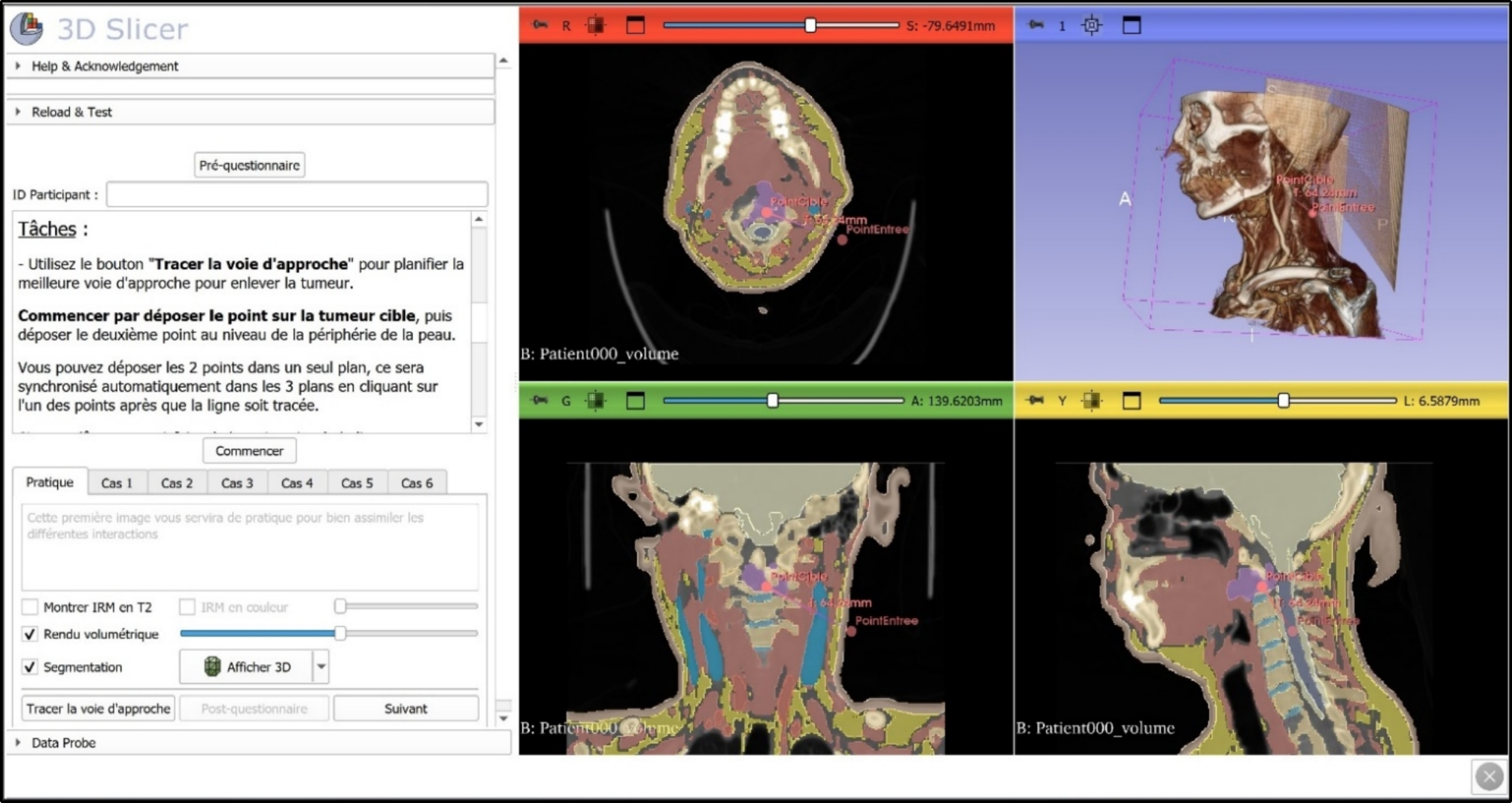
Fedorov A, Beichel R, Kalpathy-Cramer J, Finet J, Fillion-Robin JC, Pujol S, Bauer C, Jennings D, Fennessy F, Sonka M, Buatti J, Aylward S, Miller JV, Pieper S, Kikinis R (2012) 3D Slicer as an image computing platform for the quantitative imaging network. Magn Reson Imag 30(9):1323–1341
Article
Google Scholar
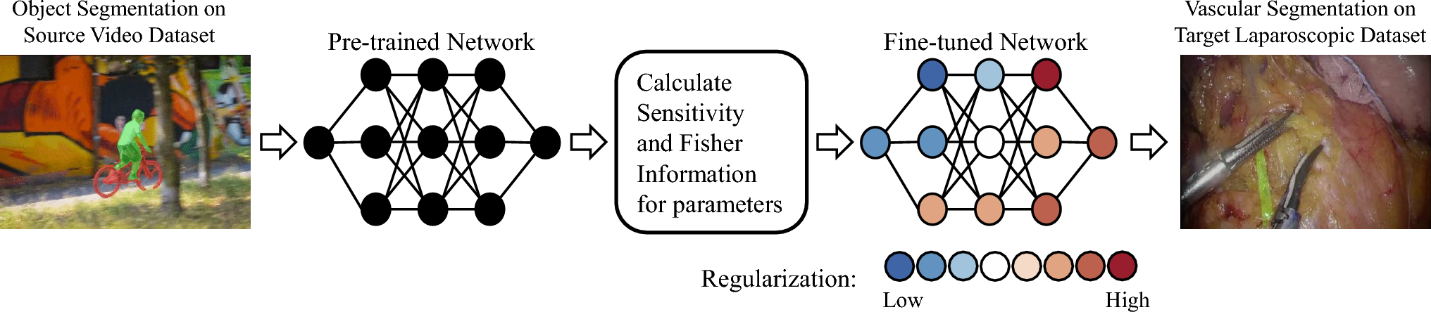
Deng J, Dong W, Socher R, Li LJ, Kai L, Li F-F (2009) ImageNet: A large-scale hierarchical image database. In: 2009 IEEE Conference on Computer Vision and Pattern Recognition
Breiman L (2001) Random forests. Mach Learn 45(1):5–32
Article
Google Scholar
Kim YS, Rhim H, Cho OK, Koh BH, Kim Y (2006) Intrahepatic recurrence after percutaneous radiofrequency ablation of hepatocellular carcinoma: analysis of the pattern and risk factors. Eur J Radiol 59(3):432–441
Article
CAS
PubMed
Google Scholar
Kim YS, Lee WJ, Rhim H, Lim HK, Choi D, Lee JY (2010) The minimal ablative margin of radiofrequency ablation of hepatocellular carcinoma (> 2 and < 5 cm) needed to prevent local tumor progression: 3D quantitative assessment using CT image fusion. AJR Am J Roentgenol 195(3):758–765
Article
PubMed
Google Scholar
Lu DS, Yu NC, Raman SS, Limanond P, Lassman C, Murray K, Tong MJ, Amado RG, Busuttil RW (2005) Radiofrequency ablation of hepatocellular carcinoma: treatment success as defined by histologic examination of the explanted liver. Radiology 234(3):954–960
Article
PubMed
Google Scholar
Vasiniotis Kamarinos N, Gonen M, Sotirchos V, Kaye E, Petre EN, Solomon SB, Erinjeri JP, Ziv E, Kirov A, Sofocleous CT (2022) 3D margin assessment predicts local tumor progression after ablation of colorectal cancer liver metastases. Int J Hyperthermia 39(1):880–887
Article
CAS
PubMed
Google Scholar
Lin YM, Paolucci I, Anderson BM, O’Connor CS, Rigaud B, Briones-Dimayuga M, Jones KA, Brock KK, Fellman BM, Odisio BC (2022) Study protocol COVER-ALL: clinical impact of a volumetric image method for confirming tumour coverage with ablation on patients with malignant liver lesions. Cardiovasc Intervent Radiol 45(12):1860–1867
Article
PubMed
PubMed Central
Google Scholar
Ablation Confirmation Study. NCT03753789 2018-2022; Available from: https://clinicaltrials.gov/ct2/show/NCT03753789.
Ruiter SJS, Tinguely P, Paolucci I, Engstrand J, Candinas D, Weber S, de Haas RJ, de Jong KP, Freedman J (2021) 3D quantitative ablation margins for prediction of ablation site recurrence after stereotactic image-guided microwave ablation of colorectal liver metastases: a multicenter study. Front Oncol 11:757167
Article
CAS
PubMed
PubMed Central
Google Scholar
Kaye EA, Cornelis FH, Petre EN, Tyagi N, Shady W, Shi W, Zhang Z, Solomon SB, Sofocleous CT, Durack JC (2019) Volumetric 3D assessment of ablation zones after thermal ablation of colorectal liver metastases to improve prediction of local tumor progression. Eur Radiol 29(5):2698–2705
Article
PubMed
Google Scholar
Zirakchian Zadeh M, Sotirchos VS, Kirov A, Lafontaine D, Gönen M, Yeh R, Kunin H, Petre EN, Kitsel Y, Elsayed M, Solomon SB, Erinjeri JP, Schwartz LH, Sofocleous CT (2024) Three-dimensional margin as a predictor of local tumor progression after microwave ablation: intraprocedural versus 4–8-week postablation assessment. J Vascular and Interv Radiol 35(4):523–532
Article
Google Scholar
Teng W, Liu KW, Lin CC, Jeng WJ, Chen WT, Sheen IS, Lin CY, Lin SM (2015) Insufficient ablative margin determined by early computed tomography may predict the recurrence of hepatocellular carcinoma after radiofrequency ablation. Liver Cancer 4(1):26–38
Article
PubMed
PubMed Central
Google Scholar
Lin YM, Paolucci I, Albuquerque Marques Silva J, O’Connor CS, Fellman BM, Jones AK, Kuban JD, Huang SY, Metwalli ZA, Brock KK, Odisio BC (2024) Intraprocedural versus initial follow-up minimal ablative margin assessment after colorectal liver metastasis thermal ablation: which one better predicts local outcomes? Invest Radiol 59(4):314–319
Article
CAS
PubMed
Google Scholar
Lee JK, Siripongsakun S, Bahrami S, Raman SS, Sayre J, Lu DS (2016) Microwave ablation of liver tumors: degree of tissue contraction as compared to RF ablation. Abdom Radiol (NY) 41(4):659–666
Article
CAS
PubMed
Google Scholar

Comments (0)