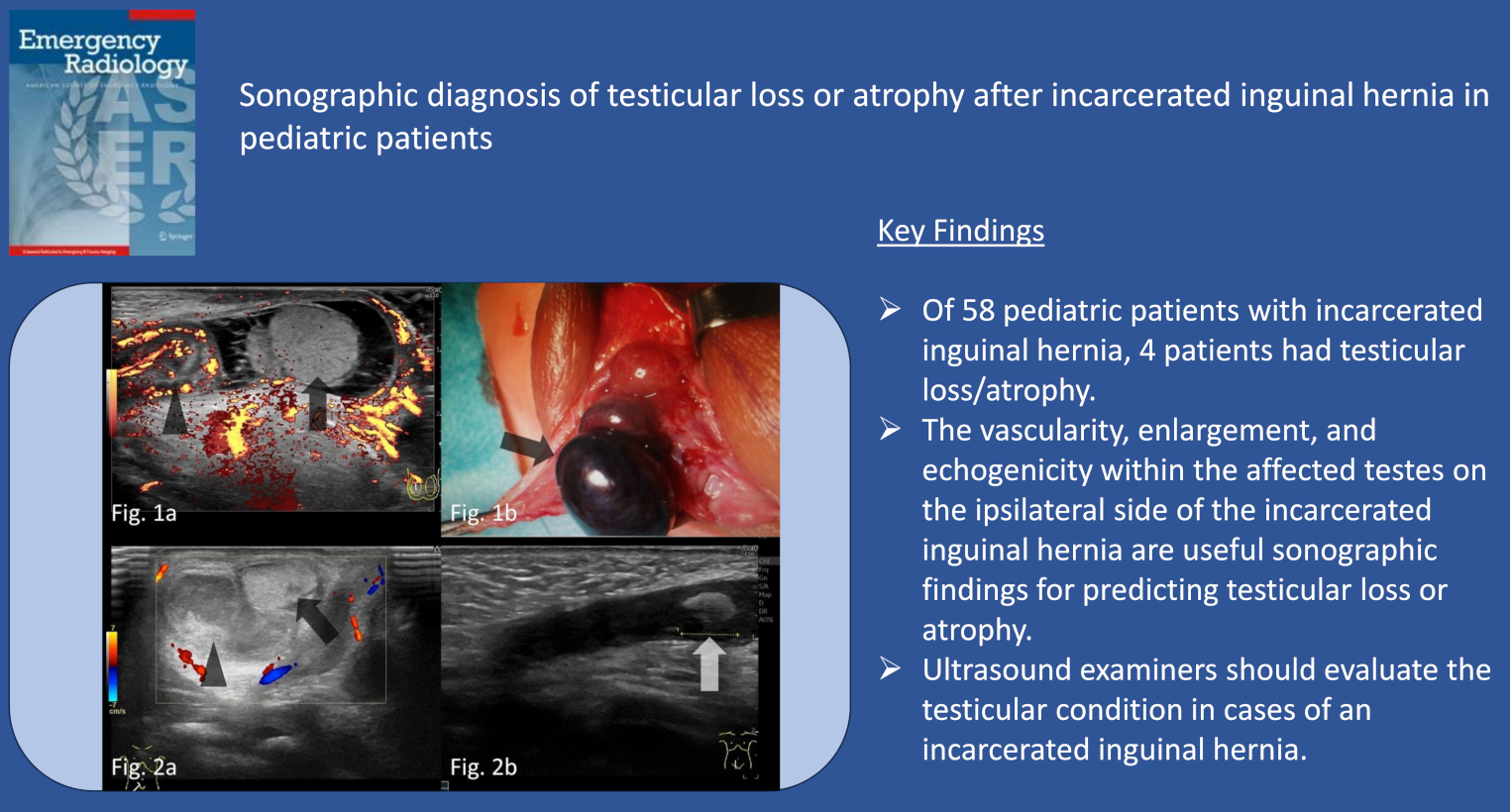
Sonographic diagnosis of testicular loss or atrophy after incarcerated inguinal hernia in pediatric patients
Purpose
Incarcerated inguinal hernia is a common condition in pediatric patients presenting to the emergency department. In addition to causing intestinal obstruction, it can lead to acute testicular venous occlusion, necessitating urgent intervention. This study aimed to demonstrate the utility of ultrasonography in predicting testicular loss/atrophy associated with incarcerated inguinal hernia in pediatric patients.
Methods
This study enrolled 58 patients with incarcerated inguinal hernia. Pre- and post-reduction sonographic findings, including the vascularity, enlargement, and echogenicity of the affected testis, were compared between patients with and without testicular loss/atrophy using Fisher’s exact and Mann–Whitney U tests. The imaging studies were reviewed by two radiologists blinded to clinical information and other imaging findings, with discrepancies resolved by consensus.
Results
All pre- and post-reduction sonographic findings were significantly different between the patients with and without testicular loss/atrophy, including vascularity (evaluated in 32 patients post-reduction; present/absent in 0/3 vs. 24/5), enlargement (evaluated in 50 patients; present/absent in 2/1 vs. 4/43), and echogenicity (evaluated in 50 patients; normal/abnormal in 0/3 vs. 47/0). The differences observed post-reduction were significant in terms of vascularity (evaluated in 38; present/absent in 0/3 vs. 30/5), enlargement (evaluated in 57 patients; present/absent in 2/1 vs. 1/53), and echogenicity (evaluated in 57 patients; normal/abnormal in 0/3 vs. 54/0).
Conclusions
The vascularity, enlargement, and echogenicity within the affected testes on the ipsilateral side of the incarcerated inguinal hernia are useful sonographic findings for predicting testicular loss/atrophy. Therefore, ultrasonologist should evaluate the testicular condition in cases of an incarcerated inguinal hernia.

Comments (0)