
Remember me

This study included 25 right-handed male participants with a normal neurology (age: 21–23 years; mean ± standard deviation: 21.4 ± 0.6 years). It was performed in accordance with the Declaration of Helsinki, and it was approved by the Ethics Committee of Niigata University of Health and Welfare (approval number: 18792–220111). In addition, all participants received comprehensive explanations about the experimental procedures, and they provided informed consent before participation.
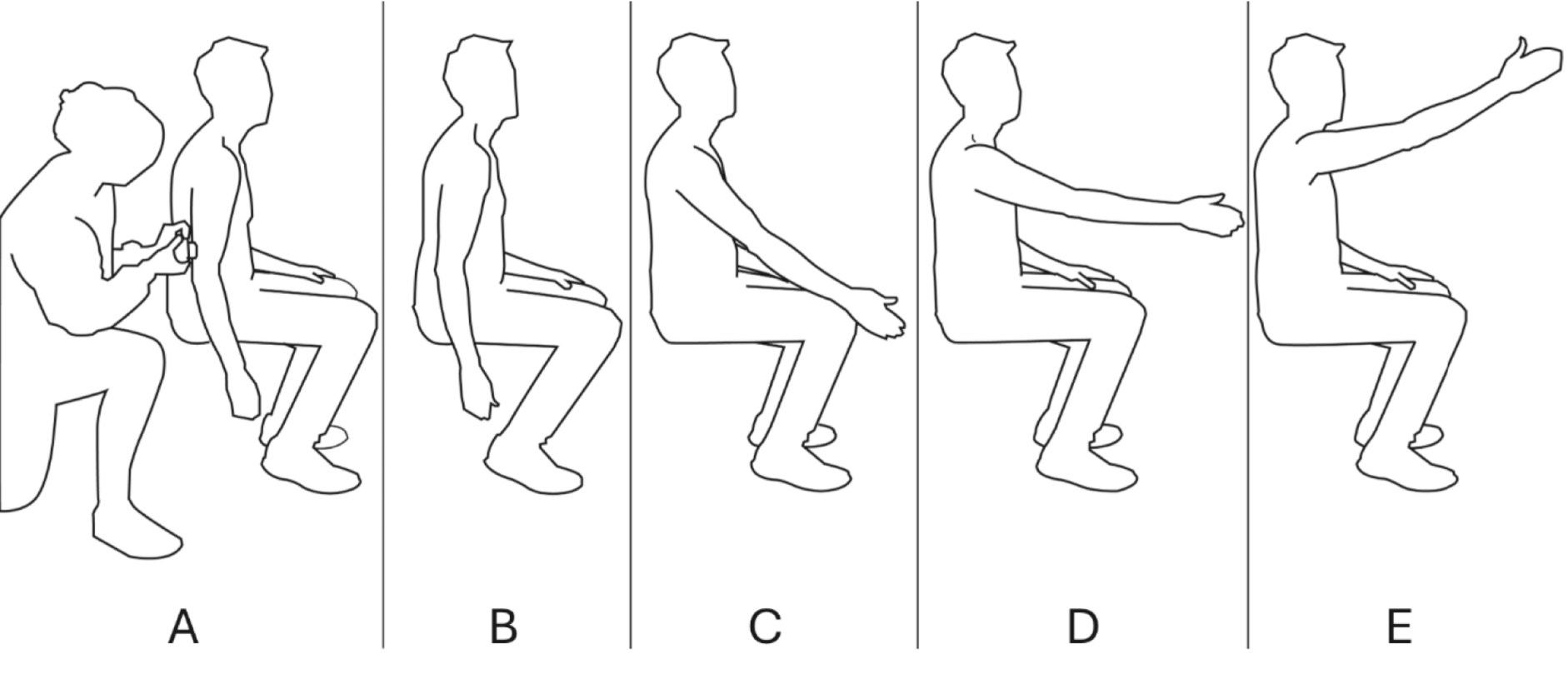
Grating orientation discrimination taskThe GOT was used to assess tactile spatial discrimination performance. Tactile stimulation was delivered to the right index finger using eight hemispherical domes of different groove widths (3.0, 2.0, 1.5, 1.2, 1.0, 0.75, 0.5, and 0.35 mm) employing a custom-made device (S-16026; Takei Scientific Instruments Co., Ltd., Niigata, Japan) (Fig. 1). The elevation speed of the hemispherical domes was set to 20 mm/s. The tactile stimulus duration was set to 1 s. The hemispherical dome was further elevated at 1.5 mm after contact of the hemispherical dome with the right index finger (Saito et al. 2022). The domes were presented five times each in the following order: 3.0, 2.0, 1.5, 1.2, 1.0, 0.75, 0.5, and 0.35 mm (8 conditions × 5 times = 40 times). Next, the procedure was repeated three times (40 times × 3 = 120 times). The participants were requested to identify the dome orientation relative to the long axis of the finger.
Fig. 1
Grating orientation discrimination task. A hemisphere-shaped stimulus block with grooves was mechanically pressed against the right index finger, and participants were instructed to indicate the orientation of the grooves (vertical or horizontal) using a button held in their left hand. The width of the grooves (stimulus width) on the stimulus block was set at 3.0, 2.0, 1.5, 1.2, 1.0, 0.75, 0.5, and 0.35 mm across eight conditions. The stimulus block was presented three times in descending order of groove size (3 repetitions × 8 conditions), totaling five sets (24 repetitions × 5 sets)
Transcranial direct current stimulationTranscranial DCS was delivered using the DC-STIMULATOR PLUS instrument (NeuroConn, Germany) via a pair of saline-soaked surface sponge electrodes (5 × 5 cm each). The stimulation intensity was set to 1 mA, and the fade in/fade out was set to 15 s (Karok and Witney 2013). For dual-hemisphere tDCS, the anodal electrode was placed at P4 and the cathodal electrode at P3 based on the international 10–20 reference system. Stimulation was delivered for 20 min. For anodal tDCS, the anodal electrode was placed at P4 and the cathodal electrode at F9 based on the international 10–20 reference system. The stimulation was delivered for 20 min. For cathodal tDCS, the anodal electrode was placed at F10 and the cathodal electrode at P4 based on the international 10–20 reference system. The stimulation was delivered for 20 min. For sham stimulation, dual-hemisphere tDCS was switched on for 30 s.
Experimental procedureFigure 2 shows the experimental flow. All participants received dual-hemisphere tDCS over the PPC, anodal tDCS over the right PPC, cathodal tDCS over the left PPC, and sham stimulation. The interval of the individual stimulation sessions was at least 3 days, and the session order was randomly counterbalanced among the participants. Tactile acuity discrimination using GOT was measured before, during, and immediately after stimulation. Subjective sensations were measured using the Numerical Rating Scale (NRS) 1.5 min after the stimulus was switched on.
Fig. 2
Experimental procedure. Evaluations using the grating orientation task was conducted before (pre), during, and immediately after (post) 20 min of transcranial direct current stimulation (tDCS). The stimulation conditions of tDCS included four conditions: dual-hemisphere tDCS, anodal tDCS, cathodal tDCS, and sham stimulation. These conditions were administered in a random order with at least 3 days between sessions
Data analysisThe proportion of correct responses in each groove width was calculated using MATLAB (R2020a). The grating width was plotted against the proportion of correct responses and the relationship fitted by a logistic regression model (Saito et al. 2022). The GOT discrimination threshold was set at 75%.
Statistical analysisStatistical analysis was performed using a linear mixed-effects model. The factors were “stimulation condition” (dual-hemisphere tDCS, anodal tDCS, cathodal tDCS, or sham stimulation) and “time” (pre, during, or post). The change in GOT discrimination threshold was analyzed using the Friedman test across the various stimulus conditions (dual-hemisphere tDCS, anodal tDCS, cathodal tDCS, or sham stimulation). The post hoc test was performed using the Dunn-Bonferroni test. Furthermore, the correlation between changes in GOT discrimination thresholds and baseline GOT discrimination thresholds before stimulation was analyzed using Spearman’s rank correlation coefficient. A significance level of P < 0.05 was considered statistically significant.
Comments (0)