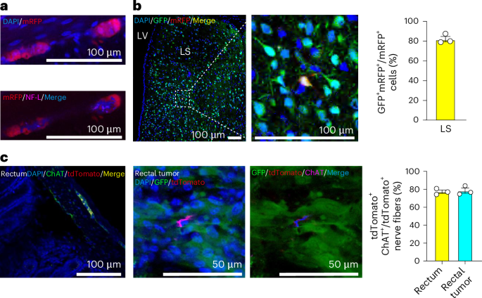
Glioblastoma’s widespread integration into neuronal networks
A few years ago, independent research groups studying brain-cancer biology demonstrated that neurons form synaptic connections with cancer cells and thereby affect tumor growth. Three recent studies published in Nature, Cell and Proceedings of the National Academy of Sciences reveal the remarkable extent and nature of neuron–glioma connections within the brain.
To map the network of neurons that innervate glioblastoma (GBM) cells, Sun et al., Tetzlaff et al. and Hsieh et al. used virus-based retrograde monosynaptic tracing systems. The authors performed intracranial xenografts of human GBM cells transduced with recombinant rabies virus and visualized presynaptic partners throughout the mouse brain. Neuronal innervation could already be detected shortly after GBM cell transplantations and comprised both local connections and long-range connections across several cortical and subcortical areas, ipsilaterally and contralaterally. Subsequent detailed imaging of mouse and human brain slices, including molecular characterization of tumor-connected neurons, together with electrophysiological recordings, Ca2+ imaging and chemogenetics in human organoids, revealed the diverse nature of the synaptic contacts. All three studies reported that GBM cells receive glutamatergic and GABAergic inputs, but Sun et al. and Tetzlaff et al. also uncovered cholinergic connections mediated via the muscarinic acetylcholine receptor M3 (CHRM3). Further experiments using GBM cells in which CHRM3 was downregulated provided functional evidence of a role for cholinergic innervation in brain-tumor invasion. Finally, Tetzlaff et al. explored possible translational interventions by disrupting neuron–glioma connections, offering a proof of concept for future therapeutic approaches.

Comments (0)