
Remember me


SenSkin™ was developed by a literature review of genes associated with cellular senescence in human skin [5, 7, 10,11,12,13,14]. Two board-certified dermatologist-scientists with research laboratories in skin biology conducted the review of peer-reviewed articles, and the genes were categorized into those that were upregulated in senescent skin cells (164 genes) and those that were downregulated in senescent skin cells (1 gene, LMNB1), comprising a total of 165 genes (shown in Table 1). SenSkin™ includes 18 genes in common with SenMayo, 36 genes in common with CellAge, 19 genes in common with GenAge, and 13 genes in common with Reactome’s Cellular Senescence pathway (shown in Fig. S2) [2,3,4,5].
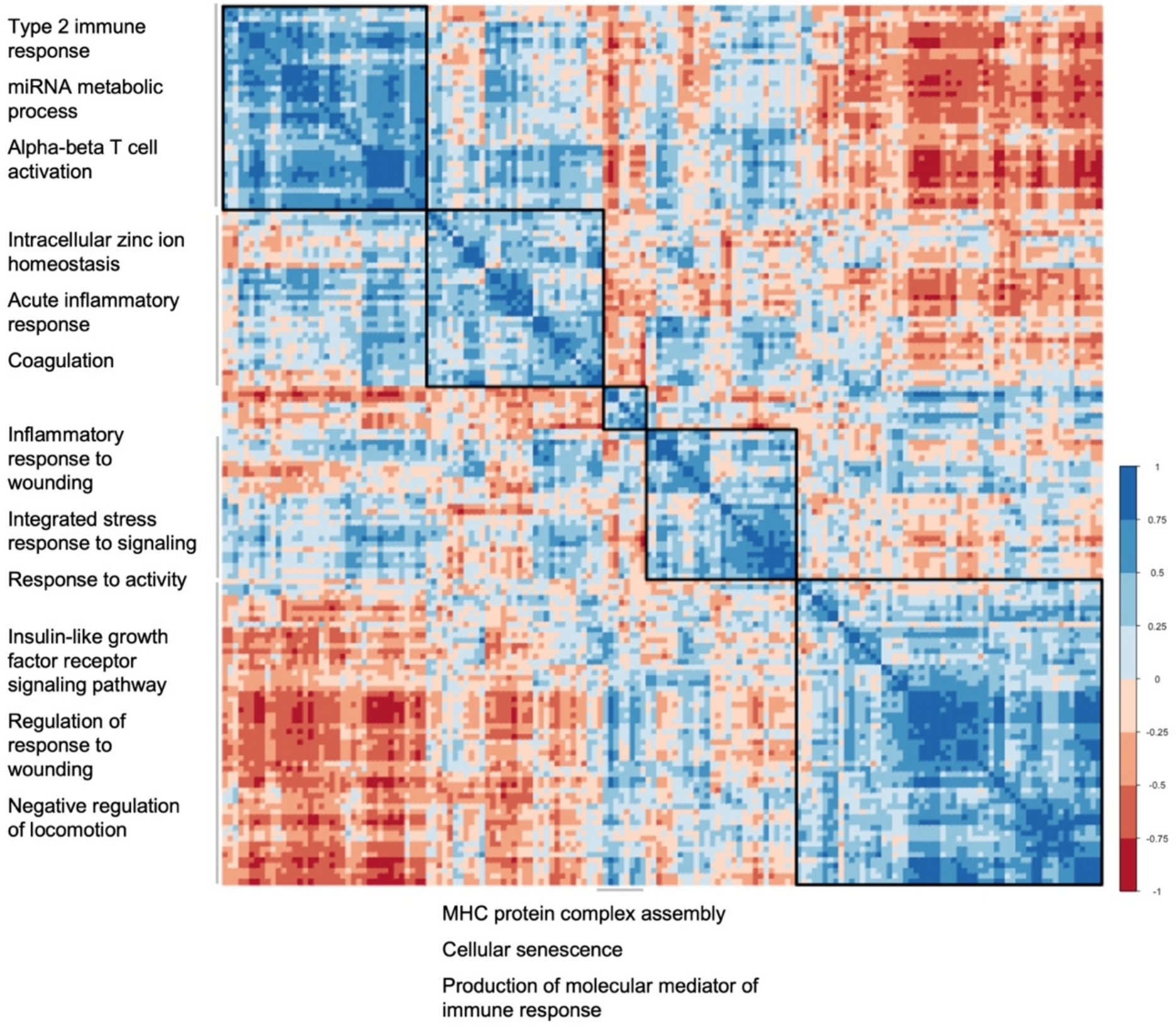
Table 1 SenSkin™ genes by gene symbolsTo identify pathways captured by SenSkin™, genes were analyzed altogether, and then, groups of highly correlated genes were analyzed as distinct groups. Many of the top Gene Ontology biological process pathways overrepresented by SenSkin™ genes as a whole were related to wound repair and immune responses, such as connective tissue development, mononuclear cell differentiation, wound healing, and regulation of hemopoiesis (shown in Fig. S1a). These pathways are consistent with findings that senescent cells affect cutaneous wound healing and modulate the immune system [15, 16]. Next, genes were hierarchically clustered based on correlations between the expression of pairs of genes in the integrated scRNA-seq datasets, and the pathways represented by each cluster were delineated (shown in Fig. 1). By examining clusters of highly correlated genes, overall mechanisms could be observed with less chances of focusing on individual genes with counter-regulatory mechanisms or genes that are associated but not directly involved in cellular senescence. Clusters of genes in SenSkin™ represent stress response, innate immunity, adaptive immunity, insulin-like growth factor pathway, and antigen presentation.
Fig. 1
Hierarchical clustering of SenSkin™ genes based on inter-gene correlations. Complete linkage hierarchical clustering of SenSkin™ genes based on correlations between pairs of genes in integrated pseudobulk RNA-seq datasets. The top three pathways represented by each cluster are listed
Genes in SenSkin™ were clustered based on known protein–protein interactions to evaluate the landscape of pathways captured by SenSkin™ (shown in Fig. S1b). The largest cluster of genes was identified as being related to cellular senescence, demonstrating concordance between SenSkin™ and other cellular senescence gene sets. Other clusters were related to features of cellular senescence, including altered lipid metabolism, immune responses, and TGF-β pathways.
High enrichment of SenSkin™ in bulk RNA-seq of chronological skin agingTo evaluate the ability of SenSkin™ to detect senescent cell accumulation with chronological aging, SenSkin™ was used to analyze bulk RNA-seq. Samples from 24 younger (20 to 25 years old) donors were compared to 24 older (55 to 66 years old) donors from Kuehne et al.’s dataset of normal human epidermis from the inner forearm [6]. Gene set enrichment analysis revealed that SenSkin™ was significantly enriched in samples from older patients, with a normalized enrichment score of 1.34 (shown in Fig. 2 and Fig. S3a).
Fig. 2
Enrichment of SenSkin™ in chronological aging in RNA-seq of epidermis. Gene set enrichment analysis normalized enrichment ratio of SenSkin™ and four commonly used cellular senescence gene sets. FDR < 0.25 is considered significant
Gene set enrichment analysis of SenSkin™ was compared to three cellular senescence gene sets (CellAge, SenMayo, and Reactome’s Cellular Senescence pathway) and one age-related gene set (GenAge) (shown in Fig. S3a-e). Of the five gene sets evaluated, three were significantly enriched in the older subjects’ samples: SenSkin™, GenAge, and SenMayo. This finding suggests that SenSkin™ effectively detected changes related to chronological aging in the skin while some of the other general cellular senescence gene sets could not, namely CellAge and Reactome’s Cellular Senescence pathway. Moreover, SenSkin™ had the highest normalized enrichment score, suggesting that SenSkin™ was more associated with chronological skin aging than other cellular senescence or aging gene sets. Although Kuehne et al.’s dataset only included epidermal tissue, SenSkin™ also had the highest normalized enrichment score when evaluating chronological aging in Solé-Boldo et al.’s dataset of full-thickness human skin as pseudobulk RNA-seq (shown in Figure S3f).
Elevated SenSkin™ score with photoaging in majority of cell types in skin scRNA-seqTo evaluate the use of SenSkin™ in scRNA-seq and photoaging, scRNA-seq datasets from Solé-Boldo et al. and Ganier et al. were integrated [7, 8]. Because the samples were obtained from UV-protected inguino-iliac skin and UV-exposed facial skin respectively, the effects of UV exposure were evaluated as an established method of cellular senescence induction. A composite single-cell gene set score based on the normalized sum of z-scores was used to calculate enrichment in SenSkin™ as previously described [17].
Overall, cell types with the highest SenSkin™ scores appeared to be fibroblasts, pericytes, and vascular endothelial cells (shown in Fig. 3a, b). SenSkin was significantly increased in most skin cell types in UV-exposed skin, including T cells, fibroblasts, macrophages, pericytes, NK cells, keratinocytes, vascular endothelial cells, innate lymphoid cells, Schwann cells, and lymphatic endothelial cells (shown in Fig. 3c). A few cell types—dendritic cells and B cells/plasma cells—showed a significant decrease in SenSkin™, which could be attributed to the complexity of immune aging, together with bidirectional relationships between senescent and immune cells. This is especially important given the large number of immune-related genes in SenSkin™. No significant differences were detected in mast cells or melanocytes with UV exposure, which may be attributed to the relatively small size of their populations.
Fig. 3
Enrichment of SenSkin™ in photoaging in scRNA-seq. a SenSkin™ composite score in UMAP labelled with cell types. b SenSkin™ composite score across skin cell types. c. Differences in SenSkin™ composite score in UV-exposed vs. UV-protected skin in each skin cell type
Comments (0)