
Remember me
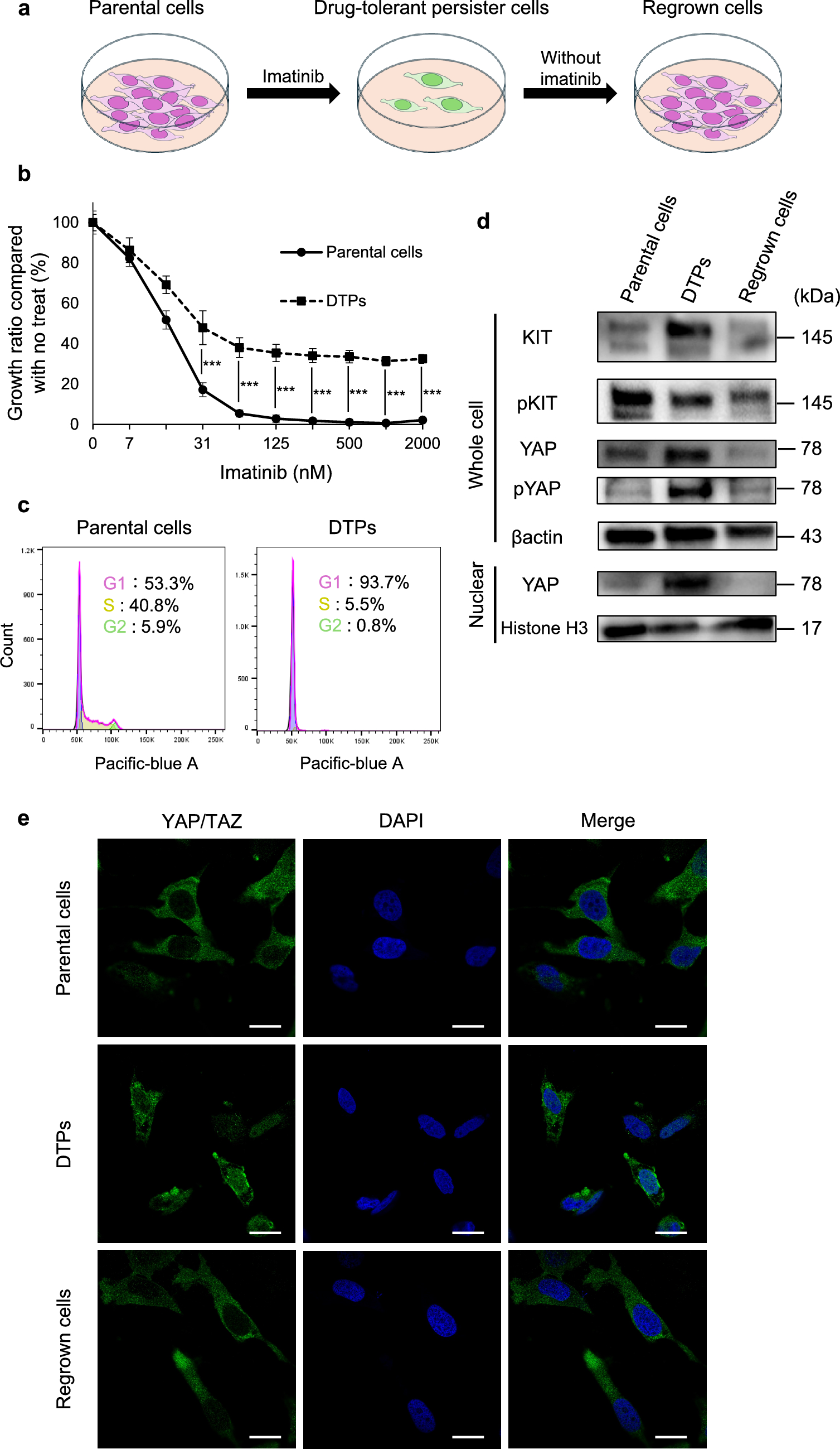

In this study, a total of 1442 samples from multiple cohorts of patients with gastric cancer were studied using three methods: immunohistochemistry (IHC, n = 977), whole transcriptome sequencing (WTS, n = 450) and digital spatial profiling (DSP, GeoMx platform, Nanostring Technologies, Inc, n = 15). Among the IHC cohorts, the South Korean CLASSIC trial contributed 549 samples, the Japanese KCCH cohort 215 samples, and the UK LTHT cohort 213. An overview of the samples is provided in Fig. 1.
Fig. 1
Summary of included samples including cohort details, method of CD20/B cell measurement and clinicopathological characteristics studied. Created with BioRender.com
CD20 density is higher in diffuse-type gastric cancerIHC staining was performed on 977 samples across three cohorts to establish the density of CD20 positive B cells, The distribution of CD20 density (% CD20 positive pixels of all pixels per core per patient) was consistent between cohorts (Fig. 2a–c). Supplementary Table S1 summarizes the baseline clinicopathological characteristics of the samples included in this study.
Fig. 2
Distribution of CD20 density across cohorts, by histological subtype and correlation with other immune cell biomarkers. A–C CD20 density per cohort. D CD20 density is significantly higher in the diffuse-type gastric cancer compared to intestinal-type GC (pooled analysis of all cohorts). E–G Per cohort analyses confirms higher CD20 density in diffuse-type GC in the CLASSIC and LTHT cohorts, but not in the KCCH cohort. H–J Correlation of CD20 density with other immune cell biomarkers
When analyzed across all cohorts, CD20 density was significantly higher in diffuse-type GC (n = 389) compared to intestinal-type GC (n = 470) (1.91% vs 1.56%, p = 0.00025) (Fig. 2d). This association remained significant when analysing the CLASSIC trial cohort (1.82% vs. 1.25%, p = 0.000014) (Fig. 2e) and the LTHT cohort (2.86% vs. 1.9%, p = 0.0004) (Fig. 2f) individually, but was not observed in the KCCH cohort (1.58% vs. 1.64%, p = 0.81) (Fig. 2g). Correlation analysis indicated that CD20 density was not significantly associated with other immune cell biomarkers, such as CD3, CD8, CD31, CD45, CD66b, CD68, and CD163 (Fig. 2h–j). CD20 density was not related to any of the other clinicopathological features (Table 1).
Table 1 Relationship between CD20 density and clinicopathological features per cohortRelationship of CD20 density and survival in patients with resectable gastric cancerAs expected, there were no significant differences in CD20 density in the resection specimen of patients who were treated with adjuvant chemotherapy after surgery compared to those who were treated by surgery alone (CLASSIC p = 0.82, KCCH p = 0.51). Patients with diffuse-type GC had poorer overall survival compared to those with intestinal-type GC in CLASSIC (HR 1.6; 95% CI: 1.1–2.3, p = 0.01)), KCCH (HR = 1.9 (95% CI = 1.3–2.9, p = 0.003)) and in LTHT (HR = 1.8 (95% CI = 1.2–2.6, p = 0.003).
Interestingly, patients with diffuse-type GC with low CD20 density (CD20-low diffuse-type) had the poorest OS when compared to all other patients. This was observed in patients from CLASSIC: CD20-low diffuse-type median OS = 49.0 months vs 62.0 months (HR = 1.9; 95% CI:1.2–3.0, p = 0.003) (Fig. 3a, Supplementary Figure S1a), and in patients from KCCH: CD20-low diffuse-type median OS = 49.1 vs 69.1 months (HR = 2.3 (95% CI = 1.2–4.2, p = 0.011)) (Fig. 3b, Supplementary Figure S1b). As the LTHT cohort only had 1 sample in the CD20-low diffuse-type group, this analysis could not be performed in LTHT (Supplementary Fig. S1c).
Fig. 3
Kaplan-Meier curves depicting OS survival analysis in IHC cohorts. A, B Survival analysis shows that patients with CD20-low diffuse-type GC have the poorest prognosis in both CLASSIC and KCCH. C Survival analysis of the surgery alone treated patients from the CLASSIC trial shows a strong difference between CD20-low diffuse-type GC and the rest of the patients. D In CLASSIC, patients with CD20-low diffuse-type GC have the same survival as the rest of the patients if treated with adjuvant chemotherapy
In the CLASSIC trial patients, we were able to perform further survival analyses of the CD20 density, stratifying patients by treatment. In patients treated with surgery alone, survival was significantly poorer in CD20-low diffuse-type patients (median OS = 46.0 vs 61.0 months (HR = 2.3 (95% CI = 1.3–4.2, p = 0.005)) (Fig. 3c, Supplementary Figure S1d). In patients treated by surgery and adjuvant chemotherapy, the difference in survival between CD20-low diffuse-type and the other patients was no longer apparent: CD20-low diffuse- type (median OS = 62.3 vs 63.0 months (HR = 1.8 (95% CI = 0.88–3.5, p = 0.108)) (Fig. 3d, Supplementary Figure S1e).
Multivariate analysis including CD20 density, histological subtype, combination of CD20-low and diffuse-type, disease stage, MSI status, sex, and treatment in the model, showed that while CD20 density alone was not an independent factor with respect to survival, a particular combination of CD20-low diffuse-type was associated with significantly poorer survival (Table 2). Similar univariate and multivariate analyses were also performed for the KCCH and the LTHT cohorts (Tables 3, 4).
Table 2 Univariate and multivariate survival analysis in the CLASSIC cohortTable 3 Univariate and multivariate survival analysis in the KCCH cohortTable 4 Univariate and multivariate survival analysis in the LTHT cohortAnalysis of Bulk-RNAseq data from TCGA STAD cohortTo assess the generalizability of our CD20 immunohistochemical findings, Bulk-RNAseq data from TCGA (n = 450) was analyzed. CIBERSORT v1.0 was used to estimate immune cell proportions based on Bulk-RNAseq data. Baseline clinicopathological characteristics are outlined in Supplementary Table S2. The proportion of B cells was significantly higher in the diffuse-type GC (n = 66) compared to intestinal-type GC (n = 189) (15% vs 7%, p < 0.001) (Fig. 4a). The proportion of B cells was not correlated with other immune cell proportions (Fig. 4b). Supplementary Table S3 summarizes the relationship of the proportion of B cells with clinicopathological features.
Fig. 4
Analysis of B-cell proportions determined by Bulk RNA-seq of TCGA STAD data. A Similar to immunohistochemically measured CD20 density, the B-cell proportion was greater in diffuse-type GC compared to intestinal-type GC. B Correlation analysis with other immune cell subtypes did not find any significant correlation
Digital spatial profiling demonstrates differences in B cell distribution within the Tumor MicroenvironmentExploratory analyses were conducted in 15 GC patients utilizing digital spatial profiling to establish the distribution of B cells within the TME. 1063 regions of interest (ROIs) were identified for analysis (Fig. 5a). Of these ROIs, 88 (8%) were from regions with intestinal metaplasia (IM), 130 (12%) from regions with lymphoid aggregates (LA), 179 (17%) from normal gastric epithelium, 138 (13%) from intratumoral stroma region, 11 (1%) from regions where lymphocytes were seen on top of tumor cells (TL), 87 (8%) from the tumor stromal interface (TSI) region and 430 (40%) from regions with tumor cells. A visual depiction of the staining and identification of these ROIs has been included in Fig. 5b.
Fig. 5
Digital spatial profiling transcriptomic analysis. A Haematoxylin/Eosin-stained TMA core with tumor outlined in blue to identify regions of interest (ROIs). B Identification of specific ROIs in one sample, NGC545. C Proportion of B cells in the tumor region is significantly higher than that of the intratumoral stroma region. D B-cell signatures are higher in tumor regions vs intratumoral stroma regions. Red lines indicate areas enriched with B cells; blue lines indicate areas with lower number of B cells. E B-cell distribution by region, stratified by histological subtype. Notably, diffuse-type samples appear to congregate in the tumor region, while intestinal-type samples are spread out more evenly between the other regions. F Immune cell proportion correlation in DSP samples does not show significant correlation with other immune cells
B cell proportions, calculated as the % of immune cells that were B cells, within each region were compared between patients (Supplementary Figure S2a) and within the same patient (Supplementary Figure S2b). The proportion of B cells was significantly higher in the tumor region compared to the intratumoral stroma (8%vs 1%, p < 0.001) (Fig. 5c, d).
The distribution of the B cells within each region and its association with clinicopathological characteristics was further analyzed. In diffuse-type GC, B cells appeared to congregate in the tumor cell ROIs, while B cells were more evenly distributed between the various ROI compartments in intestinal-type GC (Fig. 5e). The proportion of B cells was significantly higher in the intestinal-type GC compared to diffuse-type GC in the normal gastric epithelium (10.8% vs 1.4%, p < 0.05), TSI (tumor-stromal interface) (7.3% vs 2%, p = 0.026), and the intratumoral stroma (1.2% vs 0.2%, p = 0.029) regions.
B cell proportions were also significantly higher in the tumor region in stage IV GC vs stage I-III GC (Supplementary Figure S3a) (17% vs 7%, p < 0.05); and higher in distal vs proximal GC (Supplementary Figure S3b) (10% vs 6%, p < 0.05). Correlation analysis did not find any significant correlation between B cell proportion and other immune cells (Fig. 5f) (− 0.28 < r < 0.24). No significant correlations were noted when stratifying the correlation analysis by histological subtype, stage or tumor location (Supplementary Figure S3c–e).
Comments (0)