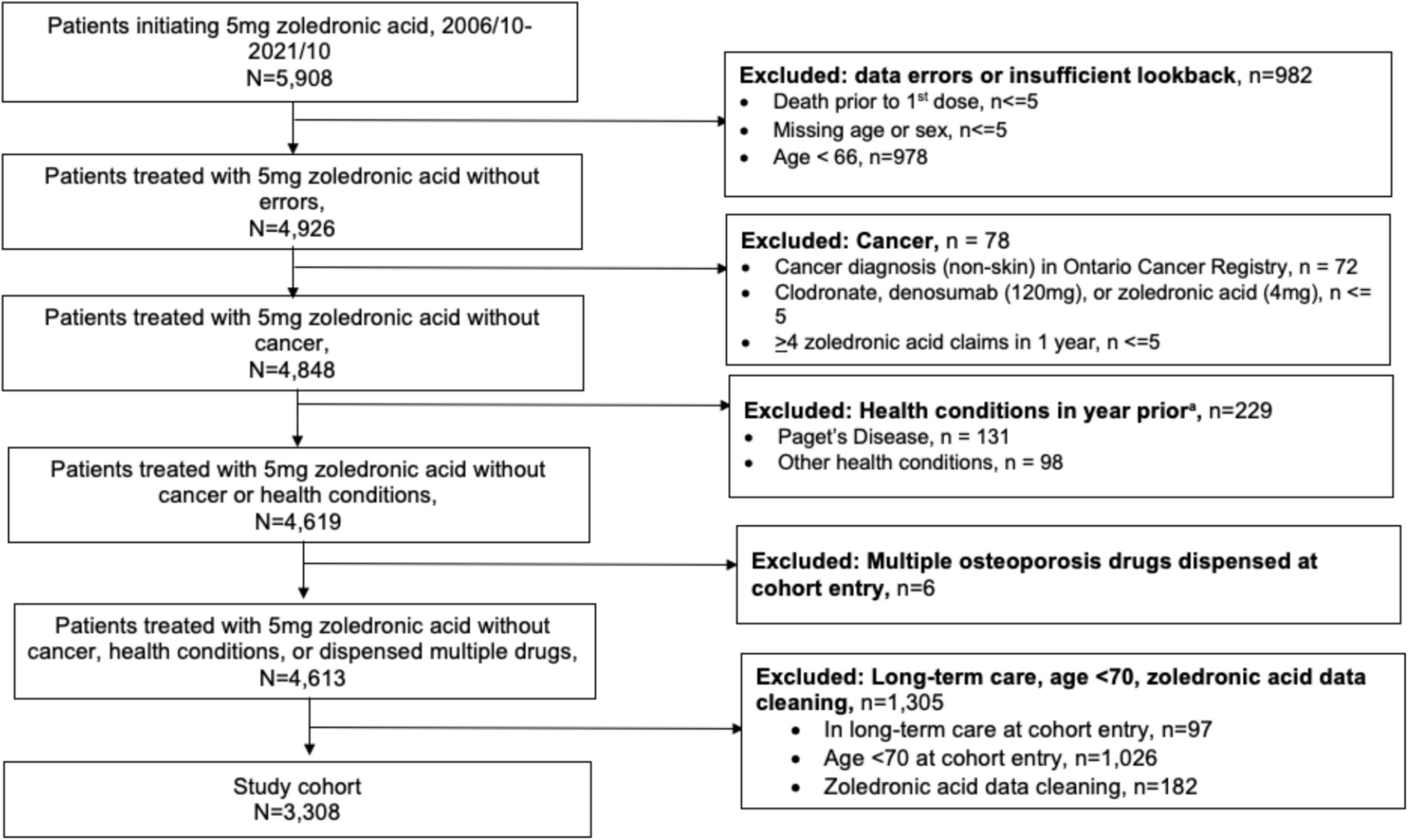
Our study found that the VF + group exhibited significantly lower GMV in a specific brain cluster compared to the VF − group. This cluster included the right hippocampus, right amygdala, right parahippocampal gyrus, and right superior temporal pole. Additionally, a significant interaction effect between sex and the presence of VF was observed for the reduction in GMV in this specific cluster.
Few studies have suggested a link between VF and brain atrophy [24]. Bae et al. investigated the relationship between osteoporotic vertebral compression fractures (OVCFs) and brain volume using MRI. They included 246 osteoporotic patients and analyzed their brain volume using semi-automated tools. They found a significant association between OVCFs and reduced brain parenchyma volume, alongside increased lateral ventricle volume. In line with their findings, our study also identified significant reductions in GMV in a specific brain cluster, including the right hippocampus, right amygdala, right parahippocampal gyrus, and right superior temporal pole in the VF + group. Both studies highlight the impact of vertebral fractures on brain structure, suggesting that these fractures may contribute to neurodegeneration. While Bae et al. focused on the overall brain parenchyma and lateral ventricles, our study provides a more detailed regional analysis, pinpointing specific areas of gray matter reduction. This complementary evidence reinforces the notion that vertebral fractures are not only a skeletal concern but also have significant implications for brain health, particularly in regions critical for cognitive and emotional processing.
There were some reports about the relationship between brain structure and BMD [12, 13]. Loskutova et al. examined BMD in early Alzheimer's disease (AD) and its relationship to brain structure and cognition [12]. They found that BMD was lower in patients with early AD compared to non-demented controls and that lower BMD was associated with reduced whole brain volume and poorer cognitive performance, particularly in memory tasks. Zhang et al. conducted a mediation analysis investigating the associations among BMD, brain atrophy, and gait variability (the stride-to-stride fluctuations in walking) [13]. They found that lower BMD, particularly in the lumbar spine, was associated with higher gait variability, and that brain atrophy in regions such as the primary motor cortex and sensorimotor cortex mediated this relationship. These studies highlight the systemic nature of bone loss and its association with global brain atrophy. However, VF differ from BMD loss alone, as they may reflect specific factors beyond systemic bone metabolism.
In our study, the observed GMV reductions in the VF + group were localized to the hippocampus, amygdala, and parahippocampal gyrus, regions known to be functionally specialized for memory, emotional processing, and visuospatial cognition. Unlike systemic BMD loss, which has been hypothesized to be associated with global brain volume reductions, the observed localization of GMV loss aligns with the concept that brain regions exhibit functional specialization. This specific association suggests that vertebral fractures may reflect not only systemic metabolic bone loss but also meaningful processes such as chronic pain, reduced physical activity, or functional mobility limitations caused by the fractures. These physical and functional consequences could be related to localized brain structure changes, highlighting the distinct nature of VF beyond being a simple surrogate marker for BMD.These studies indicate a broader connection between bone health and brain structure, extending beyond specific conditions like VF. The integration of these findings highlights the importance of considering both skeletal and neural health in managing conditions such as osteoporosis and neurodegenerative diseases. In our study, we found a significant association between the reduction in specific brain clusters and the presence of VF, even after adjusting for MMSE scores. This suggests that the reduction in brain regions responsible for cognition may be associated with VF; however, due to the cross-sectional design of this study, we cannot determine causality. It is equally possible that VF could lead to reduced mobility and physical activity, which in turn impacts brain structure by decreasing environmental interactions and stimulation.
The cluster identified in our study includes brain regions such as the right hippocampus, right amygdala, and right parahippocampal gyrus, which are crucial for memory and emotional processing. The limbic system, comprising the hippocampus and amygdala, is integral to cognitive and emotional functions [25, 26]. The hippocampus is essential for memory formation and spatial navigation [27, 28]. Age-related changes in the hippocampus are linked to declines in memory and increased susceptibility to psychosocial stress and mental health issues [29]. The amygdala is involved in emotional processing and memory, with age-related changes affecting emotional regulation [30, 31]. The parahippocampal gyrus, part of the medial temporal lobe memory system, is crucial in maintaining and updating memories [32]. It is involved in visuospatial processing, particularly in extracting scene layout information [33], and is essential for visuospatial memory functions and contextual associations [34,35,36]. Bohbot et al. showed that patients with lesions that included the right parahippocampal cortex were severely impaired on a task that required learning the spatial configuration of objects on a computer screen; these patients, however, were not impaired at learning the identity of objects [35]. The association between VF and reductions in the parahippocampal gyrus volume may suggest a bidirectional relationship. VF can lead to decreased mobility and physical activity, potentially contributing to decreased visuospatial memory function due to reduced environmental interaction. Conversely, impaired visuospatial memory function might increase the risk of falls and subsequent VF. Although this remains a hypothesis, the relationship suggests that interventions aimed at improving both physical mobility and cognitive functions, particularly visuospatial memory, could benefit individuals at risk of VF.
Our study also revealed a significant interaction between sex and VF status, showing that males in the VF + group exhibited more pronounced reductions in the specific brain cluster compared to females in the VF + group. This novel finding suggests that while postmenopausal women may experience more significant bone density loss relative to brain volume reduction due to the effects of osteoporosis, men may experience concurrent reductions in both brain volume and bone density. This indicates that the neurodegenerative and osteoporotic processes might occur simultaneously in men. Therefore, the integrated approach to patient care should consider these sex-specific differences. For females, particularly postmenopausal women, interventions might need to prioritize bone health to mitigate extensive bone density loss, whereas for males, strategies should focus on both maintaining bone health and preventing brain atrophy. This personalized approach could potentially improve patient outcomes by addressing both skeletal and neural aspects of health.
One of the strengths of our study is the use of VBM, which allows for precise measurement of gray matter volumes across the entire brain. Additionally, the large sample size of 1,751 participants enhances the generalizability of our findings. The inclusion of both male and female participants and the examination of sex-specific effects add further depth to our analysis. Furthermore, the adjustment for cognitive function using MMSE scores strengthens the validity of our findings.
However, there are several limitations in the present study. First, the participants were recruited from only two regions (coastal and mountainous), which may limit the generalizability of our findings to the broader population. However, we compared anthropometric measurements and the prevalence of smoking and alcohol consumption between our participants and the general Japanese population and found minimal significant differences [15, 37]. This suggests that our study sample may be somewhat representative of the general population. Second, the cross-sectional design of this study makes it challenging to establish causal relationships between vertebral fractures and brain volume reductions. Longitudinal studies are generally required to confirm causality. Given the long-term follow-ups involved in our study, we plan to analyze the results from longitudinal surveys in future research to better understand these relationships. Third, this study focused on simple brain volume comparisons and did not include advanced imaging techniques such as diffusion-weighted tractography or functional MRI analyses. Given that MRI scans were performed as part of health check-ups for 1,755 local residents, it was impractical to conduct more detailed MRI investigations due to cost and human resource constraints. Fourth, when we limited the analysis to participants aged 60 years and older (n = 1,212), statistical significance in some results was lost, likely due to the reduced sample size in this subgroup. Even with age included as a nuisance regressor in the analysis, the results may still have been influenced by the presence of younger participants in the full dataset. Fifth, only 1,285 participants (66.9%) completed the MMSE, primarily because the MMSE was administered mainly to participants aged 60 and over, which limits the comprehensiveness of our cognitive assessments. Sixth, there were several bone-related limitations: we lacked bone density information, which could provide further insights into the relationship between bone health and brain structure; we did not have information on whether the vertebral fractures were new or old, or whether they were traumatic or non-traumatic (pathological fractures); and information on the use of osteoporosis medications (such as antiresorptive agents, anabolic agents, vitamin D supplements, etc.) was also not available. This lack of detailed fracture and treatment information might affect the interpretation of our results. Despite these limitations, our study provides important insights into the relationship between vertebral fractures and brain structure, highlighting the need for integrated approaches to managing skeletal and neural health.


Comments (0)