
Remember me

Research on the influence of GSH on breast cancer chemotherapy sensitivity remains limited. In this study, by collecting clinicopathological data from patients receiving adjuvant chemotherapy, we found that high GSH intake increases the risk of recurrence after adjuvant chemotherapy. Additionally, our molecular experiments confirmed that chemotherapy drugs increase the expression levels of intracellular GSH and antioxidant enzymes, leading to chemotherapy resistance during neoadjuvant chemotherapy. We propose that the dosage and duration of GSH administration in clinical treatment be carefully evaluated and standardized to prevent overuse and mitigate the risk of chemotherapy resistance.
Increasing evidence indicates that elevated GSH level may contribute to chemotherapy resistance. Nevertheless, GSH is commonly used as a liver-protective drug after chemotherapy for breast cancer patients to mitigate chemotherapy-induced hepatotoxicity. Although many types of hepatoprotective drugs are available, GSH is often overused by clinicians, with its effects on tumors being overlooked. Previous studies have shown that the mechanism by which chemotherapy drugs induce tumor cell death primarily involves the promotion of ROS production [14]. An increase in ROS accelerates tumor cell death, while GSH, as a key component in redox homeostasis, plays a crucial role in scavenging ROS and neutralizing exogenous metabolites in tumor cells [15]. Thus, GSH is essential for tumor cell survival. Previous literature has demonstrated that GSH levels are elevated in various tumors, including breast, colon, laryngeal, gastric, and lung cancers [16]. Moreover, elevated GSH levels can protect tumor cells by reducing the cytotoxic effects of several chemotherapy drugs, including cisplatin, doxorubicin, melphalan, and paclitaxel, ultimately leading to drug resistance [17]. In clinical practice, during chemotherapy for patients with impaired liver function, the liver-protective effect of GSH is often prioritized, while the potential risk of chemotherapy resistance due to its non-standardized intake is frequently overlooked. This issue warrants deeper consideration. However, no systematic studies have been published to investigate the relationship between GSH intake and post-chemotherapy recurrence in breast cancer patients. Our study provides compelling speculation that GSH intake may adversely affect the prognosis of breast cancer patients undergoing chemotherapy.
After PSM, our study identified significant differences in transaminase levels, recurrence rates, and mortality after adjuvant chemotherapy between breast cancer patients between GSH group and Non-GSH group, indicating that GSH intake may have adverse effects on chemotherapy outcomes. To enhance risk stratification and optimize treatment selection, we divided the patients into two groups based on GSH intake duration: HGSH (≥ 16 days, n = 32) group and LGSH (< 16 days, n = 232) group. Patients in the HGSH group exhibited significantly higher recurrence rates, and mortality compared to the LGSH group. Kaplan–Meier survival analysis also revealed a significantly decreased survival rate in the HGSH group. These findings suggest that high GSH intake correlates with poorer prognosis in breast cancer patients who underwent adjuvant chemotherapy. Specifically, the use of chemotherapy drugs resulted in elevated levels of ALT and AST, and an increase in the severity of side effects such as nausea and vomiting, contributing to high GSH intake. This, in turn, increased the likelihood of chemotherapy resistance, recurrence, and worsened prognosis. Previous in vitro studies demonstrated that reduced GSH levels in tumor cells promoted cell death, while elevated levels inhibited cell death [18]. Our findings align with these previous studies, leading us to hypothesize that GSH levels are higher in the tumor tissues of chemotherapy-resistant breast cancer patients compared to chemotherapy-sensitive patients.
In these retrospective studies, Triple-negative status, Lymphovascular invasion, Ki67 ≥ 30%, and High GSH intake were identified as independent risk factors for poor postoperative prognosis in breast cancer chemotherapy patients. Ki67 is commonly used as a marker for breast cancer proliferation [19]. In early-stage breast cancer, high Ki67 expression is associated with poor prognosis [20]. Additionally, elevated Ki67 levels are considered high-risk factors when selecting chemotherapy regimens [21]. It is well established that triple-negative breast cancer (TNBC) is highly aggressive [22], with significantly higher rates of metastasis and recurrence compared to other breast cancer subtypes [23], especially in isceral and brain metastasis [24]. To improve the prognosis of metastatic TNBC patients, antibody–drug conjugates (ADCs), such as sacituzumab govitecan, have been utilized to specifically deliver potent chemotherapy agents directly to cancer cells expressing targeted antigens, demonstrating promising efficacy [25]. Furthermore, lymphovascular invasion is a critical step in tumor dissemination and a strong adverse prognostic factor for breast cancer survival [26]. These pathways enable tumor cells to metastasize to distant organs [27]. Our study is consistent with previous research, revealing that Triple-negative status, Lymphovascular invasion, and Ki67 ≥ 30% are risk factors for recurrence after chemotherapy. Additionally, our study confirmed that High GSH intake is an independent risk factor for poor prognosis, offering critical cautionary note for clinical interventions in postoperative breast cancer treatment strategies.
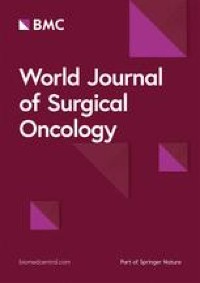
To demonstrate the role of GSH in chemotherapy resistance, we analyzed transcriptomic data from chemotherapy patient samples in the GEO database and collected post-chemotherapy samples from patients who underwent neoadjuvant chemotherapy, assessing the expression levels of GSH and GSH synthesis-related antioxidant enzymes. Our study revealed a significant upregulation of the GSH metabolic pathway in chemotherapy-resistant patients. Moreover, in fresh samples from patients who underwent neoadjuvant chemotherapy, the GSH/GSSG levels in the resistant group were significantly higher than those in the sensitive group, which is consistent with previous research [28]. Additionally, studies have shown that antioxidant enzymes GPX4 and SOD1, along with the transcription factor NRF2, are involved in intracellular GSH redox reactions [29, 30]. The upregulation of NRF2 and SOD1, along with the activation of ROS scavenging pathways, can lead to resistance to chemotherapy or targeted therapy in various tumors. For instance, activation of NRF2 promotes ROS clearance in drug-resistant liver cancer cell lines [31]. Specifically, NRF2 is a transcription factor that governs the antioxidant pathway by targeting GSH metabolism-related genes. In chemotherapy-sensitive patients, Keap1 (Kelch-like ECH-associated protein 1) binds to NRF2 and mediates NRF2 ubiquitination via the Cul3-E3 ligase complex, promoting its degradation and maintaining NRF2 at low levels [32]. However, in resistant patients, oxidative stress causes a conformational change in Keap1, leading to the release of NRF2, which translocates to the nucleus and binds to antioxidant response element (ARE), activating the expression of downstream antioxidant genes, including GPX4, NQO1, HO-1 and SOD1 [33], and promoting the synthesis of GSH, as illustrated in Fig. 5.
Fig. 5
Diagram of antioxidant mechanisms in chemotherapy-resistant and chemotherapy-sensitive patients
GSH is synthesized in two steps: First, gamma-glutamylcysteine (γ-GC) is formed from glutamate and cysteine, which is the rate-limiting step in GSH production. In the second step, glutathione synthetase (GS) adds glycine to γ-GC to produce GSH. NRF2 plays a critical role in this process by upregulating the expression of genes involved in the synthesis of γ-GC, thereby increasing the overall production of GSH [34]. Elevated GSH levels help neutralize ROS by reacting with hydrogen peroxide (H2O2), which is produced from superoxide radicals (·O2-). This reaction converts H2O2 and GSH into water and GSSG, thereby reducing ROS-induced cellular damage [35]. Through NRF2-driven GSH synthesis, tumor cells are protected from oxidative stress, which contributes to chemotherapy resistance by reducing the cytotoxic effects of ROS generated during treatment.
In addition, the high expression of NRF2 enhances the expression of downstream SOD1, an important antioxidant enzyme that plays a crucial role in protecting cells from oxidative damage [36]. Specifically, SOD1 catalyzes the conversion of ·O2- into H2O2, thereby reducing the levels of ROS and mitigating oxidative stress [37]. By activating the expression of SOD1, NRF2 helps maintain cellular redox balance and protects cells from oxidative injury [38].
GPX4 is an antioxidant enzyme that primarily reduces lipid peroxides, especially lipid hydroperoxides, into their corresponding alcohols, thus protecting cell membranes from oxidative damage [39]. This reaction is essential in lipid-rich environments, such as cellular membranes, where lipid peroxidation can lead to cell death. GPX4 works in conjunction with GSH, as it utilizes GSH as a cofactor to catalyze the reduction of lipid peroxides. By promoting the expression of GPX4 and GSH, the cellular defense mechanisms of the cell membrane are enhanced, reducing damage from lipid peroxides and maintaining cellular integrity [40].
Immune regulation within the tumor microenvironment plays a critical role in tumor progression and chemotherapy resistance. Zhang’s study demonstrated that the synthesis of GSH is intricately linked to serine metabolism, with its metabolic byproducts lactate and 2-hydroxyglutarate (2-HG) suppressing the cytotoxic function of T cells. The metabolic pathways promote the development of an immunosuppressive vascular microenvironment, thereby facilitating resistance to chemotherapy [41]. In addition, Yang et al. found that active GSH metabolism within tumors further suppresses immune responses, such as IFN-γ signaling and PD-1/PD-L1 interactions, while reducing the number of M1 macrophages and increasing the proportion of M2 macrophages. Moreover, GPX4 promotes immune evasion by inhibiting ferroptosis. The combination of GPX4 inhibitors with immune checkpoint inhibitors (ICIs) targeting the PD-1/PD-L1 axis increases the proportion of CD8 + T cells, enhances their cytotoxic activity and induces ferroptosis, thereby promoting chemotherapy resistance [42]. In addition, the incorporation of ICIs into neoadjuvant chemotherapy regimens has been shown to increase the pCR rate [43]. However, despite their significant therapeutic potential in cancer treatment, it is essential to also focus on managing immune-related adverse events (irAEs). These adverse effects, including peripheral neuropathy and hearing loss, can have a profound impact on patient quality of life and require careful monitoring throughout treatment [44, 45].
Our study also found that NRF2, GPX4 and SOD1 expression levels were higher in the neoadjuvant chemotherapy-resistant group compared to the sensitive group, further supporting our hypothesis and demonstrating the adverse effects of GSH on chemotherapy in breast cancer patients. Additionally, we discovered that a GSH threshold of 10.2 nmol/mg effectively differentiates resistant cells from sensitive cells, with an AUC of 0.913, providing a robust distinction between chemotherapy-resistant and sensitive patients. Patients were classified into IGH and IGL groups according to the cutoff value of intracellular GSH levels. Kaplan–Meier survival analysis revealed that the IGH group exhibited significantly poorer survival outcomes compared to the IGL group, suggesting that GSH may contribute to neoadjuvant chemotherapy resistance and lead to a worse prognosis in breast cancer patients. These findings offer a theoretical basis for precision treatment of breast cancer chemotherapy involving GSH.
Further investigation into the role and mechanisms of GSH and its associated antioxidant enzymes is essential for reversing chemotherapy resistance [46]. Several research teams have targeted GSH synthesis inhibitors as potential therapeutic agents. For instance, Li et al. examined the impact of GSH synthesis inhibitors on the cytotoxicity of cisplatin and gemcitabine in bile duct cancer cells [47]. Their findings revealed that low concentrations of GSH synthesis inhibitors significantly enhanced cancer cell death. This effect was similarly observed in a separate study on ovarian cancer [48]. Additionally, researchers have developed a range of glutathione-depleting nanodrugs for efficient cancer treatment [49], particularly in addressing chemotherapy resistance in TNBC [50]. Collectively, these studies underscore the significance of investigating the role of GSH in breast cancer patient prognosis, stressing the necessity of regulating GSH usage to prevent non-standardized intake and decrease post-chemotherapy recurrence.
However, our study has several limitations. As a single-center retrospective analysis, it may be subject to selection bias, and conclusions should be validated in large-scale multicenter prospective studies. Furthermore, the precise mechanisms of glutathione’s impact remain unclear, and future research should investigate its specific roles in chemotherapy resistance and cell survival. Additionally, we primarily focus on the duration of GSH intake, without examining the effect of specific dosage on the prognosis of breast cancer patients undergoing chemotherapy. Future studies should employ stratified analyses or dose–response curves to explore the influence of different GSH doses on prognosis, offering guidance for more precise clinical application.
Comments (0)