
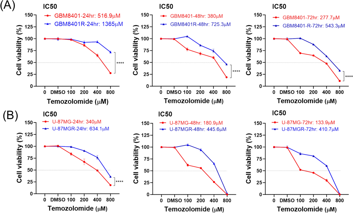
Remember me

EVs have emerged as a significant focus in recent leukemia research due to their pivotal roles in disease initiation and progression [27, 77, 78]. These small vesicles, released by cancer cells, transport a wide array of biomolecules, including proteins, miRNAs, and double-stranded DNA (dsDNA). The molecular content of EVs plays a profound role in leukemia pathogenesis, particularly within the bone marrow microenvironment (BMM), acting as key mediators of intercellular communication [79]. This interaction influences various cellular processes critical to the disease, highlighting the importance of EVs in shaping the leukemic landscape.
One of the critical functions of EVs is promoting angiogenesis, the formation of new blood vessels that supply tumors with essential nutrients and oxygen. This process is vital for tumor growth, as larger and more aggressive leukemias require an enhanced blood supply to sustain their metabolic demands. EVs not only deliver metabolic substrates and enzymes that support the survival of leukemic cells but also carry signaling molecules that stimulate cell proliferation. Consequently, these vesicles accelerate tumor growth, contributing to the aggressive nature of leukemia [80, 81].
EVs are also instrumental in the development of drug resistance, a significant challenge in leukemia treatment. They can transfer specific miRNAs that either suppress tumor suppressor genes or upregulate proteins linked to resistance mechanisms [81]. For example, certain miRNAs can inhibit the expression of pro-apoptotic genes, allowing leukemic cells to evade programmed cell death even in the presence of chemotherapeutic agents. This transfer of resistance mechanisms complicates treatment strategies, necessitating the exploration of novel interventions that target EVs to overcome resistance [82].
Moreover, EVs actively remodel the bone marrow niche, fostering an environment that supports the survival and proliferation of leukemic cells while suppressing immune responses. Thus, by altering the composition and activity of the BMM, EVs contribute to an immunosuppressive microenvironment that enables leukemic cells to evade detection and destruction by the immune system. This capacity to modulate immune responses is critical, particularly as it impedes the effectiveness of immunotherapies designed to activate the body’s immune system against cancer [83].
Nonetheless, the ability of EVs to carry genetic and epigenetic abnormalities makes them valuable for clinical applications [84]. For instance, profiling specific miRNAs in EVs may help identify patients at a higher risk of relapse or treatment failure, enabling clinicians to tailor therapeutic approaches accordingly [85]. Furthermore, the detection of unique EV markers in bodily fluids, such as blood or bone marrow, could provide insights into disease burden or treatment response, facilitating improved risk stratification. Indeed, by identifying specific genetic signatures within EVs, clinicians can better understand the molecular underpinnings of leukemia and make more informed decisions regarding patient management [81, 86].
In ALL, harnessing EVs for biomarker identification presents promising opportunities for enhancing diagnosis and personalizing therapy [78, 87]. For example, the detection of specific EV-derived miRNA profiles could help differentiate between various ALL subtypes, guiding treatment decisions [88]. Besides, utilizing blood samples to assess EV content offers a less invasive alternative to traditional bone marrow biopsies for monitoring disease status over time [86]. Overall, the integration of EV profiling into clinical practice holds great potential for advancing the understanding and management of leukemia, ultimately leading to improved patient outcomes through more effective and personalized therapeutic strategies (Fig. 1).
Fig. 1
The role of EVs in the pathogenesis and progression of ALL. In the context of ALL, EVs derived from leukemic cells are shown to influence various aspects of disease biology, such as leukemic cell proliferation, drug resistance, immune evasion, and communication with the bone marrow microenvironment. Additionally, EVs derived from stromal cells and other components of the TME are illustrated to contribute to disease progression by modulating leukemic cell behavior and altering the surrounding niche
Regulatory impact of EVs on the BMMLeukemia-derived EVs have emerged as pivotal modulators of the BMM in ALL. These small membrane-bound vesicles carry a diverse array of bioactive molecules, including proteins, miRNAs, and other signaling molecules, that significantly impact the behavior of stromal and immune cells within the bone marrow [27, 78, 81, 89].
One of the key mechanisms through which EVs influence the BMM is by transferring oncogenic miRNAs into bone marrow mesenchymal stromal cells (BM MSCs). For instance, two oncogenic miRNAs, miR-155 and miR-181, are frequently upregulated in various types of leukemia. When leukemic cells release EVs containing these miRNAs, they can fuse with or be taken up by BM MSCs, leading to the alteration of the recipient cells’ gene expression profiles. This transfer is a critical mechanism by which leukemic cells manipulate the surrounding microenvironment to support their own survival and proliferation [90].
Once inside the BM MSCs, miR-155, and miR-181 suppress the expression of crucial regulatory genes like SOCS1 and PTEN. SOCS1 (Suppressor of Cytokine Signaling 1) is a negative regulator of cytokine signaling, specifically in pathways, such as JAK/STAT, which are activated by various growth factors and cytokines. Indeed, by inhibiting SOCS1, the BM MSCs become more responsive to cytokines, resulting in enhanced signaling that promotes leukemic cell survival. On the other hand, PTEN (Phosphatase and Tensin Homolog) is a well-known tumor suppressor that counteracts the PI3K/AKT pathway, which is critical for cell survival, growth, and proliferation. Downregulation of PTEN, therefore, diminishes its regulatory influence, leading to increased activation of pro-survival signaling pathways [27, 78, 91].
The downregulation of SOCS1 and PTEN alters the secretion profile of BM MSCs, significantly increasing the production of pro-survival molecules, particularly interleukin-6 (IL-6) and stromal-derived factor 1 (SDF-1 or CXCL12). IL-6 is a pleiotropic cytokine that plays a vital role in inflammation and immune responses. Elevated levels of IL-6 in the bone marrow microenvironment contribute to a chronic inflammatory state that not only supports leukemic cell growth but also disrupts normal hematopoiesis by impairing the functionality of healthy HSCs. Specifically, IL-6 can inhibit the differentiation of HSCs into mature blood cells, allowing leukemic cells to dominate and outcompete normal hematopoiesis [92, 93].
CXCL12, also known as SDF-1, is another critical factor produced by BM MSCs in response to the altered signaling landscape created by EV-derived miRNAs. CXCL12 enhances the retention and homing of leukemic cells within the bone marrow. It does so by binding to its receptor, CXCR4, on leukemic cells, promoting their migration and retention in the bone marrow niche. This interaction fosters a protective microenvironment for leukemic cells, shielding them from therapeutic interventions and further supporting their proliferation and survival [94].
EVs disrupt normal hematopoiesis by directly targeting hematopoietic stem cells (HSCs) and progenitor cells, as seen with EVs carrying miR-486, which interfere with key differentiation pathways and inhibit healthy blood cell production [95]. This disruption allows leukemic cells to outcompete their normal counterparts for resources and space within the bone marrow, contributing to the cytopenias frequently observed in leukemia patients; by altering the balance between leukemic and healthy cells, EVs effectively tilt the microenvironment in favor of leukemia, facilitating its dominance [96]. Additionally, EVs play a critical role in remodeling the extracellular matrix (ECM) of the BMM by carrying matrix metalloproteinases (MMPs), enzymes that degrade ECM components and create physical space for leukemic cells to invade and establish themselves [97]. This degradation alters the structural integrity of the bone marrow and releases additional growth factors, such as transforming growth factor-beta (TGF-β), which further stimulates leukemic growth and survival, ultimately establishing a more favorable microenvironment for their persistence while facilitating leukemic cell expansion [98].
EVs derived from leukemia cells are pivotal in promoting angiogenesis, a process crucial for the progression of leukemia. They achieve this by delivering key pro-angiogenic factors to endothelial cells within the bone marrow. Among these factors, VEGF plays a central role. VEGF binds to its receptors on the surface of endothelial cells, triggering a cascade of signaling pathways, notably the PI3K/AKT, MAPK/ERK, and JAK/STAT pathways. The activation of the PI3K/AKT pathway enhances cell survival and growth, which is essential for the stability and longevity of newly formed blood vessels [99]. Simultaneously, the MAPK/ERK pathway is responsible for driving endothelial cell proliferation and migration, processes that are integral to the formation of new blood vessels. The cooperation of these pathways facilitates not only the structural formation of blood vessels but also ensures their functionality, which is necessary to supply nutrients and oxygen to the expanding leukemic mass [99] (Fig. 2).
Fig. 2
In ALL, several key molecular pathways contribute to cancer progression, invasion, and metastasis. The EGFR (epidermal growth factor receptor) pathway is activated upon ligand binding, triggering downstream signaling cascades, such as the Ras/Raf/MEK/ERK and PI3K/AKT/mTOR pathways. These cascades promote cell survival, proliferation, and angiogenesis. Specifically, AKT and mTOR regulate cell growth and metabolism, while ERK influences cell division. STAT3 is activated by cytokines like IL-2β, which promotes cell survival and immune evasion. AMPK, a metabolic sensor, can negatively regulate mTOR to limit excessive growth, but its dysregulation contributes to cancer progression. Together, these pathways drive leukemia cells’ ability to grow uncontrollably, invade surrounding tissues, and metastasize, making them critical therapeutic targets
In addition to VEGF, miR-210 is another critical pro-angiogenic factor transported by leukemic EVs. This miRNA plays a significant role under the hypoxic conditions frequently encountered within tumors. miR-210 stabilizes hypoxia-inducible factor 1-alpha (HIF-1α), a transcription factor that induces the expression of various pro-angiogenic genes, including VEGF itself. By upregulating these genes, miR-210 promotes angiogenesis, ensuring that the tumor microenvironment remains adequately vascularized. Furthermore, miR-210 also targets and downregulates Ephrin-A3 (EFNA3), an anti-angiogenic factor that typically inhibits blood vessel formation. Thus, miR-210 enhances the angiogenic potential of endothelial cells, thereby supporting the growth and survival of leukemic cells through improved nutrient and oxygen delivery [100, 101].
Beyond their role in angiogenesis, leukemia-derived EVs also play a crucial role in modulating immune responses, thereby facilitating leukemia progression. These EVs carry immunosuppressive molecules, such as transforming growth factor-beta (TGF-β) and programmed cell death ligand 1 (PD-L1), which directly influence the activity of immune cells within the bone marrow microenvironment [102]. TGF-β is known for its capacity to impair T-cell proliferation and function, effectively diminishing the immune system’s ability to mount a robust response against leukemic cells. On the other hand, PD-L1 binds to PD-1 receptors on T cells, inhibiting their activation and further reducing their capacity to recognize and eliminate leukemia cells. This interaction between PD-L1 and PD-1 is a critical checkpoint mechanism that tumors exploit to evade immune detection [102].
Moreover, EVs derived from leukemic cells can suppress the activity of natural killer (NK) cells, which are essential components of the innate immune system responsible for targeting and killing malignant cells. Leukemia-derived EVs can limit the secretion of cytotoxic granules, such as perforin and granzymes, from NK cells, thereby diminishing their ability to effectively eliminate leukemic cells [103]. In addition to inhibiting cytotoxicity, these EVs promote the expansion of regulatory T cells (Tregs). Tregs are immunosuppressive cells that dampen immune responses by inhibiting the activity of effector T cells, further fostering a tolerogenic environment that allows leukemia cells to thrive [104].
The collective immunosuppressive effects of leukemic EVs enable leukemia cells to evade immune surveillance effectively. This immune evasion contributes to disease progression and enhances the resistance of leukemia cells to immune-based therapies, making it challenging to achieve successful treatment outcomes [105]. Indeed, by remodeling the bone marrow microenvironment, promoting angiogenesis, and suppressing immune responses, leukemia-derived EVs create a supportive niche that facilitates leukemia progression and complicates therapeutic interventions [106]. Thus, targeting these EV-mediated pathways holds promise for developing novel therapeutic strategies aimed at disrupting the supportive microenvironment of leukemia and enhancing anti-leukemic immune responses.
Effects of EVs on leukemia cell proliferationEVs significantly influence the proliferation and apoptosis of leukemia cells through complex mechanisms involving the transfer of specific miRNAs and other signaling molecules [107]. In ALL, for instance, studies have shown that miRNA-181b-5p is highly expressed in ALL cell lines. EVs that carry this miRNA promote various aspects of leukemic cell behavior, including proliferation, migration, and invasion, while simultaneously inhibiting apoptotic processes. This suggests a pivotal role for miR-181b-5p in enhancing the survival and aggressive characteristics of ALL cells [108]. Similarly, EVs derived from patients with pediatric ALL (pALL) contain miR-181a, which upregulates several proliferation and survival genes, such as proliferating cell nuclear antigen (PCNA), Ki-67, myeloid cell leukemia-1 (MCL-1), and B-cell lymphoma 2 (BCL2). In vitro experiments have demonstrated that specific inhibition of miR-181a can reverse the proliferation induced by EVs, indicating its potential as a valuable therapeutic target in the treatment of ALL [109].
In adult T-cell leukemia/lymphoma (ATL), which is associated with infection by the human T-lymphotropic virus type 1 (HTLV-1), infected lymphocytes release EVs containing viral Tax proteins, pro-inflammatory mediators, and viral mRNA transcripts [110]. These EVs enhance the survival of leukemia cells by inhibiting the expression of Fas, a receptor that mediates apoptosis, through the activation of the AKT signaling pathway. This anti-apoptotic effect is contingent upon the presence of cFLIP (cellular FLICE-like inhibitory protein), a protein that inhibits apoptosis signaling, further underscoring the role of EVs in promoting cell survival in leukemia [111].
Moreover, leukemia-derived EVs exhibiting a stem cell phenotype not only enhance their own proliferation and survival but also bolster the survival and growth of other malignant cells [96].This creates a self-perpetuating signaling loop that contributes to chemoresistance in leukemia. Factors like fibroblast growth factor-2 (FGF2) and HMG-CoA reductase play crucial roles in this process, as increased cholesterol production supports the formation of cell membranes in rapidly dividing cells, facilitating their growth [112].
In addition, the activation of the STAT3 signaling pathway by specific microRNAs, such as miRNA-1246, involves complex cellular and molecular mechanisms that enhance the malignant behavior and aggressiveness of leukemia. MiR-1246 can be packaged into EVs or released directly by leukemia cells, facilitating its uptake into neighboring cells through endocytosis. Once inside the cell, miR-1246 associates with the RNA-induced silencing complex (RISC), where it binds to complementary sequences in the 3′ untranslated region (UTR) of target mRNAs regulating the STAT3 pathway, typically inhibiting negative regulators like SOCS proteins and promoting STAT3 activation [113]. Upon stimulation by cytokines like IL-6, JAKs associated with cytokine receptors phosphorylate STAT3, enabling its dimerization and translocation to the nucleus, where activated STAT3 binds to specific DNA response elements, inducing the transcription of oncogenic genes involved in cell proliferation, survival, and angiogenesis, such as BCL2, MCL-1, and cyclin D1. This process not only reinforces the aggressive characteristics of leukemia cells but also establishes a feedback loop where increased STAT3 activity promotes higher expression of miR-1246, creating a self-sustaining cycle that maintains elevated levels of both miR-1246 and STAT3 activation [114]. Ultimately, this enhances the survival and resistance of leukemia cells to therapeutic interventions, highlighting the potential of targeting this pathway in developing new treatment strategies for leukemia.
Despite the well-documented pro-leukemia effects of EVs, some studies revealed opposing outcomes that highlight the complexity of their role in leukemia [115,116,117]. For example, EVs derived from mesenchymal stem cells (MSCs) have been shown to reduce the chemosensitivity of ALL blasts by transferring specific miRNAs, such as miR-10a [118]. Conversely, other findings indicate that MSC-derived EVs can induce apoptosis in leukemia cells by altering the expression of pro-apoptotic proteins like BID and BAX while decreasing the expression of anti-apoptotic proteins like BCL2 [119]. This duality in the action of EVs illustrates the intricate communication network they create in leukemia, where the effects can vary significantly depending on the specific cellular context and the components involved. This complexity underscores the potential for developing targeted therapeutic strategies that can exploit the varying influences of EVs in leukemia treatment [120].
Effects of EVs on leukemia drug resistanceEVs are critical mediators of chemotherapy resistance in leukemia, acting through complex interactions between leukemia cells and the BMM. These vesicles, secreted by both leukemia cells and cells within the BMM, carry a variety of bioactive molecules that significantly influence the behavior of recipient cells. This intercellular communication allows leukemia cells to adapt to and resist chemotherapy, promoting their survival and proliferation. The mechanisms involved in this process are multifaceted, involving the transfer of resistance-conferring molecules, activation of key survival pathways, and modifications to the leukemia-supporting environment [81, 121].
One of the primary mechanisms by which EVs mediate chemotherapy resistance is through the horizontal transfer of drug resistance proteins and genetic material from chemotherapy-resistant leukemia cells to their chemotherapy-sensitive counterparts [122]. Resistant leukemia cells release EVs containing specific proteins, such as ATP-binding cassette (ABC) transporters like P-glycoprotein (MDR1). These proteins function as efflux pumps, actively transporting chemotherapeutic agents out of cells, thus reducing their cytotoxic effects. When sensitive leukemia cells take up these EVs, they begin expressing these resistance proteins, which enables them to pump out drugs like doxorubicin or vincristine, thereby reducing the drug’s intracellular concentration and promoting survival. This transfer of resistance traits leads to a heterogeneous population of leukemia cells, where even those previously vulnerable to chemotherapy become resistant, complicating treatment outcomes [123].
In addition to directly transferring drug-resistant proteins, EVs carry other molecular cargo that activates signaling pathways central to leukemia cell survival. Galectin-3, a lectin protein packaged into EVs by BM MSCs, plays a significant role in activating key signaling cascades that contribute to chemotherapy resistance. Galectin-3 has been shown to engage the Wnt/β-catenin signaling pathway in leukemia cells [124]. The Wnt/β-catenin pathway is crucial for regulating cell proliferation, differentiation, and survival, making it an attractive target for cancer cells. Once activated, β-catenin translocates into the nucleus, where it promotes the transcription of genes that enhance leukemia cell growth and survival, even in the presence of chemotherapy [125]. This pathway is frequently dysregulated in leukemia and its activation through EVs-mediated Galectin-3 delivery further fortifies the leukemia cells’ resistance to treatment [124]. Pro-inflammatory cytokines, which can create a more favorable microenvironment for leukemia cells. In pre-B-ALL, Galectin-3-containing EVs have also been shown to activate the nuclear factor kappa-light-chain-enhancer of activated B cells (NF-κB) signaling pathway [126]. NF-κB is a key transcription factor involved in regulating immune responses, inflammation, and cell survival. In the context of leukemia, NF-κB activation leads to the transcription of anti-apoptotic genes, such as BCL-2 and BCL-XL, which help leukemia cells evade apoptosis, induced by chemotherapeutic agents. The sustained activation of the NF-κB pathway in leukemia cells, triggered by EVs-mediated Galectin-3 transfer, thus plays a crucial role in their resistance to chemotherapy, making it more challenging to induce remission [127].
Another key aspect of Evs-mediated drug resistance in ALL is the transfer of miRNAs, which regulate gene expression in recipient cells. Extracellular vesicle miRNAs (EV-miRNAs) can modulate pathways involved in cell survival, apoptosis, and drug metabolism [128]. For instance, miR-181a, commonly observed in EVs from ALL cells, has been shown to confer resistance to glucocorticoids, a class of drugs widely used in ALL therapy. miR-181a achieves this by downregulating pro-apoptotic proteins, such as BCL-2-interacting mediator of cell death (BIM), thereby preventing the induction of apoptosis in response to glucocorticoid treatment [129]. Similarly, miR-19 can activate the PI3K/AKT signaling pathway, which regulates cell survival, growth, and metabolism. This activation enhances leukemia cell survival and resistance to chemotherapy by phosphorylating downstream targets that inhibit apoptosis and promote cell proliferation, allowing the leukemia cells to thrive despite the presence of cytotoxic drugs [130]. Likewise, exosomes containing miR-21, an oncogenic miRNA, can suppress tumor suppressor genes like PTEN, promoting leukemia cell survival and resistance to chemotherapy [131].
In addition to these local effects within the bone marrow, EVs facilitate long-range communication between leukemia cells and distant components of the immune system, contributing to immune evasion and systemic resistance. For instance, EVs have been shown to modulate the activity of immune cells, including T cells and dendritic cells, by delivering immunosuppressive molecules like TGF-β and PD-L1. This immune modulation suppresses anti-leukemia immune responses, allowing leukemia cells to escape immune surveillance and persist despite chemotherapy. By altering the immune landscape, EVs further ensure the survival of residual leukemia cells, which can lead to relapse after the initial response to treatment [102].
Another important factor is the role of hypoxia in promoting EVs release. In ALL, rapidly proliferating leukemia cells can create a hypoxic environment, particularly within the bone marrow. This hypoxia stimulates the release of EVs, which carry miRNAs and proteins that contribute to drug resistance. These hypoxia-induced EVs further modulate the local environment to support leukemia cell survival and reduce the efficacy of chemotherapeutic agents [132].
Understanding these mechanisms highlight the critical roles that EVs play in leukemia chemotherapy resistance and emphasizes the need for innovative therapeutic strategies. Targeting EVs production, release, or uptake, as well as the key molecules they transport, presents new opportunities to combat chemotherapy resistance and improve treatment outcomes for leukemia patients [121].
Implications of EVs in leukemia immunotherapyEVs have a dual role in leukemia immunotherapy, serving as mediators of immune suppression and as therapeutic tools. Leukemia-derived EVs promote immune evasion by delivering immunosuppressive molecules that impair NK cell function and suppress T-cell activity, enabling leukemia cells to survive [96]. Conversely, immune cell-derived EVs can be engineered to enhance anti-tumor responses, acting as cancer vaccines and targeted drug delivery systems. These therapeutic EVs contain bioactive molecules that interact with the immune system, and ongoing research aims to improve their efficacy through genetic modifications and surface engineering. Further, EVs may serve as biomarkers for monitoring treatment efficacy and predicting patient outcomes [120].
EVs in cancer vaccinesEVs have gained considerable attention for vaccine development due to their unique ability to transport and deliver bioactive molecules directly to target cells. This natural delivery system enables EVs to play a critical role in modulating immune responses by presenting antigens to immune cells, making them particularly attractive for designing cancer vaccines [120]. Additionally, EVs can be engineered to carry specific tumor antigens, further enhancing their ability to stimulate robust and targeted immune responses [133].
A promising strategy to improve the efficacy of dendritic cell (DC)-based vaccines involves the use of tumor-derived EVs to deliver antigens directly to DCs [134]. For instance, EVs derived from TGF-β1-silenced leukemia cells (LEXTGF-β1si) have demonstrated improved effectiveness in DC-based leukemia vaccines. In this approach, a lentiviral shRNA vector was employed to silence TGF-β1 expression in leukemia cells, and EVs were subsequently isolated from these modified cells. These EVs were then used to pulse dendritic cells (DCLEX-TGF-β1si), resulting in enhanced DC maturation, immune function, and anti-tumor response. Findings revealed that DCLEX-TGF-β1si significantly boosted CD4+ T-cell proliferation, promoted Th1 cytokine secretion, and induced a robust tumor-specific cytotoxic T lymphocyte (CTL) response in vitro. Additionally, in animal models, the modified DC vaccine (DCLEX-TGF-β1si) effectively inhibited tumor growth compared to control formulations, demonstrating efficacy as both a preventive and therapeutic vaccine [135]. Similarly, another study aimed to improve anti-leukemia immune responses using leukemia cell-derived EVs loaded onto DCs modified to express co-stimulatory molecules CD80 and CD86 (LEX-8086). These molecules are essential for enhancing DC function and boosting immune efficacy. The modified DCs (DCsLEX-8086) were found to significantly enhance DC maturation, improve antigen presentation, and activate CD8+T cells, leading to stronger CTL responses. In animal models, DCsLEX-8086 not only inhibited tumor growth but also increased levels of CD4+ and CD8+T cells, along with M1 macrophages in the tumor microenvironment, while reducing Tregs [136]. Moreover, modifications to EVs derived from leukemia cells, such as downregulating PD-L1 expression using short hairpin RNA, have shown additional promise in leukemia immunotherapy. EVs from PD-L1-silenced leukemia cells (LEXPD-L1si) enhanced DC maturation, stimulated T helper cell proliferation, increased cytokine release, and boosted CTL activity. These results were further validated in vivo, where vaccination with LEXPD-L1si significantly inhibited tumor growth and extended the survival of immunized mice [117]. These studies collectively highlight the essential role of EVs in enhancing dendritic cell-mediated immune responses, while also shaping the broader immune response against tumor cells.
In parallel, EVs derived from immune cells, particularly dendritic cell-derived EVs (Dex), have also shown great potential in leukemia immunotherapy. Dex can be engineered to carry leukemia-specific antigens, proteins, or peptides derived from leukemia cells, making them ideal vehicles for cancer vaccines [
Comments (0)