
Remember me
The innate immune system is the first line of defence against infection (Strauss and Strauss 2007). A part of this system includes mannose-binding lectins (MBLs), which are a superfamily of collagen-containing C-type lectins (Lau 2020). Mannose-binding lectins are proteins produced by the liver and secreted into the serum where they can trigger an immune response (Lau 2020). Mannose-binding lectins have carbohydrate recognition domains (CRDs) which facilitate their binding to infectious agents such as bacteria and viruses, thereby activating the complement cascade of the immune system (Lau 2020). Mannose-binding lectins enhance phagocytosis by stimulating binding and complement activation as well as acting as opsonins via the lectin pathway (Fig. 1) (Lau 2020). Complement activation also serves as a bridge to the adaptive immune system for the production of antigen-specific antibodies, through which acquired immunity can be attained (Lau 2020). The molecular interaction between a lectin and its carbohydrate substrate is highly specific, allowing for the recognition of both monomeric sugars as well as oligosaccharides that constitute branched high-mannose or complex glycans (Mitchell et al. 2017). Given that oligosaccharide-based post-translational modifications of proteins are ubiquitous through all forms of life, lectins have evolved to perform manifold roles in organismal biology such as self-recognition, protein folding, and cell movement and adherence (Mitchell et al. 2017). Glycosylated envelope proteins (GEPs) are the key viral proteins involved in recognition and entry into the host (Ahmed et al. 2022). They exhibit strong affinities for the cell-surface proteins of host cells (Ahmed et al. 2022). Glycosylated envelope proteins reflect significant levels of high-mannose-type glycans (HMGs), which buffers them from antibody neutralization and enables them to interact with cell entry receptors (Nabi-Afjadi et al. 2022). By reacting with high-mannose glycans, antiviral lectins can also trigger the glycosylation of viral GEPs (Ahmed et al. 2022). Such structures aid in viral evasion of the immune system and entry into host cells, which is also mediated by recognition of the CD4 + trigger (Ahmed et al. 2022). Antiviral lectins also inhibit the conformational reorganization of the GEP complex, which results in the suppression of viral entry into host cells (Ahmed et al. 2022).
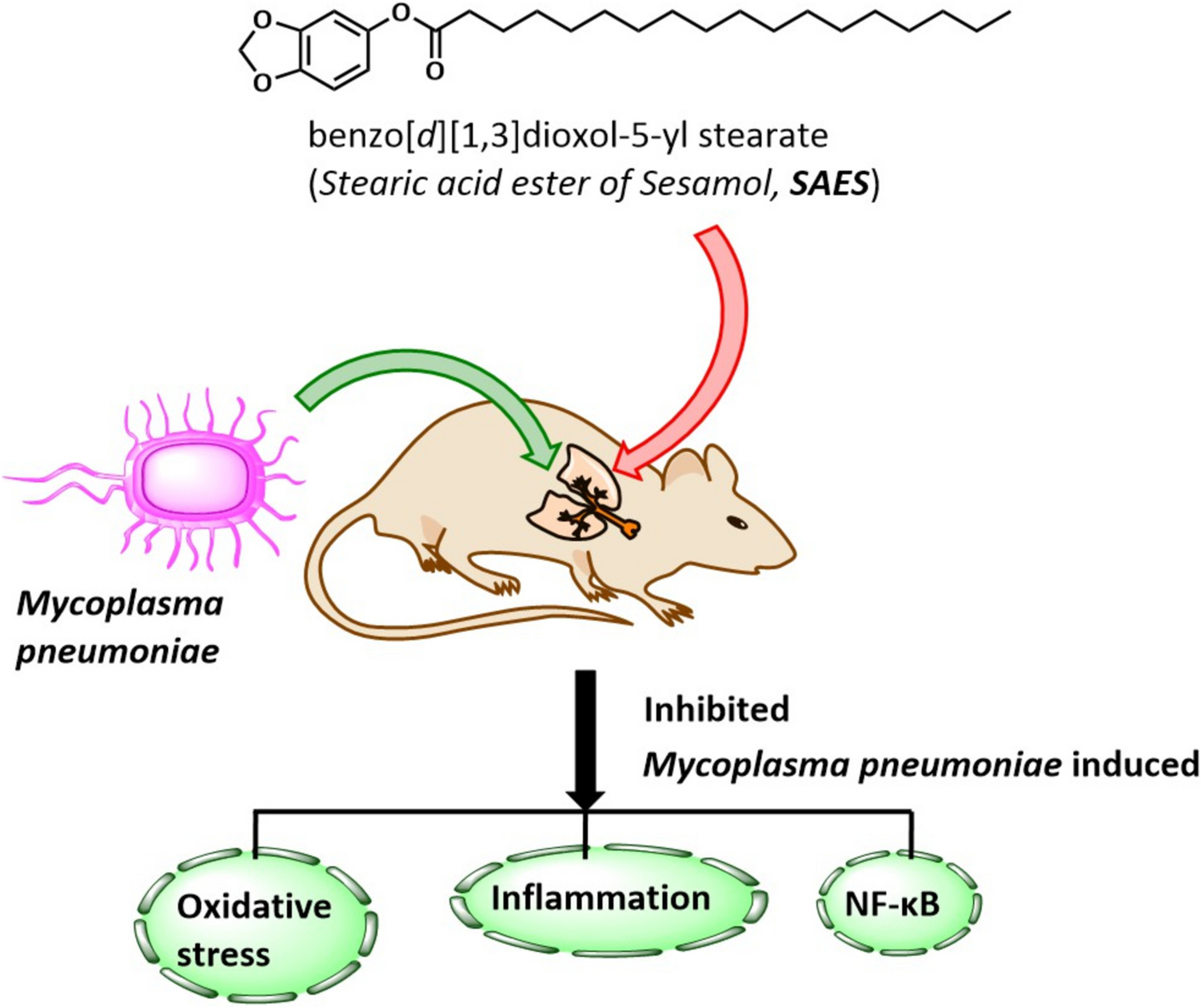
Fig. 1
Reproduced from Lau (2020)
The lectin pathway. Mannose-binding lectins, key molecules in the innate immune response, contribute to the host defence against coronaviruses such as SARS-CoV. A Opsonization, from the Greek opsönein meaning to prepare for eating. MBLs use their carbohydrate recognition domains to bind to the exposed mannose glycons on SARS-CoV-2. B Phagocytosis, from ancient Greek phagein meaning to eat. Phagocytes recognize MBL-bound SARS-CoV-2, ingest the pathogen and digest it.
BioavailabilityA significant hurdle to overcome in the clinical development of antiviral lectins is the assessment of their bioavailabilities (Mitchell et al. 2017). Considering they are proteins, it stands to reason that lectins could not follow an oral route of administration as they would not survive the enzymes of the digestive system (Mitchell et al. 2017). Surprisingly few studies have attempted to assess the systemic bioavailability of lectins (Mitchell et al. 2017). For example, the cyanobacterial lectin cyanovirin-N (CV-N) was biologically available following subcutaneous injection into mice, with doses up to 5.6 mg/kg/day shown to be well tolerated (Mitchell et al. 2017). Understandably less information is available on such effects for the Amaryllidaceae lectins. However, one such study involved a screen of NMRI mice for susceptibilities to GNA and HHA (Balzarini et al. 2004a). When injected intravenously into the tail with GNA (100 mg/kg) or HHA (50 mg/kg) as a bolus injection, none of the animals displayed signs of acute toxicity or visible toxic side effects for at least 5 days post-treatment (Balzarini et al. 2004a).
Toxicity and ImmunogenicityA serious concern in the development of any clinical agent is the possibility that toxicities may arise that are unrelated to the target of the active agent. Although the oligosaccharides on viral GEPs are the targets of antiviral lectins, they are invariably synthesized by and attached to host cell enzymes (Mitchell et al. 2017). This suggests that they can also be present in human cells. Potential deleterious effects from the systemic use of lectins could be borne out of their renown abilities to agglutinate cells (Mitchell et al. 2017). In regard to such effects for Amaryllidaceae lectins, both GNA and NPA were capable of agglutinating trypsin-exposed rabbit erythrocytes but not human red blood cells (RBCs), a reaction that was reversible by mannose (Van Damme et al. 1987, 1988). Further toxicities could potentially be manifested via acute immunological responses to the administration of a foreign protein (anaphylaxis) (Mitchell et al. 2017). For example, concanavalin A was shown to be responsible for acute liver damage in mice due to it binding to sinusoidal endothelial cells, which caused them to be attacked by CD4 + T cells (Mitchell et al. 2017). Nonetheless, there are examples for the successful use of heterologous proteins in the management of human conditions, such as the cone snail peptide ziconotide for chronic pain, the Gila monster saliva peptide exenatide for type II diabetes and the microbial product botulinum toxin in cosmetic therapy (Mitchell et al. 2017). Heterologous proteins also have the potential to stimulate the immune system, such as the activation of T cells or PBMCs (Mitchell et al. 2017). This can, in HIV for example, stimulate CD4 + HIV target cells close to sites of administration, which can have the reverse effect of actually raising the probability of infection (Mitchell et al. 2017; Naik and Kumar 2022). This should be of some concern for Amaryllidaceae lectins since their greatest potential appears to reside in the area of HIV therapeutics.
AdministrationOral means of administration of lectins have been precluded by virtue of their proteinaceous characteristics (Mitchell et al. 2017). Efficacies of antiviral proteins against various respiratory infections have nevertheless been demonstrated via deliveries made topically, systemically (by subcutaneous or intraperitoneal injection) and by intranasal presentation (Mitchell et al. 2017). Vaginal administration has also been shown to be a viable route; for example, in the delivery of genetically engineered lactobacilli secreting CV-N (Mitchell et al. 2017). It remains to be clarified what mode of administration would suit the delivery of Amaryllidaceae lectins.
Galanthus nivalis AgglutininGalanthus nivalis agglutinin (GNA) was the first lectin to be described from the Amaryllidaceae (Van Damme et al. 1987). It was identified as a tetrameric protein composed of four identical subunits (Fig. 2), each with a relative molecular weight Mr of 13 kDa (Van Damme et al. 1987; Hester et al. 1996). Labelling experiments indicated that GNA was synthesized in the endoplasmic reticulum as a precursor with an Mr of 15 kDa and converted post-translationally into the authentic lectin polypeptide (Mr 13 kDa) (Van Damme and Peumans 1988). The molecular structure of GNA is pH-dependent as it was shown to exist as a dimer at pHs of 5 and 9, respectively (Van Damme et al. 1988). Amino acid analysis indicated significant representation of asparagine/aspartic acid, glycine, serine and leucine residues, without the presence of methionine (Van Damme et al. 1987). GNA exhibited exclusive mannose-binding specificity (Van Damme et al. 1987). It readily agglutinated trypsin-treated rabbit erythrocytes, but not human red blood cells (RBCs), which could be reversed by mannose (Van Damme et al. 1987). Agglutination was maintained after reduction with β -mercaptoethanol, suggesting that the binding of GNA subunits did not involve disulphide bridges (Van Damme et al. 1987). Furthermore, it was shown that GNA precipitated branched yeast mannans, without affecting any glucans, and that d-mannose (but not d-glucose) was an inhibitor of the GNA-mannan interaction (Shibuya et al. 1988). Inhibition experiments with various sugars revealed that GNA required the presence of equatorial hydroxyl groups at the C-3 and C-4 positions and an axial group at the C-2 position of the d-pyranose ring (Shibuya et al. 1988). A non-reducing terminal d-mannose residue was necessary for the interaction of oligosaccharides, of which those with terminal man(α-1,3)man units were the most inhibitory (10–30 times more so than d-mannose) (Shibuya et al. 1988).
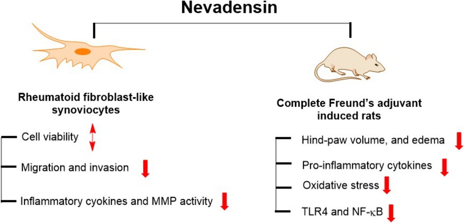
Fig. 2
Crystal structure of GNA (resolved at 2.3 Å) complexed with methyl α-d-mannose showing the four independent subunits of the tetramer as well as selected amino acid residues. Structure available free of charge at the Protein Data Bank via the PDB http://doi.org/10.2210/pdb1MSA/pdb; see also Hester et al. (1996)
Following this, GNA was examined for antiviral effects against the retrovirus HIV, where it was active against both HIV-1(IIIB) and HIV-2(LAV-2) (EC50s 0.4 and 0.3 μg/ml, respectively) cultured in human MT-4 cells (Table S1), proving to be as effective as dextran sulphate 10,000 (DS-10000) (EC50 0.4 μg/ml) (Balzarini et al. 1991). A further feature of its reactivity towards HIV was the fact that GNA suppressed syncytium formation between HIV-1- or HIV-2-infected HuT-78 lymphocytes and uninfected Molt-4 lymphoblasts (EC50s 2.7 and 3.0 μg/ml, respectively), much more so than DS-5000 was able to (EC50 25 μg/ml) (Balzarini et al. 1991). Whilst GNA did not inhibit OKT4A from binding to MT-4 cells, it did inhibit anti-gp120 binding to HuT-78/HIV-1 cells (47% inhibition after 15 min) (Balzarini et al. 1991). The monoclonal antibody OKT4A is used as a phenotypic marker for CD4 expression (Tubo and Jenkins 2014). Found on the surface of immune cells such as T-helper cells, monocytes, macrophages and dendritic cells, CD4 functions as a co-receptor for the T cell receptor (Tubo and Jenkins 2014). The glycoprotein gp120 is found on the surface of the HIV envelope (Env) and undergoes distinct conformational changes upon binding to CD4, which lead to the fusion of the viral membrane with the host cell membrane (Tubo and Jenkins 2014). MT-4 cells that adsorbed HIV-1 particles were distinguishable from those that did not via immunofluorescence with polyclonal anti-HIV antibodies (Balzarini et al. 1991). Whilst DS-5000 (at 25 μg/ml) completely blocked HIV-1 adsorption, GNA only became active at a concentration of 100 μg/ml (or higher) suggesting that it did not markedly inhibit HIV-1 binding to CD4 + cells (Balzarini et al. 1991). Overall, the data suggested that GNA interfered with the HIV replicative cycle via an event that was subsequent to the attachment (i.e. the fusion process) of the virions to the cells (Balzarini et al. 1991).
MT-4 cells were also exploited as hosts to probe the effects of GNA on two clinical isolates of HIV-1: the lymphadenopathy-associated virus (LAV) and nucleoside diphosphate kinase-associated (NDK) isolates (Hammar et al. 1994). Pretreatment of the virus with GNA (5 μg/ml-5 ng/ml) resulted in the neutralization of both isolates to the same extent (Hammar et al. 1994). Pretreatment of MT-4 cells also offered significant protection from infection, but in which case the NDK isolate was about 100-fold more sensitive to treatment than the LAV isolate (Hammar et al. 1994). The fact that α-mannosidase-treated cells were not protected confirmed the requirement of terminal mannose residues for GNA to exert its effects, also suggesting that such residues were not receptors for the virus (Hammar et al. 1994). From the overall effects of GNA in MT-4 cells it was suggested that the lectin may prevent infection by binding to glycoproteins on the surface of infected cells (Hammar et al. 1994). However, the observations made with the pretreatment of non-infected cells suggested that there could be other factors at play, such as the binding of structures in the vicinity of essential receptors which would affect the interaction of the virus with the receptor (Hammar et al. 1994).
Further examination of the effects of GNA on HIV-1(IIIB) involved the use of C8166 T cell leukaemia cells as host (Animashaun et al. 1993). Syncytium formation relative to 3′-azido-2′,3′-dideoxythymidine (AZT) was inhibited by GNA at both 10 and 100 μg/ml, with the lectin shown to be ineffective at the lowest tested dosage (1 μg/ml) (Animashaun et al. 1993). GNA significantly depleted viral multiplication, with new colonies (50%) only observable at the lowest tested concentration (Animashaun et al. 1993). Uninfected C8166 cells were fully viable at all three tested dosages, which was reduced to 63% in infected cells only at the lowest dosage, suggesting that GNA posed no cytotoxic threat to the host (Animashaun et al. 1993). GNA markedly inhibited p24 production in HIV-infected cells with 107% of the antigen detected at the lowest tested dosage (0.016 μg/ml), which fell in a dose-dependent manner to 12% at 2 μg/ml and 0% with the two remaining concentrations (50 and 100 μg/ml, respectively) (Animashaun et al. 1993). The HIV antigen p24 is a protein that makes up most of the HIV capsid (Animashaun et al. 1993). The onset of AIDS correlates with a reduction in the number of CD4 + T cells and increased levels of virus and p24 in the blood, making it a useful handle for diagnostic purposes (Animashaun et al. 1993). Analysis of bound glycoproteins from GNA-sepharose beads that were mixed with either infected or uninfected C8166 cell lysates indicated (via Western blotting and gp120-specific antibodies) the binding of gp120 (Animashaun et al. 1993). GNA has also been used in lectin affinity chromatography to purify the envelope glycoproteins of HIV-1, HIV-2 and SIV (simian immunodeficiency virus), which were subsequently shown to exhibit CD4-binding and antigenic reactivities (Gilljam 1993). Following immunization with these preparations, strong immune responses to envelope proteins and peptides were observed in mice and primate subjects (Gilljam 1993).
Galanthus nivalis agglutinin (in CEM lymphoblastic cells) was also noted for its response to several drug-resistant HIV strains including non-nucleoside reverse transcriptase inhibitor NNRTI-resistant HIV-1 (EC50 0.85 μg/ml), lamivudine-resistant HIV-1 (EC50 0.40 μg/ml), zidovudine-resistant HIV-1 (EC50 1.0 μg/ml) and protease inhibitor-resistant HIV-1 (EC50 0.33 μg/ml) (Balzarini et al. 2004a). The mean EC50 established for tenofovir over these four cases was 8.45 μg/ml (Balzarini et al. 2004a). Further studies of the virus in CEM cells involved the HIV-1(IIIB) strain against which GNA displayed a potent effect (EC50 0.013 μM), over 30 times more so than DS-5000 (EC50 0.45 μM) (Bertaux et al. 2007). An interesting observation made here was that the effect could be attenuated by the inclusion of the mannose polysaccharide mannan (2.5 mg/ml) to the culture medium, so that the half effective concentration dropped to as low as 0.54 μM (Bertaux et al. 2007). This reinforced the notion for the affinity of CBAs (carbohydrate-binding agents) with viral envelope–associated glycans (Bertaux et al. 2007). The EC50 drop for DS-5000 in the same instance was only by 0.15 μM (Bertaux et al. 2007). HIV-2 strain ROD was also strongly inhibited by GNA (EC50 0.011 μM) relative to DS-5000 (EC50 0.16 μM), with negligible effects in each case manifested on CEM cells (CC50s > 2 and > 100 μM, respectively) (Bertaux et al. 2007). GNA efficiently prevented infection of MT-4 cells by HIV-1 strains that were resistant to the virus adsorption inhibitor DS-5000 and the virus entry inhibitors AMD-3100 and SDF-1, with EC50s of 0.13, 0.59 and 0.60 μg/ml, respectively (Balzarini et al. 2004a). The corresponding wild-type HIV-1 strain NL4.3 also succumbed to exposure to GNA (EC50 0.57 μg/ml), but at a markedly higher level than by DS-5000 (EC50 0.14 μg/ml) or AMD-3100 (EC50 0.005 μg/ml) (Balzarini et al.
Comments (0)