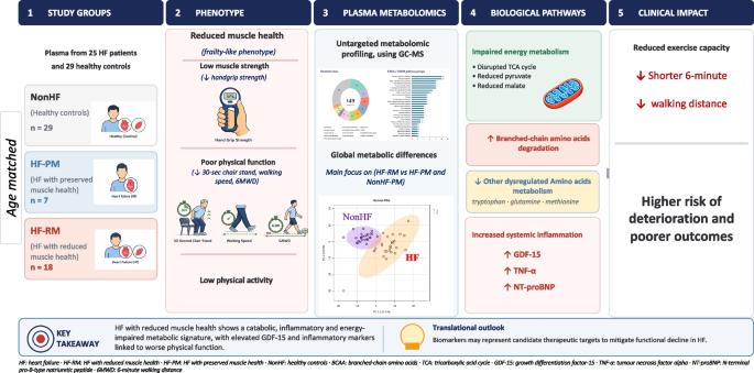
Findings from this study provide novel insights into the biological crosstalk between muscle weakness, HF, and circulating molecular pathways that may drive muscle catabolism in HF. Using an untargeted metabolomic profiling approach, we identified a distinct catabolic metabolic signature that parallels reductions in muscle strength among patients with HF. The concurrent increase in BCAA degradation, altered TCA intermediates, and elevations in TNF-α, NT-proBNP, and GDF-15 may reflect a state of systemic cardiac stress and coordinated alterations in metabolic, inflammatory, and skeletal muscle-derived pathways that could contribute to HF-RM. Moreover, metabolomic analyses revealed disruptions across fatty acid–related metabolites, amino acid metabolism, and glycolytic pathways in both HF-PM and HF-RM groups compared with NonHF controls, indicating impaired energy production and a sustained catabolic phenotype. Two interpretive caveats are important. First, the HF-RM label denotes an integrated cardiac–metabolic–muscular functional phenotype rather than a primary myopathic state. The performance-based tests used in our screening definition (30CST, 6MWD, gait speed) are partially determined by cardiac output reserve, and NT-proBNP was directionally highest in the HF-RM stratum (median 137 vs 86 pg/mL in NonHF-PM, ~ 1.6-fold; Supplementary Table S4), and rose markedly in the stricter sensitivity subgroup defined by low IPAQ + low HGS only (median 522 pg/mL, n = 1; Supplementary Table S5), indicating that this subgroup represents a more advanced HF state. Our findings are therefore best read as the metabolic and inflammatory correlates of advanced HF in patients who also display reduced physical-functional capacity. Second, absolute appendicular lean mass and absolute strength are preserved or even elevated in our HF cohort, while body-load-normalised indices (ALSTI/BMI, HGS/BMI) are reduced in HF-RM. This pattern is consistent with a sarcopenic-obesity/cardiometabolic phenotype (mean BMI ~ 30 kg/m2, body fat 37–42%) rather than classic atrophic sarcopenia, in line with the body size–adjusted indices recommended by the FNIH sarcopenia project and validated for HF by Tinggaard et al. The metabolic perturbations we report should accordingly be interpreted as the signature of sarcopenic-obesity superimposed on HF, not as evidence of overt muscle wasting. Noteworthy that the pre-specified sensitivity analysis omitting 30CST (low IPAQ + low HGS only) confirmed that the inflammatory and cardiac-stress signal persisted even within this stricter subgroup, indicating that this signature is not an artefact of including a cardiac-output-dependent functional test in the classifier.
In our cohort, HGS/BMI consistently predicted ALSTI and muscle strength in NonHF, whereas in HF, it predicted muscle strength but not ALSTI, suggesting that HF-related metabolic impairments may weaken the link between muscle quantity and function. These observations indicate that HF could disrupt both structural and functional muscle relationships, making HGS/BMI a more robust functional marker than ALSTI/BMI. Tinggaard et al. previously showed that ALSTI/BMI predicts walking capacity and muscle weakness; our findings suggest this relationship may be bidirectional and that HGS/BMI may be a stronger predictor. Despite our smaller sample, this implies that handgrip strength assessment can offer meaningful clinical value at the individual level, though larger studies are needed to confirm these observations. Importantly, the interpretation of HGS/BMI as a body-load-normalised functional marker is supported by sensitivity analyses using alternative normalisations. When HGS was expressed per kilogram of body weight, a body size adjustment that does not introduce a height redundancy, the negative correlations with NT-proBNP and TNF-α were stronger than those obtained with HGS/BMI, and the positive correlation with the activin A/follistatin-3 ratio reached significance only when body-weight normalisation was applied. Absolute HGS, by contrast, was associated only with NT-proBNP. The body-load-normalised relationship between strength and the cardiac–inflammatory axis is therefore not a mathematical artefact of dividing strength by BMI in an obese cohort: it is robust across normalisation choices and is in fact strongest with the cleanest body size adjustment, supporting the biological interpretation that mass-relative force generation, rather than absolute strength, is what couples to cardiac stress and systemic inflammation in HF.
Collectively, these biomarker–function associations indicate a potential link between elevated inflammatory and cardiac-stress markers and diminished physical function in HF. Among them, the inverse coupling of GDF-15 with 30CST stands out as a candidate indicator of functional decline and sarcopenia risk, in line with observations from the PROTECT cohort of older medical patients. The biological magnitude of these elevations (reported in Results and Supplementary Tables S4–S5) suggests clinically meaningful shifts in the cardiac-stress and inflammatory axis, with the most cardiac-decompensated patients falling within the HF-RM phenotype. These findings align with the hypothesis that chronic inflammation and cardiac stress may share a bidirectional relationship with muscle weakness. However, the cross-sectional nature of the data limits causal inference, warranting longitudinal studies to elucidate these correlations. A specific concern is that the HF-PM and HF-RM sub-strata differed in HF subtype distribution, raising the possibility that the inflammatory–metabolic signature attributed to reduced muscle health might instead reflect HFrEF-specific biology. As shown in the sensitivity analyses (Results; Supplementary Tables S6–S7), HF subtype was not a major driver of the biomarker differences observed between HF-PM and HF-RM, and EF-adjusted linear models indicated that GDF-15 and insulin tracked the HF-RM phenotype independently of EF, whereas NT-proBNP did not. This pattern is consistent with the interpretation that NT-proBNP elevation reflects myocardial wall stress (i.e. HF severity), while the GDF-15 and metabolic/inflammatory signal is at least partially independent of EF. We acknowledge that disentangling muscle-health phenotype from HF subtype definitively will require a larger, balanced HFrEF/HFpEF cohort.
Metabolic alterations in heart failure with reduced muscle health
Our findings regarding the HF-RM phenotype revealed several differentially expressed metabolites, including galacturonic acid-1-phosphate, glutamic acid, indole-3-propionic acid, methionine, phenylalanine, and 2-hydroxy-glutaric acid. These findings, although based on raw p-values, suggest that HF-RM may amplify metabolic perturbations compared to patients with HF-PM. However, elevation of amino acids such as glutamic acid and phenylalanine in HF-RM may yield controversial results regarding their link to frailty [1, 2]. Regarding HF, higher phenylalanine has been linked to increased mortality and HF (re)hospitalisation [3, 4], while glutamic acid may reflect HF severity [5], which may be exacerbated by frailty. Furthermore, the higher levels of galacturonic acid-1-phosphate, a carbohydrate metabolite, may reflect altered glycosaminoglycan metabolism, potentially contributing to impaired tissue remodelling, considering that proteomics have demonstrated it as a top pathway associated with frailty [6]. It is worth noting that the HF-RM cohort exhibited lower 30CST performance, reduced weekly moderate physical activity, and lower MET and MNA scores compared to HF-PM.
These changes were accompanied by higher levels of glucose, TNF-α, and GDF-15, but lower levels of activin A in HF-RM vs. HF-PM. Elevated GDF-15 is a biomarker of inflammation and oxidative stress that has been previously linked to sarcopenia and frailty. The elevated levels of TNF-α in our study are in line with evidence on its link with frailty in cardiovascular diseases [7], while increased GDF-15 may not only serve as a marker of HF hospitalisation, mortality [8], inflammation, or malnutrition [9], but also of frailty and/or impaired muscle health severity in HF. In addition, higher activin A has been linked to impaired cardiac remodelling [10], and myocardial damage [11] in humans and murine models, which contradict our results on the detrimental impact muscle weakness may confer in HF. Our data showed that the ratio of activin A to follistatin-3 was lower in both the total HF and HF-RM groups compared to controls, suggesting that follistatin-3 may mask activin A action more effectively in the HF-RM population.
HF-RM exhibited a broad range of metabolic alterations compared to NonHF-PM. For instance, elevated amino acids (e.g. alanine, methionine, isoleucine, glutamic acid, ornithine, and cystine) and carbohydrate metabolites (e.g. galactose, galactonic acid, and glycerolaldopyranosid) suggest increased metabolic stress. Conversely, reduced levels of energy metabolism intermediates (e.g. pyruvic acid, malic acid, and 2-oxo-glutaric acid) may indicate compromised mitochondrial energy production, a feature consistent with the relationship between frailty and impaired energy homeostasis, which was also observed in the total HF vs. NonHF. The lower concentrations of tryptophan, histidine, serine, cysteine, and asparagine further highlight the catabolic state in HF-RM, linking these to potentially accelerated muscle wasting and reduced production of serotonin, kynurenines, and NAD+ as shown previously [12]. Thus, patients with HF-RM may exhibit extensive metabolic perturbations, including elevated amino acids and carbohydrate metabolites, and reduced energy intermediates compared to NonHF-PM.
Clinical and research implications
The notable muscle weakness in HF-RM was apparent at the similar age with total NonHF and HF-PM counterparts, suggesting that muscle weakness may start at a younger age in this population, which is an established risk factor for worsened prognosis in HF. The distinct metabolic profiles identified in this study could have significant implications for the management of HF. The elevation of GDF-15 and TNF-α in HF-RM highlights the role of inflammation and oxidative stress in this population, suggesting that potential therapies (pharmacological and non-pharmacological) could be explored to mitigate impaired muscle health–related complications. Notably, GDF-15 has been recently emerged as a therapeutic target in cancer-associated cachexia, a condition characterised by severe muscle and weight loss [13]. In addition, the observed metabolic alterations, particularly in BCAAs, glycolytic metabolites, and the tryptophan-indole pathway, suggest potential therapeutic targets to improve energy metabolism and muscle health in this HF-RM phenotype. For example, interventions aimed at enhancing mitochondrial function or optimising amino acid metabolism could help alleviate symptoms of muscle weakness and improve functional outcomes [14, 15]. Specifically, nutritional status, as indicated by lower MNA scores in HF-RM, may be a critical factor, given its link to lower energy and protein intake that may amplify sarcopenia risk or sarcopenia progression. Additionally, the lower 30CST performance and low physical activity levels in the HF-RM group mark further the need for tailored nutritional and exercise programmes to improve physical function. Furthermore, larger sample sizes or more sensitive analytical approaches may be needed in studies to fully elucidate muscle health–specific metabolic signatures. Future studies could employ longitudinal designs to explore how these metabolic changes evolve over time and evaluate their prognostic impact on clinical outcomes such as hospitalisation or mortality.
Study limitations
The relatively small sample size and cross-sectional nature preclude causal interpretations and to investigate the physical frailty bidirectionally to HF, particularly for muscle health and/or HF phenotype-specific (HFrEF and HFpEF) analyses, where statistical significance was based on raw p-values rather than adjusted models. Additionally, the consideration of comorbidities and different prescribed medications may have an impact on metabolomic analyses. MSI level 2 annotations are not definitive metabolite identifications. Future work would be to run authentic standards to confirm the ones not already in CMR library, although hits in Mummichog give extra confidence. Despite these constraints, the robust application of GC–MS in clinical and community-dwelling cohorts provides further mechanistic insights into HF and muscle health. The HF-RM screening definition is based on Fried physical-frailty (low IPAQ) plus EWGSOP2 “probable sarcopenia” criteria (low HGS) and/or low 30CST; because performance-based tests are partly determined by cardiac output reserve in HF, the HF-RM phenotype is best interpreted as integrated cardiac–metabolic–muscular dysfunction rather than as primary skeletal myopathy. Confirmatory sarcopenia (low ALSTI per EWGSOP2) was not used to define groups, and absolute appendicular lean mass was preserved or elevated in our HF cohort, identifying a sarcopenic-obesity rather than atrophic-sarcopenia phenotype. Group sizes were modest (n = 21, 8, 7, 18) and the HF-PM (n = 7) and HF-RM (n = 18) strata were imbalanced for HF subtype (3 HFpEF/4 HFrEF and 4 HFpEF/14 HFrEF, respectively); although our sensitivity analyses suggest that HF subtype is not a major confounder of the GDF-15, TNF-α, and BCAA signal (Supplementary Tables S6–S7), confirmation in a larger, balanced HFrEF/HFpEF cohort is required. Finally, body-load-normalised strength indices (HGS/BMI) are interpretively useful but mathematically sensitive to body composition; we therefore report sensitivity analyses using HGS/body weight and BMI-adjusted residuals (Supplementary Table S8), which show a directionally consistent pattern. These findings highlight the need for integrated approaches to manage HF-RM, incorporating nutritional, exercise, and potentially pharmacological interventions to address metabolic and functional deficits.

Comments (0)