
Remember me
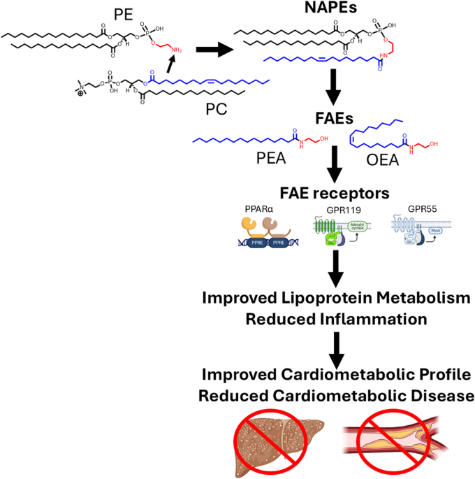
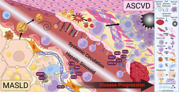
In 2001, Rodríguez de Fonseca et al. reported that ingestion of dietary fat triggered oleoylethanolamide (OEA) production in the small intestine and that OEA inhibited food intake [1]. In the 25 years since this discovery, fatty acid ethanolamides (FAEs) including OEA and palmitoylethanolamide (PEA) have emerged as key bioactive lipids produced in the intestine, liver, and adipose tissues that signal through PPARα, GPR119, and GPR55 to regulate the metabolism of triglycerides and cholesterol and promote the resolution of inflammation that may therefore protect against the development and progression of cardiometabolic diseases including atherosclerosis (Fig. 1). This review will summarize the key studies elucidating the mechanisms by which FAEs act and emerging pre-clinical and clinical evidence supporting their therapeutic potential as lipid lowering and anti-atherosclerotic agents. It will also highlight key gaps in our current knowledge including how the biosynthesis of these bioactive lipids is regulated and why FAE biosynthesis becomes dysregulated during development of cardiometabolic diseases.
Fig. 1 The alternative text for this image may have been generated using AI.
Biosynthesis of fatty acid ethanolamides protects against cardiometabolic diseases. Metabolic stimuli that cause an increase in free fatty acids (FFA) and subsequent increases in phosphatidylcholine (PC) and phosphatidylethanolamine (PE) trigger increased formation of fatty acid ethanolamides (FAEs) including oleoylethanolamide (OEA) and palmitoylethanolamide (PEA). These metabolic stimuli may increase the activity of the N-acyltransferases that form NAPEs. OEA and PEA then act on receptors including PPARα, GPR119, and GPR55 to regulate triglyceride (TAG) and cholesterol (CHOL) metabolism and inflammation. The integrated effects of FAE receptor activation protect against cardiometabolic diseases including metabolic-dysfunction associated steatotic liver disease (MASLD) and atherosclerotic cardiovascular disease (ASCVD). Hollow arrows indicate biochemical synthesis pathways and filled arrows represent signaling pathways,
Fatty acid Ethanolamide BiosynthesisBesides OEA and PEA, other major species of FAE (also known as N-acyl-ethanolamines) include stearoylethanolamide (SEA), linoleoylethanolamide (LEA), and arachidonoylethanolamide (AEA, also known as anandamide). The overall framework for FAE biosynthesis has been elucidated, but important details related to the regulation of their biosynthesis in response to stimuli remain uncharacterized. In vertebrates, biosynthesis of FAEs requires at least two steps: the formation of N-acyl-phosphatidylethanolamines (NAPEs) and then enzymatic hydrolysis of these NAPEs to generate FAEs. Phosphatidylethanolamine N-acyltransferases (NATs) transfer an O-acyl chain from phosphatidylcholine (PC) or phosphatidylethanolamine (PE) to the headgroup nitrogen of an acceptor PE to form NAPEs (Fig. 2). Humans express at least six different NATs: a family of five calcium-independent NATs, the phospholipase A/acyl transferases (PLAAT-1 to -5) that differ significantly in terms of their NAT activity [2] and a calcium-dependent NAT, phospholipase A2 group 4 epsilon (PLA24ε) [3]. Three known pathways hydrolyze NAPEs to FAEs. NAPE-hydrolyzing phospholipase D (NAPE-PLD) hydrolyzes the distal phosphodiester bond of NAPE to directly generate FAE and phosphatidic acid. In mice, genetic deletion of Napepld globally or in specific tissues reduced levels of individual FAEs by 20–70% [4,5,6,7,8], revealing that NAPE-PLD-independent pathways also contributed to FAE biosynthesis. Subsequently, ABDH4 was shown to hydrolyze NAPEs to form glycerophospho-N-acyl-ethanolamines [9] which can then be hydrolyzed by GDE1 to form FAE [10]. NAPE were also shown to be hydrolyzed to phospho-FAEs by an as yet unidentified NAPE-hydrolyzing phospholipase C, with the phospho-FAE then hydrolyzed by PTPN22 to form FAE [11]. The intestine, liver, adipose tissue, and skeletal muscle of humans each express multiple NATs and NAPE-hydrolyzing enzymes (Fig. 2).
Fig. 2 The alternative text for this image may have been generated using AI.
Distribution and relative expression of the enzymes of fatty acid ethanolamide biosynthesis and catabolism in metabolically active tissue. In humans, six known PE N-acyltransferases (NATs) biosynthesize NAPEs by transferring an acyl chain from phosphatidylcholine (PC) to the nitrogen of phosphatidylethanolamine (PE). The atoms of each molecule which eventually form the final FAE are highlighted in grey. The relative intrinsic NAT activity of each enzyme is illustrated by number of + symbols based on Uyuma et al. [2] and Ogura et al. [3]. Three independent pathways convert NAPEs to FAEs. FAEs are inactivated by catabolism by Fatty Acid Amide Hydrolase (FAAH), FAAH2, or N-acylethanolamine hydrolyzing acid amidase (NAAA). The relative mRNA expression (median transcripts per million, TPM) of each gene encoding a biosynthetic or catabolizing enzyme is indicated by number of * symbols as follows: not shown TPM ≤ 1; *1 < TPM ≤ 5; **5 < TPM ≤ 15; ***15 < TPM ≤ 45; ****45 < TPM ≤ 145; *****TPM > 145, based on data exported from the Genotype-Tissue Expression (GTEx) Portal on 11/02/2025. The GTEx project is supported by the Common Fund of the Office of the Director of the National Institutes of Health, and by NCI, NHGRI, NHLBI, NIDA, NIMH, and NINDS
Individual FAEs vary significantly from one another in their levels in each tissue and these levels differentially change in response to metabolic stimuli. For instance, Petersen et al. reported that intestinal NAEs in fasted rats were as follows: OEA 1.4 ± 0.4, PEA 3.9 ± 0.22, SEA 4.2 ± 0.15, LEA 2.8 ± 0.4, and AEA 0.46 ± 0.15 pmol / µmol phospholipid [12]. Refeeding increased levels of intestinal OEA 5.6-fold, PEA 2.0-fold, SEA 1.1-fold, LEA 11.7-fold, and reduced levels of AEA 2.6-fold [12]. The relatively low levels of AEA compared to other FAEs in peripheral tissue likely results from all NATs preferentially utilizing O-acyl chains from the sn-1 position of PC where the acyl group is typically palmitate, stearate, or oleate, rather than arachidonate. Differences between individual FAE levels is also the result of differential expression of the two major FAE catabolizing enzymes, Fatty Acid Amide Hydrolyase (FAAH) and N-acyl-ethanolamine hydrolyzing acid amidase (NAAA) in various tissues (Fig. 2). Unlike rodents, humans also express a second FAAH enzyme, FAAH-2. FAAH preferentially catabolizes AEA over OEA and PEA [13], while NAAA preferentially catabolizes saturated FAEs like PEA [14]. However, the extent to which individuals NATs, NAPE-hydrolyzing enzymes, and FAE catabolizing enzymes contribute to individual FAE levels in each tissue is poorly characterized. In macrophages, LPS appears to stimulate AEA biosynthesis by upregulating the NAPE-PLC/PTPN22 pathway [11] while inhibiting PEA biosynthesis by reducing NAPE-PLD expression [15]. Because individual FAE species differ in their physiological effects, a better understanding of how levels of each FAE are differentially regulated may be critical to a complete understanding of how FAEs regulate triglyceride and cholesterol metabolism.
Changes in Intracellular Levels of free Fatty Acids Triggers FAE BiosynthesisFeeding-induced increases in intestinal FAEs have been observed in all vertebrate species where it has been examined [1, 12, 16,17,18,19]. Fat ingestion drives the feeding-induced increase in intestinal FAEs [20, 21] with free fatty acids (FFAs) released from ingested triglycerides (TAGs) being transferred into enterocytes by the fatty acid translocase CD36 and the newly acquired FFAs incorporated into NAPEs [20], presumably after the FFA is first incorporated into PCs. In liver and adipose tissue, FAE levels are at their apex during fasting and at their nadir after feeding [16, 22], consistent with fasting increasing intracellular FFA in adipocytes and hepatocytes. Thus, rising intracellular FFA levels trigger FAE production, positioning them to initiate functional responses that help restore lipid homeostasis.
Increased intestinal biosynthesis of N-oleoyl-PE, N-linoleoyl-PE, and N-palmitoyl-PE appear to be required for the increases in their respective FAEs during fasting and refeeding [12, 16]. Unfortunately, the specific NATs in the mammalian intestine, liver, and adipose tissue that generate NAPEs in response to rising FFAs levels are unknown. We have shown that genetic deletion of plaatl1 in zebrafish blocks the feeding-induced rise in intestinal NAPEs and FAEs and leads to weight gain and fat accumulation [19]; however, plaat1l1 is not a direct ortholog of human PLAAT1 or mouse Plaat1, so another PLAAT/Plaat may be responsible in mammals. Furthermore, precisely how elevated FFA levels trigger increased NAPE biosynthesis remains poorly understood. The substrates for NATs are PC and PE, not FFA or FFA-CoA [2, 23]. Even though FFA influxes increase PC and PE synthesis, both phospholipids would already seem to be available to NATs at saturating concentrations as they are the primary components of membranes and are present at concentration far above their critical micellar concentration. Thus, the mechanism(s) whereby intracellular FFA levels increase NAPE/FAE biosynthesis still requires elucidation.
Deletion of Endogenous FAE Biosynthetic Enzymes Induces Dyslipidemia.Support for the notion that FAE biosynthesis is required for appropriate regulation of triglyceride (TAG) and cholesterol (CHOL) levels comes from studies where a key FAE biosynthetic enzyme, NAPE-PLD, has been genetically deleted in three key sites of lipid homeostasis—the liver, intestine, and adipose tissue. Liver-specific deletion of Napepld (NapepldΔHep) only reduced liver levels of OEA and LEA ~ 20% and did not change levels of AEA or PEA compared to wild-type (WT) mice [6]. Even so, NapepldΔHep mice had increased total body fat mass compared to WT mice [6]. Average lipid droplet size was increased in the liver of NapepldΔHep mice, although total liver fat mass did not. NapepldΔHep mice had significant reductions in both liver bile acid and oxysterols, but not CHOL [6]. Plasma total or lipoprotein CHOL were not reported, but NapepldΔHep showed greater insulin resistance [6], which is often correlated with an increase in plasma TAG to HDL-C ratio.
More compelling evidence for FAE biosynthesis regulating lipid homeostasis comes from mice where Napepld is specifically deleted in intestinal epithelial cells (NapepldΔIEC mice). Intestinal OEA, PEA, SEA, and AEA levels are reduced by ~ 50% in NapepldΔIEC mice compared to WT mice [4]. On high-fat diet (HFD), NapepldΔIEC mice show increased liver TAGs and lipid droplet size, but no significant changes in liver CHOL levels [4]. Immediately following an oral lipid challenge, NapepldΔIEC mice show modest increases in plasma TAGs, but not FFAs, compared to WT mice [4]. On first exposure to HFD, NapepldΔIEC mice also eat more than WT mice and after 8 weeks of HFD NapepldΔIEC mice show significantly increased adipose tissue weight and total body weight compared to WT mice [4]. NapepldΔIEC mice showed no significant changes in insulin resistance and glucose tolerance [4].
The most compelling evidence for FAE biosynthesis regulating lipid homeostasis comes from studies of mice with adipose tissue-specific deletion of Napepld (NapepldΔAdipo mice). Adipose OEA, PEA, and SEA levels in NapepldΔAdipo mice are ~ 50% of those found in WT mice, with no change in AEA levels [5]. Even when fed low-fat diet, NapepldΔAdipo mice had 20–25% higher plasma TAG and CHOL than WT mice [5]. Food intake in NapepldΔAdipo mice was not increased compared to WT mice on either a low-fat or high-fat diet; nevertheless, total body weight increased ~ 25%, total body fat increased ~ 50%, and glucose tolerance was impaired [5]. Of note, adipose-specific deletion of Abhd4 in mice did not alter FAE levels in adipose tissue or increase adiposity or total body weight compared to WT mice [24].
Determining the full extent to which endogenous FAE biosynthesis regulates lipid homeostasis will require a more complete ablation of FAE biosynthesis, such as by concomitantly deleting NAT genes (i.e. Plaat1-5 and Pla2g4e) and NAPE-PLD.
Administering FAEs Normalizes Triglyceride and Cholesterol Homeostasis and Inhibits Atherosclerosis ProgressionFurther evidence for FAEs playing an important role in the regulation of TAGs and CHOL and in the prevention of cardiometabolic diseases including metabolic-dysfunction associated steatotic liver disease (MASLD) and atherosclerotic cardiovardiovascular disease (ASCVD) comes from studies where various FAEs have been administered to rodents or humans. In mice, intraperitoneal (i.p.) administration of OEA administration (5 mg/kg/day) reduced tissue levels of TAGs in liver and adipose tissues [25], while oral OEA administration (100 mg/kg/day) reduced plasma TAG by 59% and plasma CHOL by 17% [26]. In Zucker obese rats, i.p. OEA administration (5 mg/kg/day) reduced both TAG and CHOL levels in serum and hepatocytes [27], while in Sprague-Dawley rats fed a high-fat diet (HFD), it reduced both their plasma TAG and their plasma CHOL by > 50%, lowered circulating levels of liver enzymes and inflammatory cytokines, and reduced hepatosteatosis [28]. In rats given valproic acid to induce steatohepatitis, i.p. OEA administration (10 mg/kg/day) reduced serum TAG, low density lipoprotein-cholesterol (LDL-C), high density lipoprotein-cholesterol (HDL-C), and total CHOL, and reversed their steatohepatitis as well as reduced ALT/AST levels [29]. Other FAEs besides OEA also exert clear lipid-lowering effects. Administering PEA (30 mg/kg s.c. daily) to ovariectomized rats reduced their serum CHOL levels, but not serum TAG levels [30]. This treatment also reduced food intake, body weight and fat mass, and improved glucose tolerance [30]. In mice fed HFD, oral PEA administration (30 mg/kg/day) reduced serum levels of TAG, total CHOL, liver enzymes, insulin and glucose, and resulted in improved glucose tolerance and insulin sensitivity [31]. In Spraque-Dawley rats previously made obese by feeding a cafeteria diet for 12 weeks, administering LEA (10 mg/kg i.p.) for 14 days resulted in large reductions in plasma TAGs and CHOL, as well as uric acid, AST, IL-6, and TNFα [32].
Pilot clinical trials show similar lipid-lowering effects. Giving oral OEA (300 mg/day) to twenty stroke patients reduced their plasma TAG, HDL-C, total CHOL, urea, IL-6, and hsCRP compared to patients receiving placebo [33]. Giving oral OEA (250 mg/day) to thirty obese MASLD patients reduced their plasma TAG, BMI and fat mass compared to similar patients receiving placebo [34]. Oral OEA (250 mg/day) significantly reduced serum TAG levels in thirty obese individuals compared to placebo-treated individuals, but did not significantly alter their LDL-C, HDL-C or total CHOL [35]. Giving PEA (700 mg daily, orally) to 29 obese adults reduced plasma TAG and IL-2 levels compared to placebo treatment (15 adults), without changing HDL-C, LDL-C, fasting glucose, or various other cytokines and markers of inflammation [36]. While these small-scale studies support FAEs playing a key role in the regulation of lipid homeostasis in humans, whether pharmacologic administration of FAEs is an effective clinical treatment for humans with significant dyslipidemia awaits large scale randomized control trials.
A major goal of cholesterol-lowering therapies is to inhibit the development of ASCVD, and several studies in mice support the potential efficacy of administering FAEs in this regard. For instance, administration of OEA (5 mg/kg daily i.p.) visibly reduced the amount of atherosclerotic plaque in balloon-aorta denudation mice or in Apoe−/− mice fed a Western diet, although this was not quantified [37]. Administration of PEA (3 mg/kg daily i.p.) reduced atherosclerotic lesion area in Apoe−/− mice when PEA administration began with the start of Western diet [38]. If mice were fed Western diet for 12 weeks prior to beginning PEA administration, PEA did not reduce atherosclerotic lesion area but did reduce necrotic core area and increased fibrous cap thickness [38], consistent with plaque stabilization. We showed that raising tissue FAE level by orally administering bacteria genetically engineered to secrete NAPEs significantly reduced serum CHOL levels and atherosclerotic lesion necrotic core area in Ldlr−/− mice fed a Western diet [39]. Despite these promising results in mouse models, no randomized clinical trials examining the efficacy of PEA or OEA in the treatment of ASCVD have been undertaken.
Potential Mechanisms for the Anti-Atherosclerotic Effects of FAEsMany of the protective effects of FAEs against cardiometabolic diseases can be rationalized by their activation of three key receptors, PPARα, GPR119, and GPR55 (Fig. 1) as synthetic agonists of these receptors reduce fat intake, increase fat oxidation, reduce fat synthesis, decrease secretion of ApoB-containing lipoproteins, reduce inflammation, and enhance the resolution of inflammation.
Peroxisome Proliferator-Activated Receptor alpha (PPARα) is a nuclear hormone receptor that heterodimerizes with the retinoid X receptor to drive expression of a large number of genes related to lipid metabolism. OEA is one of the most potent endogenous agonists of PPARα (EC50 120 nM) [40], with PEA also being a PPARα agonist (EC₅₀ 3.1 µM) [41] (Fig. 1). AEA has been reported to either not activate PPARα [41] or to do so only at high concentration [42]. While several actions of SEA and LEA require PPARα, to best of our knowledge their binding affinity for PPARα has not been reported. FAEs also activate G-protein coupled receptors including GPR119 which primarily couples to Gαs, GPR55 that primarily couples to Gα12/13, and endocannabinoid receptor-1 and − 2 (CNR1 and CNR2, respectively) that primarily couple to Gαi/o. OEA was the first endogenous GPR119 agonist identified (EC50 0.2–3.2 µM, depending on downstream readout) [43,44,45]. To the best of our knowledge, concentration response curves for GPR119 activation by other FAEs have not been reported, but in cAMP-reporter based assay, 30 µM of OEA, PEA, SEA, and AEA were shown to induce 37-fold, 17-fold, 5-fold, and 2-fold increases in reporter activity, respectively [43]. PEA is a highly potent GPR55 agonist (EC50 4 nM), with AEA and OEA being somewhat less potent (EC50 18 nM and 440 nM, respectively) [14]. However, not all studies have found PEA or other FAEs to activate GPR55 signaling [46, 47] and GPR55 activation by FAE may require concomitant integrin clustering [48]. Activation of CNR1 has many well-established neurological effects including increasing appetite [49], while activation of CNR2 modulates immune cell function. AEA is a robust partial agonist of CNR1 (EC50 31 nM) and CNR2 (EC50 27 nM) [50, 51]. Importantly, saturated and monounsaturated FAEs such as PEA, SEA, and OEA do not have any meaningful efficacy for CNR1 or CNR2 [50, 52].
Administering OEA significantly reduces food intake [1]. Since intestinal OEA biosynthesis is triggered by fat ingestion, OEA’s satiation effect presumably helps prevent an overload of dietary fats. The satiating effects of OEA require Ppara [40]. PEA, SEA, and LEA reduce food intake by rodents less potently than OEA [1, 53,54,55], consistent with their lower potency as PPARα agonists. AEA promotes food intake [1], consistent with agonism of CNR1 being the primary action of AEA [49] and highlights to importance of the finding that intestinal AEA levels drop in response to fat ingestion in contrast to other FAEs. Activation of PPARα by OEA induces satiation and satiety by multiple mechanisms that require oxytocin or histamine [56,57,
Comments (0)