Granulovacuolar degeneration bodies protect neurons from tau pathology
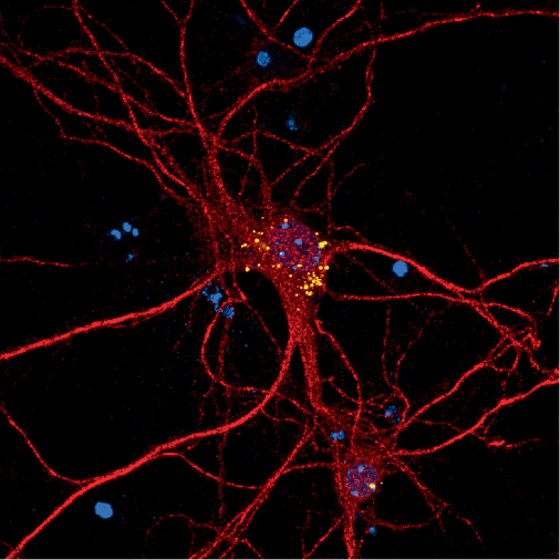
GVBs are a widely recognized hallmark of AD that can be observed in postmortem brain tissue from humans. A 2019 study led by Wiep Scheper demonstrated that GVBs are proteolytically active lysosomal structures that form in response to pathological tau. Building on their previous work, Scheper and colleagues used their previously validated primary neuron model of seed-independent tau pathology to investigate the function of GVBs.
The analysis showed that during tau-induced neurodegeneration, the fraction of tau+GVB+ neurons increased and tau+GVB− neurons decreased, which indicates that GVB+ neurons are more resistant to tau-dependent neuronal loss. Treatment with an inhibitor of CK1δ, a kinase that accumulates in GVBs, significantly reduced the number of GVB+ neurons after 48 h, which demonstrates that CK1δ is a rate-limiting factor.

Comments (0)