
Remember me
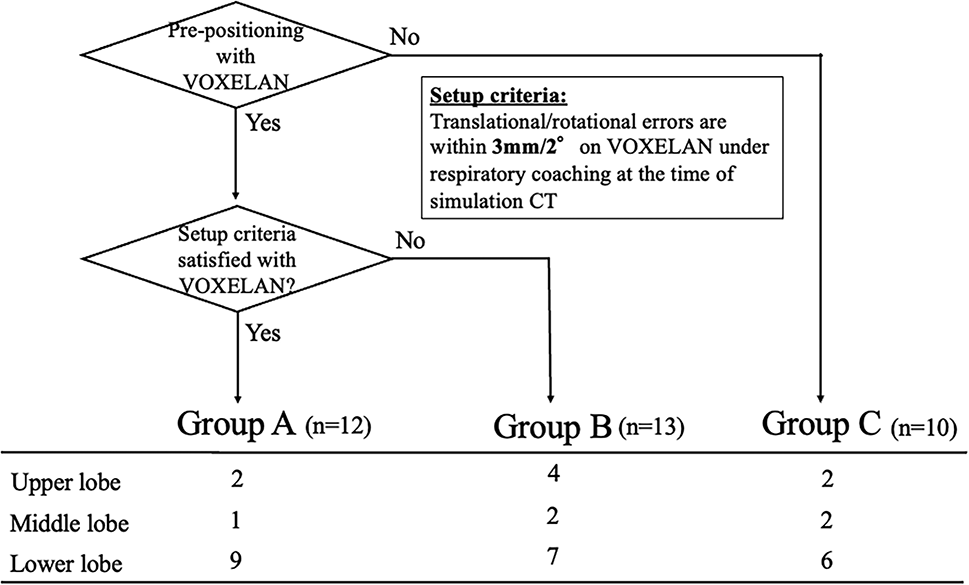
In this study, we evaluated target localization reproducibility in patients treated with SBRT using the DIBH protocol, both with and without the use of VOXELAN in prepositioning, and with and without respiratory hold coaching using VOXELAN. We divided participants into three groups, analyzed them, and found that the variation in the long direction was large in all groups. After prepositioning with VOXELAN and implementing breath-hold coaching, Group A satisfied the setup criteria and exhibited the smallest variation in the longitudinal direction; however, there was no statistically significant difference among the three groups. Although the quantitative improvement was modest, the results indicate that combining SGRT-based prepositioning with respiratory coaching can stabilize patient setup reproducibility under clinical conditions. This may be because there were cases in Group A that did not match sufficiently in the longitudinal direction. Therefore, we analyzed the results of the cases that were outliers in the longitudinal boxplot in detail. Among these, cases G and H were outliers, exhibiting lower internal target matching than did the other cases. Thus, cases G and H were investigated in detail.
Figures 4 and 5 display a comparison of tumor positions among the diagnostic CT, simulation CT, and CBCT images on the sagittal and axial planes, respectively. For patient G, the tumor position on diagnostic CT and CBCT was almost the same as that during the deep inspiration breath-hold; nonetheless, on simulation CT, the tumor position was superior to that on diagnostic CT and CBCT. Conversely, for patient H, the tumor position on the diagnostic CT and CBCT was nearly the same as that during the deep inspiration breath-hold; nevertheless, on the simulation CT, it was located inferiorly to those images. This indicates that even with VOXELAN-assisted respiratory management, the patient was breathing shallower or deeper than they did during simulation CT. This may be due to patient anxiety during simulated CT or differences between thoracic and abdominal breathing. Previous studies comparing thoracic-DIBH and abdominal-DIBH have shown that abdominal breath-hold can produce larger cranio-caudal diaphragm excursion and greater lung expansion, often resulting in lower heart and lung doses than thoracic DIBH, although reproducibility may vary by patient [18]. In addition, SGRT-based DIBH stability studies have shown that the external thoracic surface may still move several millimeters depending on breathing pattern, highlighting the clinical relevance of distinguishing thoracic from abdominal breath-holds [19]. Such physiological variations underline the importance of standardized respiratory coaching and patient relaxation during simulation. Even when using VOXELAN for respiratory management, the technologist involved in the simulation CT scan could not detect any differences in the normal respiratory state of the patient. When performing simulated CT, it is essential to ensure that the patient does not experience excessive tension and that differences between thoracic and abdominal breathing do not occur.
Fig. 4 The alternative text for this image may have been generated using AI.
Comparison of tumor positions among diagnostic CT, simulation CT, and CBCT images in the sagittal plane. Red lines denote the positions of the vertebral bodies used as reference points. For patients G and H, Th11 and Th9 served as the reference points. The lower boundary of the tumor is represented by orange (diagnostic CT), yellow (simulation CT), and green (CBCT) lines. CBCT cone-beam computed tomography, CT computed tomography, Th thoracic vertebra
Fig. 5 The alternative text for this image may have been generated using AI.
Comparison of tumor positions among diagnostic CT, simulation CT, and CBCT images in the axial plane. Red lines denote the positions of the vertebral bodies used as reference points. For patients G and H, Th11 and Th9 served as the reference points. The lower boundary of the tumor is represented by orange (diagnostic CT), yellow (simulation CT), and green (CBCT) lines. CBCT cone-beam computed tomography, CT computed tomography, Th thoracic vertebra
Prado et al. evaluated intra-fractional target shift comparisons using two breath-hold systems in lung stereotactic body radiotherapy [20]. They compared spirometry- and surface-guided systems for target detection and reported that the intrafraction shift was reduced when using spirometry-based DIBH rather than surface-guided DIBH, although both methods were equivalent in accuracy for intrafraction control. Additionally, higher superior-inferior shifts were observed in patients with inferior lobe tumors. Their findings demonstrated that the SB system offered slightly better reproducibility, particularly for lower-lobe tumors, which exhibited greater shift. However, both systems maintained an average submillimeter accuracy. Prado et al. concluded that although SGRT is clinically viable, careful consideration is required for lower-lobe cases because of greater diaphragm-related movement. Our findings are consistent with this observation, as the longitudinal direction showed the largest variability even under VOXELAN-guided control. In a similar study, the reproducibility of surface-guided DIBH for lung SBRT was evaluated using a ring-mounted SGRT system with a closed-bore linear accelerator [21]. Seventy-three SGRT-guided abdominal DIBH treatment sessions were conducted, and the tumor and surface positions were assessed using kV-CBCT and SGRT reports. Tumor motion remained within the submillimeter range, and a strong correlation was found between the surface and internal motion in several directions. DIBH significantly reduced the planning target volumes and lung doses. These results, together with our data, demonstrate that SGRT provides clinically acceptable reproducibility for most directions, while residual longitudinal discrepancies require further management.
Fu et al. reported intra-fractional tumor motion in lung stereotactic radiotherapy with DIBH using CBCT images [22]. They analyzed 28 patients who underwent posttreatment CBCT to qualify for tumor shifts, demonstrating that most shifts remained within the applied PTV margins (5 mm vertical/lateral; 8 mm longitudinal), with a slightly larger variation in the longitudinal direction. They mentioned that it was possible to reduce it further in the lateral direction, even though this was not performed. Their reported magnitude of longitudinal variation aligns with the present study, confirming that longitudinal motion remains a key limiting factor for safe margin reduction.
Naumann et al. assessed the feasibility of SGRT for position verification and intra-fraction monitoring during DIBH in SBRT [23]. In 20 patients with lung and liver tumors, SGRT was compared with CBCT and fluoroscopy, exhibiting a high match between the surface and internal positions, with mean deviations of less than 2 mm. The authors concluded that SGRT reliably detected breath-hold reproducibility and intra-fractional motion. Similar to their findings, our study confirmed that VOXELAN effectively maintained positional reproducibility in vertical and lateral directions, reinforcing its clinical reliability.
Sarudis et al. reported surface-guided frameless positioning for lung SBRT [24]. They evaluated CBCT data with free breathing using an SGRT device and compared the dosimetric parameters in radiotherapy planning. According to their report, the intra-fractional shifts of the patients were larger in the longitudinal direction. They stated that monitoring during irradiation with SGRT enabled them to set appropriate margins for the target in the treatment plan as a countermeasure to these problems. The authors concluded that frameless immobilization using SGRT for motion management and respiration monitoring is a feasible approach for lung SBRT. Importantly, our findings corroborate this conclusion. We performed respiratory management on patients when acquiring simulated CT images to ensure the reproducibility of their breathing and referred to the body surface data and respiratory waveforms obtained by VOXELAN installed in the simulation CT room to perform prepositioning in the treatment room. After completing image guidance by matching the tumors with the CBCT images, we performed radiation management during irradiation using VOXELAN in the treatment room. This process considers not only the position of the patient but also the respiratory waveform, contributing to the reproducibility of the position and waveform from the simulation CT to the treatment room on the same coordinate scale using VOXELAN. Furthermore, a European team reported on patterns of practice for respiratory motion management [25], stating that breath-holding is a common technique used in the treatment of lung cancer and concluding that motion-management techniques are important. VOXELAN is based on the light-section method and accurately acquires surface shape. Our findings extend these reports by providing quantitative evidence that integrating surface-based prepositioning and respiratory coaching in a unified workflow improves reproducibility and supports safer margin definition. We conclude that the implementation of motion management and prepositioning using VOXELAN during simulated CT acquisition, as well as body surface monitoring during irradiation, is effective.
The case group examined in this study consisted of cases that were generally similar and eligible for SBRT. Nonetheless, because not all cases were identical, random errors due to differences between cases were included. Additionally, in this analysis, the vertebral structure was assumed fixed and not subjected to respiratory motion when the marker was set. However, in the middle thoracic spine, the vertebrae may have bent slightly during deep breathing compared to the treatment planning phase. Therefore, uncertainty resulting from this effect cannot be ruled out. Furthermore, the number of patients in this study was 35, which may not have been sufficient for statistical analysis. Nevertheless, by ensuring that the sample sizes in each group were similar, statistical errors in the analysis were excluded. Future studies with a larger cohort and integrated intra-fraction monitoring are warranted to validate these findings and to explore potential dosimetric benefits of margin reduction based on SGRT reproducibility.
Comments (0)