Fan J, Watanabe T. Atherosclerosis: known and unknown. Pathol Int. 2022;72:151–60. https://doi.org/10.1111/pin.13202.
Article
PubMed
Google Scholar
Chen W, Li Z, Zhao Y, Chen Y, Huang R. Global and national burden of atherosclerosis from 1990 to 2019: trend analysis based on the Global Burden of Disease Study 2019. Chin Med J (Engl). 2023;136:2442–50. https://doi.org/10.1097/CM9.0000000000002839.
Article
PubMed
PubMed Central
Google Scholar
Hansson GK, Hermansson A. The immune system in atherosclerosis. Nat Immunol. 2011;12:204–12. https://doi.org/10.1038/ni.2001.
Article
PubMed
CAS
Google Scholar
Gisterå A, Hansson GK. The immunology of atherosclerosis. Nat Rev Nephrol. 2017;13:368–80. https://doi.org/10.1038/nrneph.2017.51.
Article
PubMed
CAS
Google Scholar
Falk E. Pathogenesis of atherosclerosis. J Am Coll Cardiol. 2006;47:C7-12. https://doi.org/10.1016/j.jacc.2005.09.068.
Article
PubMed
CAS
Google Scholar
Słomka T, Drelich-Zbroja A, Jarząbek M, Szczerbo-Trojanowska M. Intima-media complex thickness and carotid atherosclerotic plaque formation in Lublin’s population in the context of selected comorbidities. J Ultrason. 2018;18:133–9. https://doi.org/10.15557/JoU.2018.0019.
Article
PubMed
PubMed Central
Google Scholar
Khulusi SA. Arab medicine and circulation of the blood. Lancet. 1978;1:1314. https://doi.org/10.1016/s0140-6736(78)91302-8.
Article
PubMed
CAS
Google Scholar
Zhang C, Shi J. 7T MRI for intracranial vessel wall lesions and its associated neurological disorders: a systematic review. Brain Sci. 2022. https://doi.org/10.3390/brainsci12050528.
Article
PubMed
PubMed Central
Google Scholar
DiFrancesco MW, et al. Comparison of SNR and CNR for in vivo mouse brain imaging at 3 and 7 T using well matched scanner configurations. Med Phys. 2008;35:3972–8. https://doi.org/10.1118/1.2968092.
Article
PubMed
PubMed Central
CAS
Google Scholar
Soulat G, McCarthy P, Markl M. 4d flow with MRI. Annu Rev Biomed Eng. 2020;22:103–26. https://doi.org/10.1146/annurev-bioeng-100219-110055.
Article
PubMed
CAS
Google Scholar
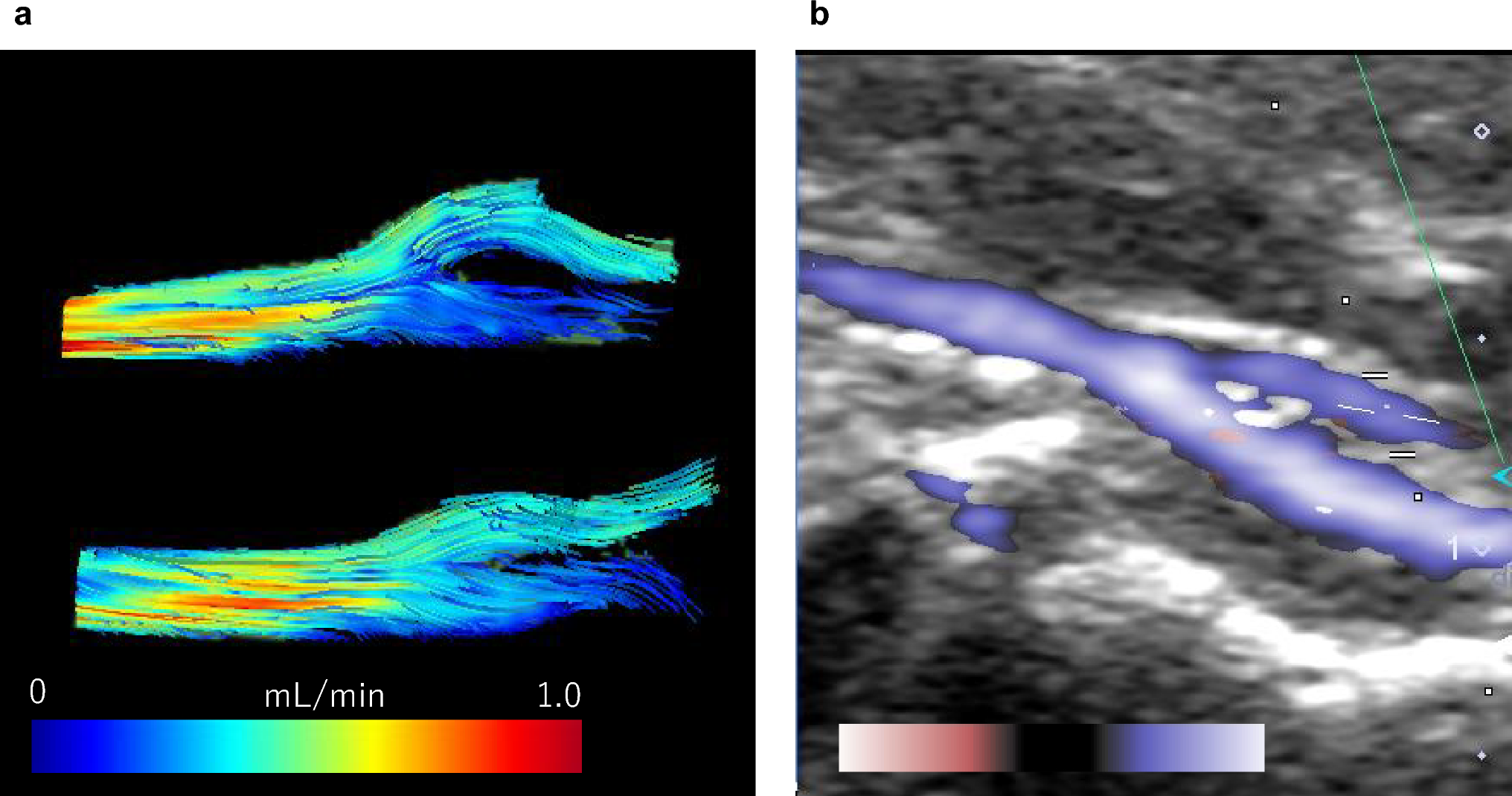
Yasuda S, et al. In-vivo assessment of vascular endothelial injury in a rat model of unilateral carotid artery injury using 4D-flow MRI. Sci Rep. 2025;15:18571. https://doi.org/10.1038/s41598-025-03721-1.
Article
PubMed
PubMed Central
CAS
Google Scholar
Harloff A, et al. 3D blood flow characteristics in the carotid artery bifurcation assessed by flow-sensitive 4D MRI at 3T. Magn Reson Med. 2009;61:65–74. https://doi.org/10.1002/mrm.21774.
Article
PubMed
CAS
Google Scholar
Seitz J, et al. Quantification of blood flow in the carotid arteries: comparison of Doppler ultrasound and three different phase-contrast magnetic resonance imaging sequences. Invest Radiol. 2001;36:642–7. https://doi.org/10.1097/00004424-200111000-00003.
Article
PubMed
CAS
Google Scholar
Oktar SO, et al. Blood-flow volume quantification in internal carotid and vertebral arteries: comparison of 3 different ultrasound techniques with phase-contrast MR imaging. AJNR Am J Neuroradiol. 2006;27:363–9.
PubMed
PubMed Central
CAS
Google Scholar
Karwatowski SP, et al. Mitral valve flow measured with cine MR velocity mapping in patients with ischemic heart disease: comparison with Doppler echocardiography. J Magn Reson Imaging. 1995;5:89–92. https://doi.org/10.1002/jmri.1880050116.
Article
PubMed
CAS
Google Scholar
Saito S, Takahashi Y, Ohki A, Shintani Y, Higuchi T. Early detection of elevated lactate levels in a mitochondrial disease model using chemical exchange saturation transfer (CEST) and magnetic resonance spectroscopy (MRS) at 7T-MRI. Radiol Phys Technol. 2019;12:46–54. https://doi.org/10.1007/s12194-018-0490-1.
Article
PubMed
Google Scholar
Saito S, Ueda J. Preclinical magnetic resonance imaging and spectroscopy in the fields of radiological technology, medical physics, and radiology. Radiol Phys Technol. 2024;17:47–59. https://doi.org/10.1007/s12194-024-00785-y.
Article
PubMed
PubMed Central
Google Scholar
Likittanasombut P, Reynolds P, Meads D, Tegeler C. Volume flow rate of common carotid artery measured by Doppler method and color velocity imaging quantification (CVI-Q). J Neuroimaging. 2006;16:34–8. https://doi.org/10.1177/1051228405001523.
Article
PubMed
Google Scholar
Lotz J, Meier C, Leppert A, Galanski M. Cardiovascular flow measurement with phase-contrast MR imaging: basic facts and implementation. Radiographics. 2002;22:651–71. https://doi.org/10.1148/radiographics.22.3.g02ma11651.
Article
PubMed
Google Scholar
Winkler AJ, Wu J. Correction of intrinsic spectral broadening errors in Doppler peak velocity measurements made with phased sector and linear array transducers. Ultrasound Med Biol. 1995;21:1029–35. https://doi.org/10.1016/0301-5629(95)00047-u.
Article
PubMed
CAS
Google Scholar
Hawkes RA, et al. Uterine artery pulsatility and resistivity indices in pregnancy: comparison of MRI and Doppler US. Placenta. 2016;43:35–40. https://doi.org/10.1016/j.placenta.2016.04.002.
Article
PubMed
CAS
Google Scholar
Brahim JS, Thut PD. Hemodynamic changes during isoflurane anesthesia. Anesth Prog. 1984;31:207–12.
PubMed
PubMed Central
CAS
Google Scholar
Atkinson P, Wells PN. Pulse-doppler ultrasound and its clinical application. Yale J Biol Med. 1977;50:367–73.
PubMed
PubMed Central
CAS
Google Scholar
Yazici B, Erdogmus B, Tugay A. Cerebral blood flow measurements of the extracranial carotid and vertebral arteries with doppler ultrasonography in healthy adults. Diagn Interv Radiol. 2005;11:195–8.
PubMed
Google Scholar
Vdoviaková K, Askin SJ, Krešáková L, Vrabec V, Vrzgula M, Danková M. The head and neck vascular anatomical variability in the laboratory rat and its significance to medical science. Folia Vet. 2022;66:9–18.
Article
Google Scholar

Comments (0)