Optimal elevation distance of the X-ray focus in vertical dual-exposure panoramic radiography for mitigating ghost images of cervical vertebrae in the upper incisor region
Objectives
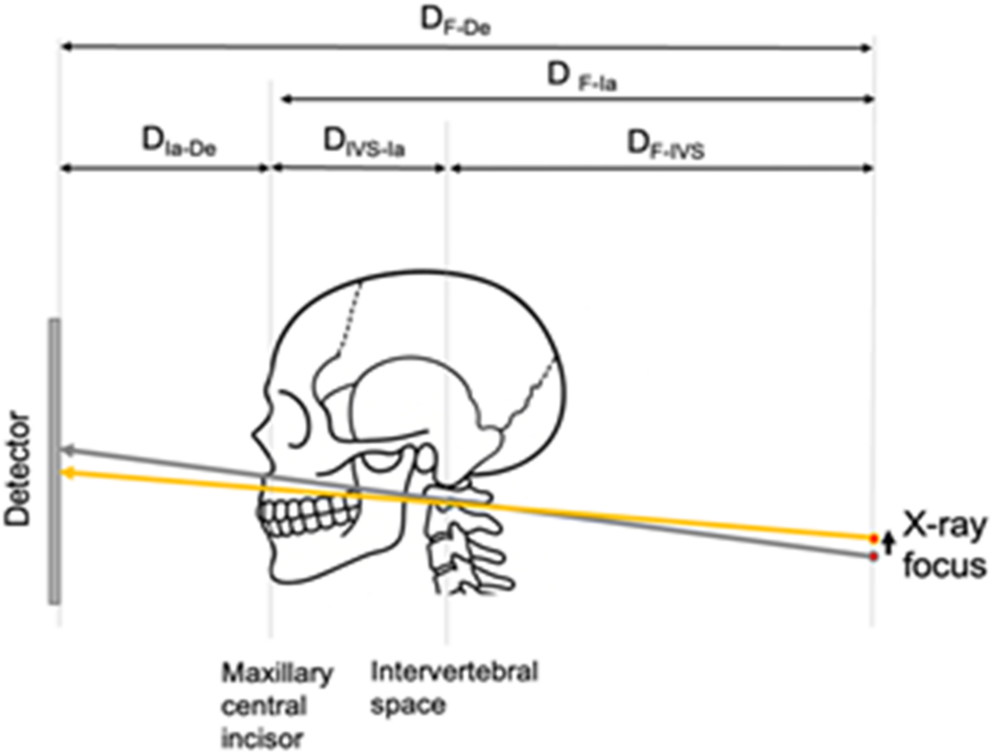
Panoramic radiography (PR) often produces ghost images from cervical vertebrae (CVe) in the upper incisor region. Vertical dual-exposure panoramic radiography (VDPR) reduces these artifacts by merging two scans at different X-ray focus heights, but excessive elevation introduces incisor distortion. This study aimed to determine the optimal elevation distance that minimizes ghost images while limiting distortion.
Methods
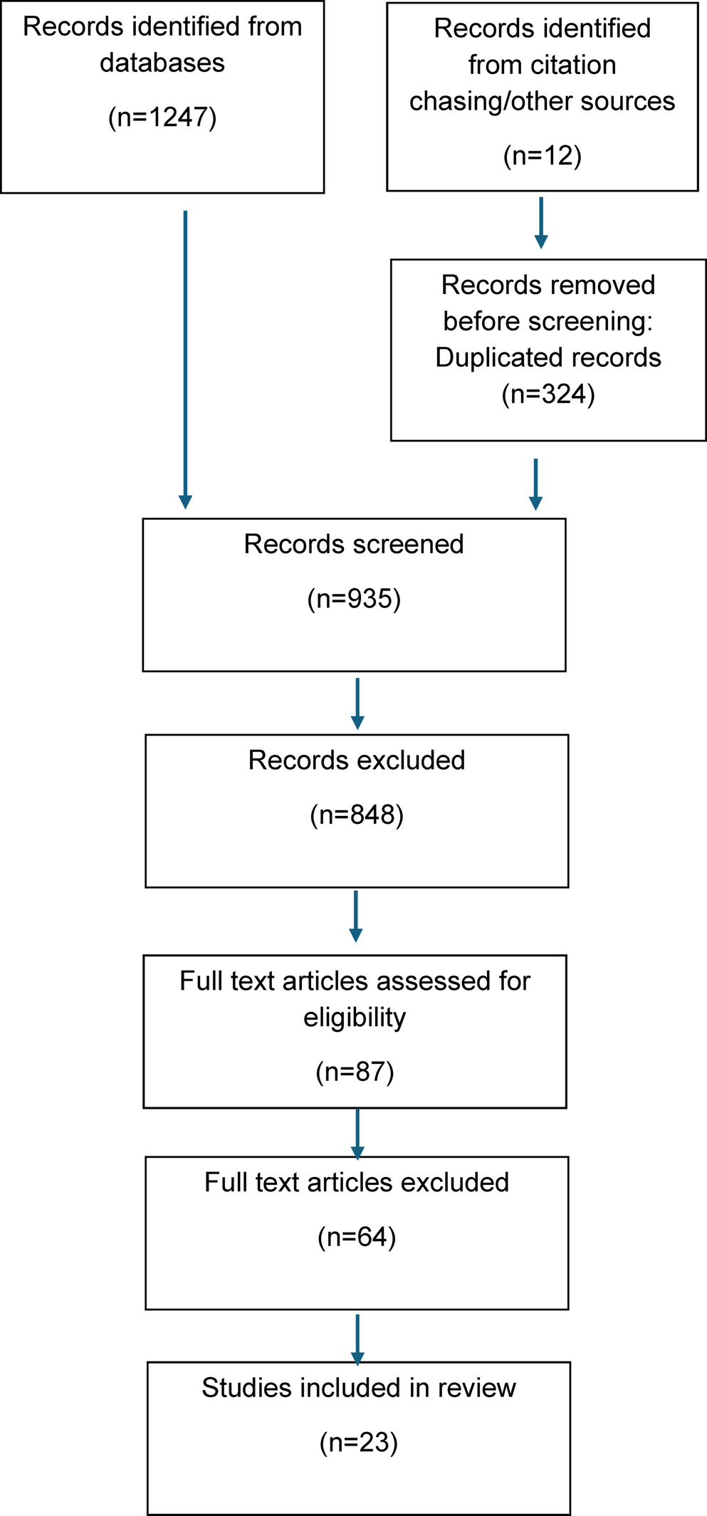
Three experiments were performed. (1) A theoretical model calculated the minimum elevation required to displace CV ghost images beyond their vertical width using cone-beam computed tomography (CBCT)-derived measurements of a cranial phantom. (2) Ghost images of CVe were simulated with CBCT data, with merged-CV images created as if the X-ray focus had been raised from 0 to 20 mm in 5-mm increments. Ghost image intensity was quantified by spatial bootstrapping of pixel variability. (3) Phantom experiments were performed with identical increments, and merged VDPR images were analyzed. Image quality was assessed using the standard deviation of brightness within defined regions of interest.
Results
(1) Theoretical analysis indicated an optimal elevation of 10 mm. (2) Simulation results confirmed that merged-CV image combining 0 mm and 10 mm produced the greatest reduction in ghost image intensity compared with other condition. (3) Phantom experiments demonstrated significant reduction at 5, 10, and 15 mm, with 10 mm providing the most consistent improvement. Elevations ≥ 15 mm introduced incisor distortion.
Conclusions
VDPR with an X-ray focus elevation of 10 mm effectively mitigates CV ghost images in the upper incisor region while minimizing distortion, representing the optimal setting for clinical application.

Comments (0)